Downloaded 316 times


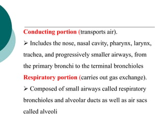


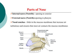


The respiratory system allows for oxygen intake and carbon dioxide removal. It consists of the nose, pharynx, larynx, trachea, bronchi, lungs and muscles. The nose and mouth allow air intake and filter it. The pharynx and larynx direct air to the proper passages. The trachea and bronchi form the conducting airways to the lungs. In the lungs, gas exchange occurs between the air in alveoli and blood in capillaries, transferring oxygen to blood and carbon dioxide out of blood.