The respiratory system allows for oxygen to enter the body and carbon dioxide to exit through a series of major organs. Air enters through the nose or mouth and passes through the pharynx, larynx, trachea, bronchi and into the lungs where gas exchange occurs in the alveoli. Oxygen then passes into the bloodstream and carbon dioxide passes out of the bloodstream and is exhaled. Breathing is facilitated by the contraction and relaxation of the diaphragm and rib cage which expands and contracts the chest cavity to inhale and exhale air.
Human digestive system structure and function
overview
Major organs
Mouth
Esophagus
Stomach
small intestine
large intestine
Acessory organs:
Liver
gall bladder
Pancreas.
Human digestive system
Major organs
Mouth
Esophagus
Stomach
small intestine
large intestine.
Acessory organs:
Liver
Gall bladder
Pancreas.
MAJOR ORGANS The Mouth
pH: 7
The first part of the digestive system
the entry point of food.
Structures in the mouth that aids digestion
Teeth – cut, tear, crush and grind food.
Salivary glands – produce and secrete saliva into the oral cavity.
saliva
moistens the food
contains enzymes (ptyalin or salivary amylase)
begins digestion of starch into smaller polysaccharides.
Function:
Mechanical digestion.
increasing surface area for faster chemical digestion.
The Esophagus
a tube connecting the mouth to the stomach
running through the Thoracic cavity.
Location:
lies behind windpipe (Trachea).
The trachea has as an epiglottis
preventing food from entering the windpipe,
moving the food to the esophagus while swallowing.
Food travels down the esophagus, through a series of involuntary rhythmic contractions (wave-like) called peristalsis.
Function:
The lining of the esophagus secretes mucus
lubricating
to support the movement of food.
Esophageal sphincter:
bolus reaches the stomach
must pass through a muscular ringed valve called the esophageal sphincter (Cardiac Sphincter).
Function:
prevent stomach acids from back flowing into the esophagus.
Stomach
J-shaped muscular sac
Has inner folds (rugae)
Increasing surface area of the stomach.
Function:
Stomach performs mechanical digestion
HOW By churning the bolus and mixing it with the gastric juices
secreted by the lining of the stomach.
GASTRIC JUICES HCl, salts, enzymes, water and mucus)
HCL helps break down of food and kills bacteria that came along with the food.
The bolus is now called Chyme.
Enzymes in stomach:
Acidic environment
HCl secreation
kill any microbes that are found in the bolus,
creating a pH of 2.
Mucus prevents the stomach from digesting itself.
Pepsin secreation
responsible for initiating the breakdown of proteins (in )food.
hydrolyzes proteins to yield polypeptides.
pH is 2, the enzyme from the salivary glands stops breaking down carbohydrates.
Pyloric sphincter:
chyme moves from the stomach to the small intestine.
It passes through a muscular ringed sphincter called the pyloric sphincter.
stomach does not digest itself Why ?
Protective Mechanism:
three protective mechanisms.
First the stomach only secretes small amounts of gastric juices until food is present.
Second the secretion of mucus coats the lining of the stomach protecting it from the gastric juices.
The third mechanism is the digestive enzyme pepsin is secreted in an inactive protein c
This is about the general physiology of sense organs for medical and paramedical professional beginners who choose pharmacy, nursing and physiotherapy to study.
The urinary system, components, the urine formation process, The gross structure of the kidney, Microscope structure of the kidney, Renin-Angiotensin Aldosterone System
he sense organs — eyes, ears, tongue, skin, and nose — help to protect the body. The human sense organs contain receptors that relay information through sensory neurons to the appropriate places within the nervous system.
Each sense organ contains different receptors.
General receptors are found throughout the body because they are present in skin, visceral organs (visceral meaning in the abdominal cavity), muscles, and joints.
Special receptors include chemoreceptors (chemical receptors) found in the mouth and nose, photoreceptors (light receptors) found in the eyes, and mechanoreceptors found in the ears.
The respiratory system is the network of organs and tissues that help you breathe. It includes your airways, lungs, and blood vessels. The muscles that power your lungs are also part of the respiratory system. These parts work together to move oxygen throughout the body and clean out waste gases like carbon dioxide.
The content in the slide are solely depended upon the syllabus of Purbanchal University for third-semester students. This content of the respiratory system will be enough for B.Pharmacy students studying anatomy and physiology
TEST BANK for Operations Management, 14th Edition by William J. Stevenson, Ve...kevinkariuki227
TEST BANK for Operations Management, 14th Edition by William J. Stevenson, Verified Chapters 1 - 19, Complete Newest Version.pdf
TEST BANK for Operations Management, 14th Edition by William J. Stevenson, Verified Chapters 1 - 19, Complete Newest Version.pdf
Explore natural remedies for syphilis treatment in Singapore. Discover alternative therapies, herbal remedies, and lifestyle changes that may complement conventional treatments. Learn about holistic approaches to managing syphilis symptoms and supporting overall health.
Title: Sense of Taste
Presenter: Dr. Faiza, Assistant Professor of Physiology
Qualifications:
MBBS (Best Graduate, AIMC Lahore)
FCPS Physiology
ICMT, CHPE, DHPE (STMU)
MPH (GC University, Faisalabad)
MBA (Virtual University of Pakistan)
Learning Objectives:
Describe the structure and function of taste buds.
Describe the relationship between the taste threshold and taste index of common substances.
Explain the chemical basis and signal transduction of taste perception for each type of primary taste sensation.
Recognize different abnormalities of taste perception and their causes.
Key Topics:
Significance of Taste Sensation:
Differentiation between pleasant and harmful food
Influence on behavior
Selection of food based on metabolic needs
Receptors of Taste:
Taste buds on the tongue
Influence of sense of smell, texture of food, and pain stimulation (e.g., by pepper)
Primary and Secondary Taste Sensations:
Primary taste sensations: Sweet, Sour, Salty, Bitter, Umami
Chemical basis and signal transduction mechanisms for each taste
Taste Threshold and Index:
Taste threshold values for Sweet (sucrose), Salty (NaCl), Sour (HCl), and Bitter (Quinine)
Taste index relationship: Inversely proportional to taste threshold
Taste Blindness:
Inability to taste certain substances, particularly thiourea compounds
Example: Phenylthiocarbamide
Structure and Function of Taste Buds:
Composition: Epithelial cells, Sustentacular/Supporting cells, Taste cells, Basal cells
Features: Taste pores, Taste hairs/microvilli, and Taste nerve fibers
Location of Taste Buds:
Found in papillae of the tongue (Fungiform, Circumvallate, Foliate)
Also present on the palate, tonsillar pillars, epiglottis, and proximal esophagus
Mechanism of Taste Stimulation:
Interaction of taste substances with receptors on microvilli
Signal transduction pathways for Umami, Sweet, Bitter, Sour, and Salty tastes
Taste Sensitivity and Adaptation:
Decrease in sensitivity with age
Rapid adaptation of taste sensation
Role of Saliva in Taste:
Dissolution of tastants to reach receptors
Washing away the stimulus
Taste Preferences and Aversions:
Mechanisms behind taste preference and aversion
Influence of receptors and neural pathways
Impact of Sensory Nerve Damage:
Degeneration of taste buds if the sensory nerve fiber is cut
Abnormalities of Taste Detection:
Conditions: Ageusia, Hypogeusia, Dysgeusia (parageusia)
Causes: Nerve damage, neurological disorders, infections, poor oral hygiene, adverse drug effects, deficiencies, aging, tobacco use, altered neurotransmitter levels
Neurotransmitters and Taste Threshold:
Effects of serotonin (5-HT) and norepinephrine (NE) on taste sensitivity
Supertasters:
25% of the population with heightened sensitivity to taste, especially bitterness
Increased number of fungiform papillae
These simplified slides by Dr. Sidra Arshad present an overview of the non-respiratory functions of the respiratory tract.
Learning objectives:
1. Enlist the non-respiratory functions of the respiratory tract
2. Briefly explain how these functions are carried out
3. Discuss the significance of dead space
4. Differentiate between minute ventilation and alveolar ventilation
5. Describe the cough and sneeze reflexes
Study Resources:
1. Chapter 39, Guyton and Hall Textbook of Medical Physiology, 14th edition
2. Chapter 34, Ganong’s Review of Medical Physiology, 26th edition
3. Chapter 17, Human Physiology by Lauralee Sherwood, 9th edition
4. Non-respiratory functions of the lungs https://academic.oup.com/bjaed/article/13/3/98/278874
ARTIFICIAL INTELLIGENCE IN HEALTHCARE.pdfAnujkumaranit
Artificial intelligence (AI) refers to the simulation of human intelligence processes by machines, especially computer systems. It encompasses tasks such as learning, reasoning, problem-solving, perception, and language understanding. AI technologies are revolutionizing various fields, from healthcare to finance, by enabling machines to perform tasks that typically require human intelligence.
Pulmonary Thromboembolism - etilogy, types, medical- Surgical and nursing man...VarunMahajani
Disruption of blood supply to lung alveoli due to blockage of one or more pulmonary blood vessels is called as Pulmonary thromboembolism. In this presentation we will discuss its causes, types and its management in depth.
Couples presenting to the infertility clinic- Do they really have infertility...Sujoy Dasgupta
Dr Sujoy Dasgupta presented the study on "Couples presenting to the infertility clinic- Do they really have infertility? – The unexplored stories of non-consummation" in the 13th Congress of the Asia Pacific Initiative on Reproduction (ASPIRE 2024) at Manila on 24 May, 2024.
Title: Sense of Smell
Presenter: Dr. Faiza, Assistant Professor of Physiology
Qualifications:
MBBS (Best Graduate, AIMC Lahore)
FCPS Physiology
ICMT, CHPE, DHPE (STMU)
MPH (GC University, Faisalabad)
MBA (Virtual University of Pakistan)
Learning Objectives:
Describe the primary categories of smells and the concept of odor blindness.
Explain the structure and location of the olfactory membrane and mucosa, including the types and roles of cells involved in olfaction.
Describe the pathway and mechanisms of olfactory signal transmission from the olfactory receptors to the brain.
Illustrate the biochemical cascade triggered by odorant binding to olfactory receptors, including the role of G-proteins and second messengers in generating an action potential.
Identify different types of olfactory disorders such as anosmia, hyposmia, hyperosmia, and dysosmia, including their potential causes.
Key Topics:
Olfactory Genes:
3% of the human genome accounts for olfactory genes.
400 genes for odorant receptors.
Olfactory Membrane:
Located in the superior part of the nasal cavity.
Medially: Folds downward along the superior septum.
Laterally: Folds over the superior turbinate and upper surface of the middle turbinate.
Total surface area: 5-10 square centimeters.
Olfactory Mucosa:
Olfactory Cells: Bipolar nerve cells derived from the CNS (100 million), with 4-25 olfactory cilia per cell.
Sustentacular Cells: Produce mucus and maintain ionic and molecular environment.
Basal Cells: Replace worn-out olfactory cells with an average lifespan of 1-2 months.
Bowman’s Gland: Secretes mucus.
Stimulation of Olfactory Cells:
Odorant dissolves in mucus and attaches to receptors on olfactory cilia.
Involves a cascade effect through G-proteins and second messengers, leading to depolarization and action potential generation in the olfactory nerve.
Quality of a Good Odorant:
Small (3-20 Carbon atoms), volatile, water-soluble, and lipid-soluble.
Facilitated by odorant-binding proteins in mucus.
Membrane Potential and Action Potential:
Resting membrane potential: -55mV.
Action potential frequency in the olfactory nerve increases with odorant strength.
Adaptation Towards the Sense of Smell:
Rapid adaptation within the first second, with further slow adaptation.
Psychological adaptation greater than receptor adaptation, involving feedback inhibition from the central nervous system.
Primary Sensations of Smell:
Camphoraceous, Musky, Floral, Pepperminty, Ethereal, Pungent, Putrid.
Odor Detection Threshold:
Examples: Hydrogen sulfide (0.0005 ppm), Methyl-mercaptan (0.002 ppm).
Some toxic substances are odorless at lethal concentrations.
Characteristics of Smell:
Odor blindness for single substances due to lack of appropriate receptor protein.
Behavioral and emotional influences of smell.
Transmission of Olfactory Signals:
From olfactory cells to glomeruli in the olfactory bulb, involving lateral inhibition.
Primitive, less old, and new olfactory systems with different path
Ethanol (CH3CH2OH), or beverage alcohol, is a two-carbon alcohol
that is rapidly distributed in the body and brain. Ethanol alters many
neurochemical systems and has rewarding and addictive properties. It
is the oldest recreational drug and likely contributes to more morbidity,
mortality, and public health costs than all illicit drugs combined. The
5th edition of the Diagnostic and Statistical Manual of Mental Disorders
(DSM-5) integrates alcohol abuse and alcohol dependence into a single
disorder called alcohol use disorder (AUD), with mild, moderate,
and severe subclassifications (American Psychiatric Association, 2013).
In the DSM-5, all types of substance abuse and dependence have been
combined into a single substance use disorder (SUD) on a continuum
from mild to severe. A diagnosis of AUD requires that at least two of
the 11 DSM-5 behaviors be present within a 12-month period (mild
AUD: 2–3 criteria; moderate AUD: 4–5 criteria; severe AUD: 6–11 criteria).
The four main behavioral effects of AUD are impaired control over
drinking, negative social consequences, risky use, and altered physiological
effects (tolerance, withdrawal). This chapter presents an overview
of the prevalence and harmful consequences of AUD in the U.S.,
the systemic nature of the disease, neurocircuitry and stages of AUD,
comorbidities, fetal alcohol spectrum disorders, genetic risk factors, and
pharmacotherapies for AUD.
Prix Galien International 2024 Forum ProgramLevi Shapiro
June 20, 2024, Prix Galien International and Jerusalem Ethics Forum in ROME. Detailed agenda including panels:
- ADVANCES IN CARDIOLOGY: A NEW PARADIGM IS COMING
- WOMEN’S HEALTH: FERTILITY PRESERVATION
- WHAT’S NEW IN THE TREATMENT OF INFECTIOUS,
ONCOLOGICAL AND INFLAMMATORY SKIN DISEASES?
- ARTIFICIAL INTELLIGENCE AND ETHICS
- GENE THERAPY
- BEYOND BORDERS: GLOBAL INITIATIVES FOR DEMOCRATIZING LIFE SCIENCE TECHNOLOGIES AND PROMOTING ACCESS TO HEALTHCARE
- ETHICAL CHALLENGES IN LIFE SCIENCES
- Prix Galien International Awards Ceremony
2. Respiratory System: Oxygen Delivery System
The respiratory system is the set of organs that allows a person to
breathe and exchange oxygen and carbon dioxide throughout the
body.
The integrated system of organs involved in the intake and
exchange of oxygen and carbon dioxide between the body and
the environment and including the nasal passages, larynx,
trachea, bronchial tubes, and lungs.
The respiratory system performs two major tasks:
Exchanging air between the body and the outside
environment known as external respiration.
Bringing oxygen to the cells and removing carbon dioxide
from them referred to as internal respiration.
4. 1. Supplies the body with oxygen and disposes of carbon dioxide
2. Filters inspired air
3. Produces sound
4. Contains receptors for smell
5. Rids the body of some excess water and heat
6. Helps regulate blood pH
Breathing
Breathing (pulmonary ventilation).
consists of two cyclic phases:
Inhalation, also called
inspiration - draws gases into
the lungs.
Exhalation, also called
expiration - forces gases out of
the lungs.
5. Air from the outside environment enters the nose or mouth
during inspiration (inhalation).
Composed of the nose and nasal cavity, paranasal sinuses,
pharynx (throat), larynx.
All part of the conducting portion of the respiratory system.
Nostril
Mouth
Nasal Cavity
Throat
(pharynx)
Voice box(Larynx)
6. Nose Also called external nares.
Divided into two halves by the nasal septum.
Contains the paranasal sinuses where air is warmed.
Contains cilia which is responsible for filtering out foreign
bodies.
Nose and Nasal Cavities
Nasal concha Sphenoid sinus
Internal naris
Nasopharynx
External naris
Frontal sinus
Middle nasal concha
Inferior nasal
concha
7. Internal nares - opening to exterior
External nares - opening to pharynx
Nasal conchae - folds in the mucous membrane that
increase air turbulence and ensures that most air contacts
the mucous membranes
Provides and airway for respiration
Moistens and warms entering air
Filters and cleans inspired air
Resonating chamber for speech
- detects odors in the air stream
8. Pharynx Common space used by both the respiratory and digestive
systems.
Commonly called the throat.
Originates posterior to the nasal and oral cavities and extends
inferiorly near the level of the bifurcation of the larynx and
esophagus.
Common pathway for both air and food.
Walls are lined by a mucosa and contain skeletal muscles that
are primarily used for swallowing.
Flexible lateral walls are distensible in order to force swallowed
food into the esophagus.
9. Three Sections of the Pharynx
Nasopharynx
contains the pharyngeal tonsils (adenoids) which aid in the
body’s immune defense.
Oropharynx
back portion of the mouth that contains the palatine tonsils
which aid in the body’s immune defense.
Laryngopharynx
bottom section of the pharynx where the respiratory tract
divides into the esophagus and the larynx.
Nasopharynx
Oropharynx
Laryngopharynx
10. Larynx
Voice box is a short,
somewhat cylindrical
airway ends in the trachea.
Prevents swallowed
materials from entering the
lower respiratory tract.
Conducts air into the lower
respiratory tract.
Produces sounds.
Supported by a framework
of nine pieces of cartilage
(three individual pieces and
three cartilage pairs) that
are held in place by
ligaments and muscles.
Hyoid Bone Epiglottis
Thyrohyoid
Membrane
Cricothyroid
Ligament
Cricothyroid
Muscles
Cricothyroid
Cartilage
Trachea
Thyroid
Cartilage
11. Trachea
A flexible tube also called windpipe.
Extends through the mediastinum
and lies anterior to the esophagus
and inferior to the larynx.
Cartilage rings reinforce and
provide rigidity to the tracheal wall
to ensure that the trachea remains
open at all times.
At the level of the sternal angle, the
trachea bifurcates into two smaller
tubes, called the right and left
primary bronchi.
Each primary bronchus projects
laterally toward each lung.
Trachea
Bronchi
Larynx
12. Lungs
Each lung has a conical shape. Its wide, concave base rests
upon the muscular diaphragm.
Its superior region called the apex projects superiorly to a
point that is slightly superior and posterior to the clavicle.
Both lungs are bordered by the thoracic wall anteriorly,
laterally, and posteriorly, and supported by the rib cage.
Toward the midline, the lungs are separated from each
other by the mediastinum.
The relatively broad, rounded surface in contact with the
thoracic wall is called the costal surface of the lung.
13. Left lung
– divided into 2 lobes by oblique fissure
– smaller than the right lung
– cardiac notch accommodates the heart
Right lung
– divided into 3 lobes by oblique and horizontal fissure
– located more superiorly in the body due to liver on right side
Lungs
14. Pleura
The outer surface of each lung and the adjacent internal
thoracic wall are lined by a serous membrane called pleura.
The outer surface of each lung is tightly covered by the
visceral pleura.
while the internal thoracic walls, the lateral surfaces of the
mediastinum, and the superior surface of the diaphragm are
lined by the parietal pleura.
The parietal and visceral pleural layers are continuous at the
hilus of each lung
Pleural Cavities
The potential space between the serous membrane layers is a
pleural cavity.
The pleural membranes produce a thin, serous pleural fluid that
circulates in the pleural cavity and acts as a lubricant, ensuring
minimal friction during breathing.
Pleural effusion – pleuritis with too much fluid
15.
16. Air enters your lungs through a system of pipes called the bronchi.
The alveoli are where the important work of gas exchange takes place
between the air and your blood. Covering each alveolus is a whole
network of little blood vessel called capillaries,
It is important that the air in the alveoli and the blood in the capillaries
are very close together, so that oxygen and carbon dioxide can move (or
diffuse) between them.
When you breathe in, air comes down the trachea and through the
bronchi into the alveoli.
This fresh air has lots of oxygen in it, and some of this oxygen will travel
across the walls of the alveoli into your blood stream.
Travelling in the opposite direction is carbon dioxide, which crosses from
the blood in the capillaries into the air in the alveoli and is then breathed
out.
In this way, you bring in to your body the oxygen that you need to live,
and get rid of the waste product carbon dioxide.
17. How Lungs work?
Branch of
Pulmonary vein
Branch of
Pulmonary artery
Bronchiole
Terminal
Bronchiole
Respiratory Bronchiole
Capillary beds
Alveoli
18. Breathing
Lungs are sealed in
pleural membranes
inside the chest cavity.
At the bottom of the
cavity is a large, flat
muscle known as the
diaphragm.
19. Breathing
During inhalation, the
diaphragm contracts and the
rib cage rises up.
This expands the volume of
the chest cavity.
The chest cavity is sealed, so
this creates a partial vacuum
inside the cavity.
Atmospheric pressure fills
the lungs as air rushes into
the breathing passages.
20. Breathing
Often exhaling is a passive
event.
When the rib cage lowers and
the diaphragm relaxes,
pressure in the chest cavity is
greater than atmospheric
pressure.
Air is pushed out of the lungs.
Exhalation
Rib cage
lowers
Air Exhaled
21. The End
Call us for more
information
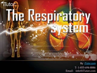
www.iTutor.com
1-855-694-8886
Visit