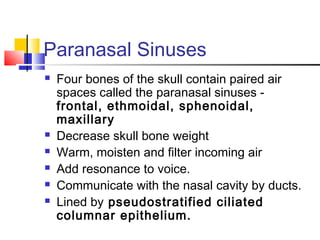
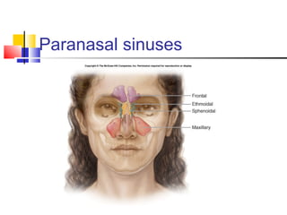
Download to read offline


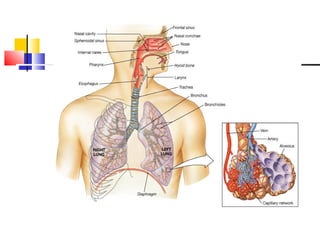
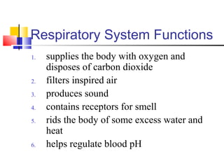
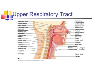
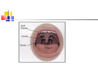
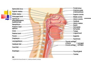

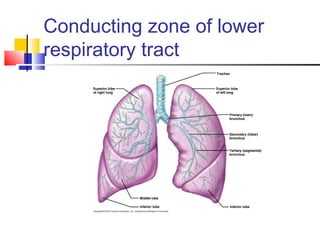

The respiratory system consists of an upper respiratory tract and lower respiratory tract. The upper tract includes the nose, nasal cavity, pharynx and larynx. The lower tract includes the trachea, bronchi and lungs. The conducting portion transports air through the nose, pharynx and into the lungs. The respiratory portion, including alveoli in the lungs, performs gas exchange between the air and blood. Breathing involves inhalation that draws air into the lungs and exhalation that forces air out. The document describes the anatomy and functions of the different parts of the respiratory system.