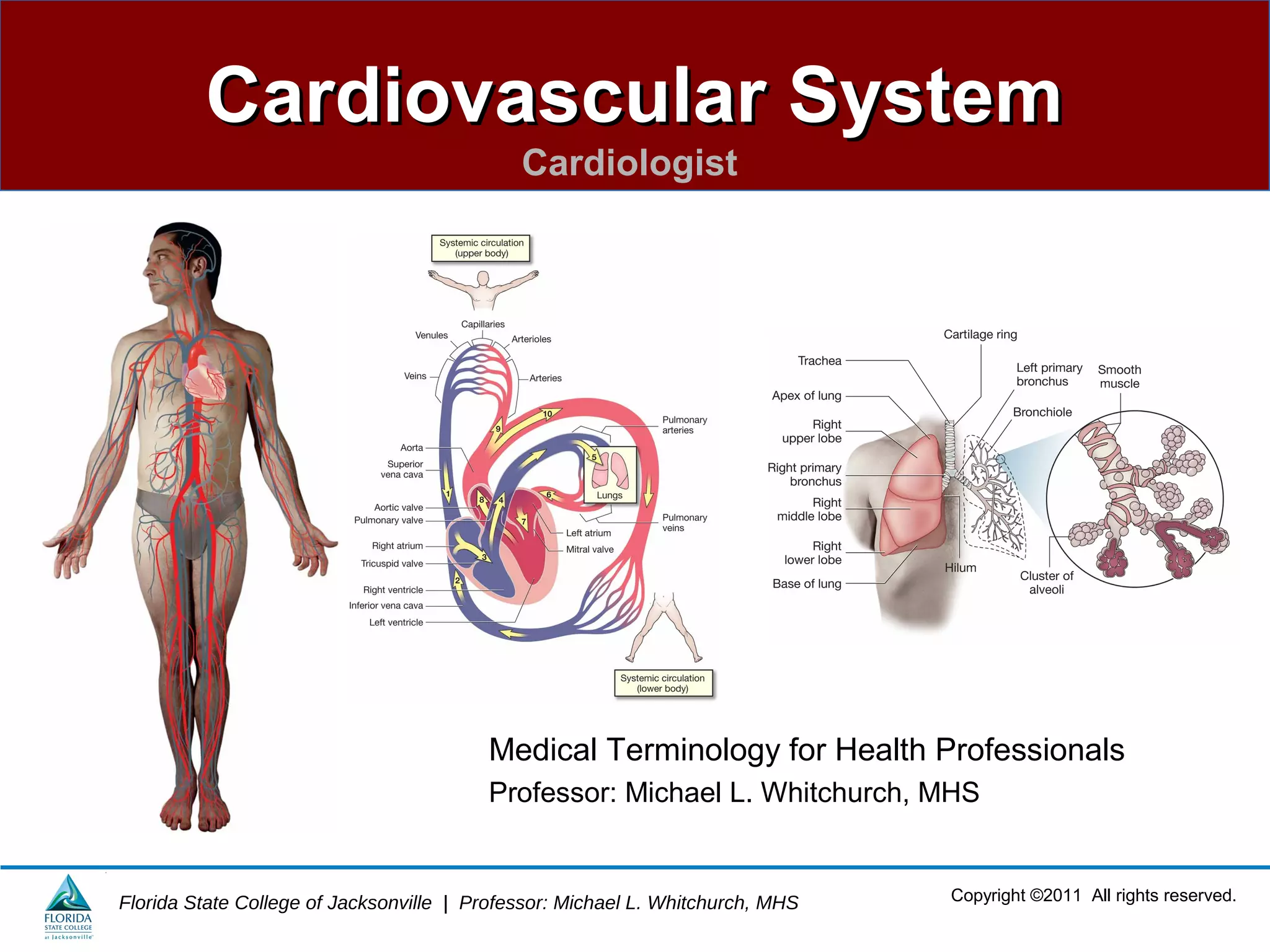
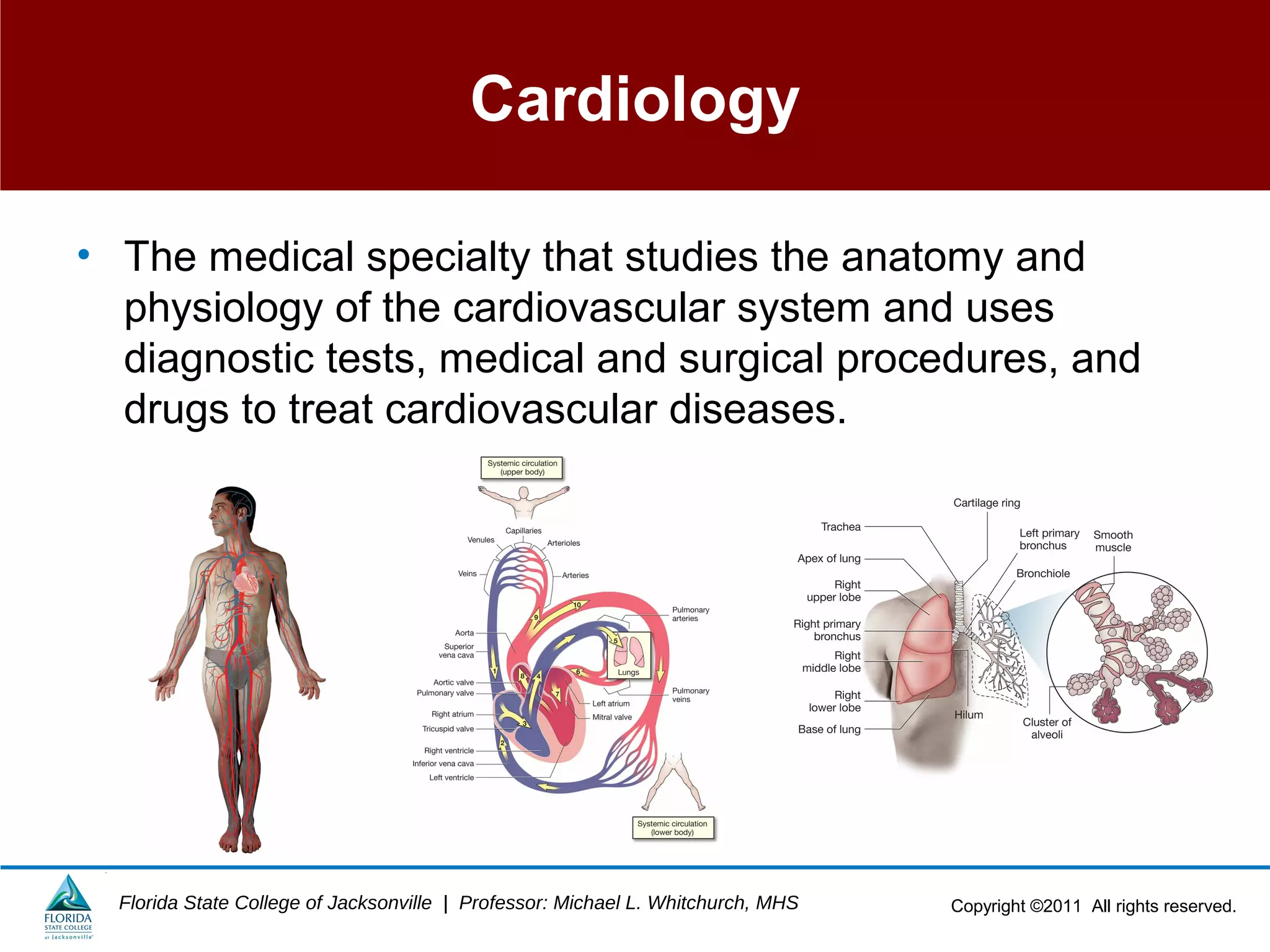
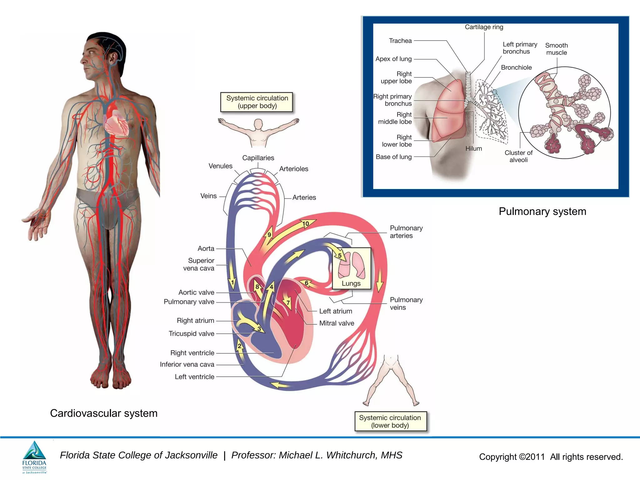
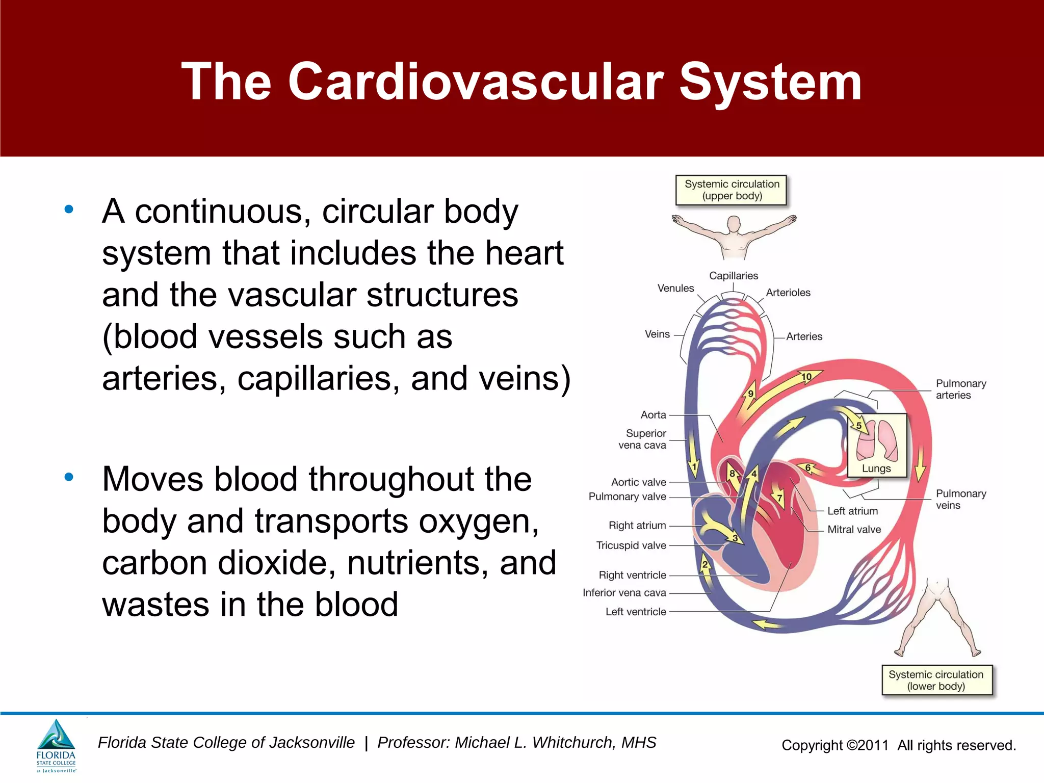
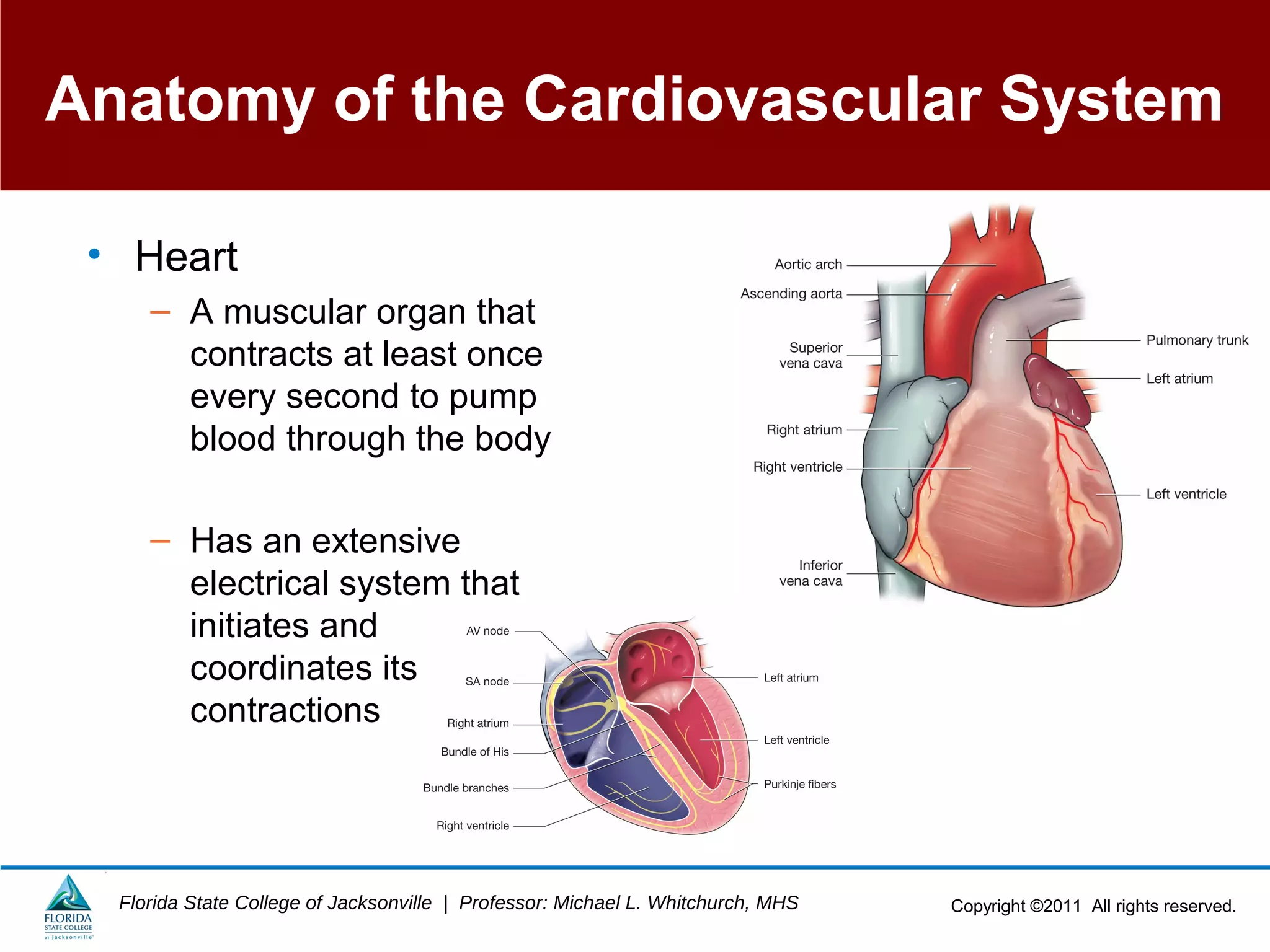
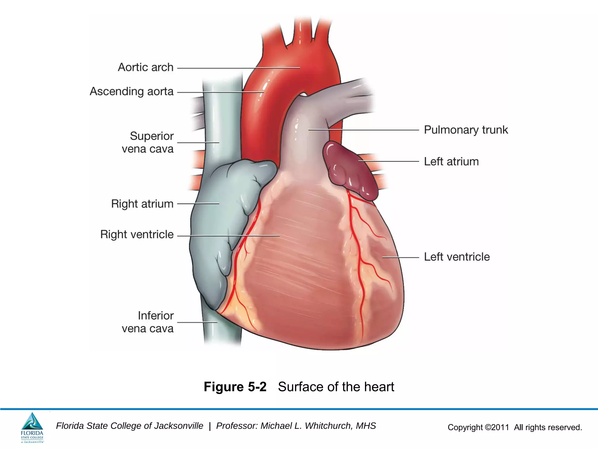
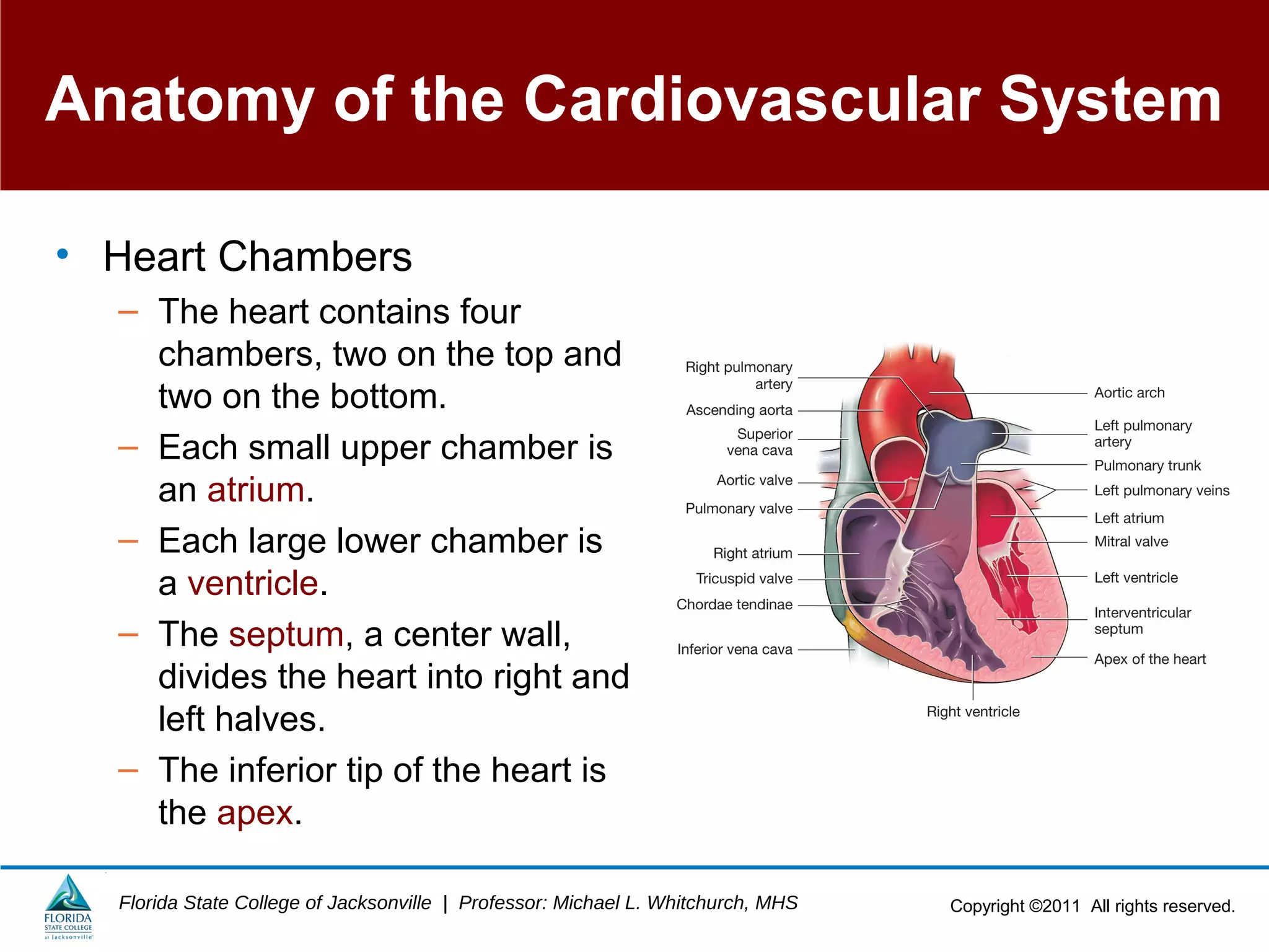
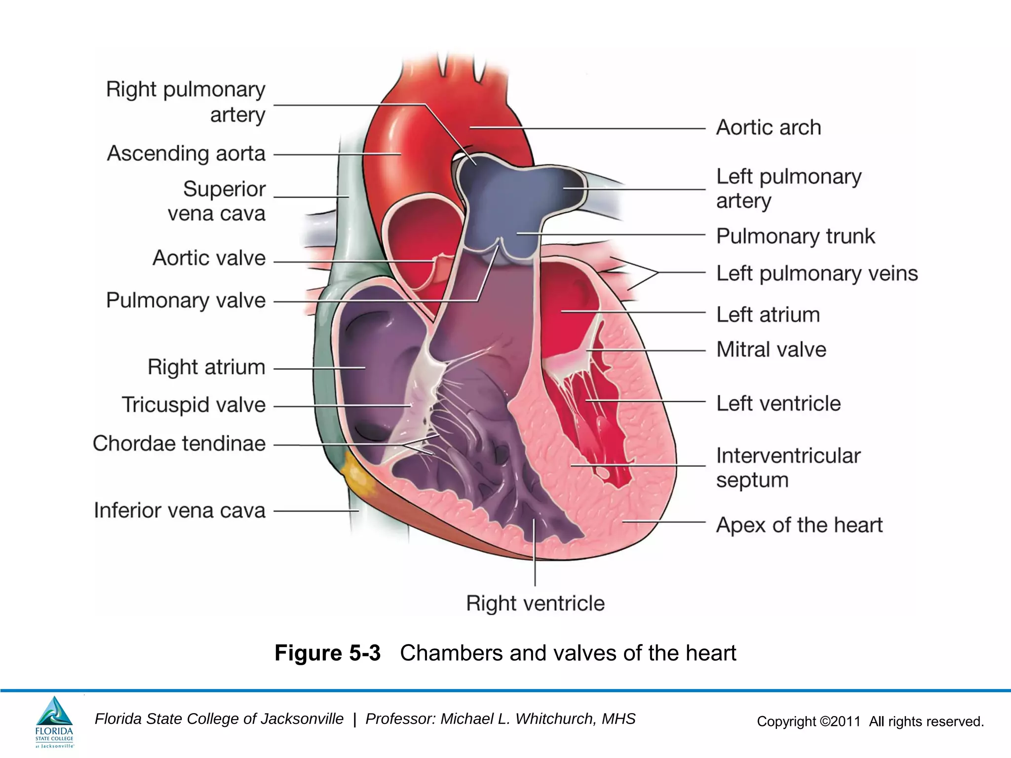
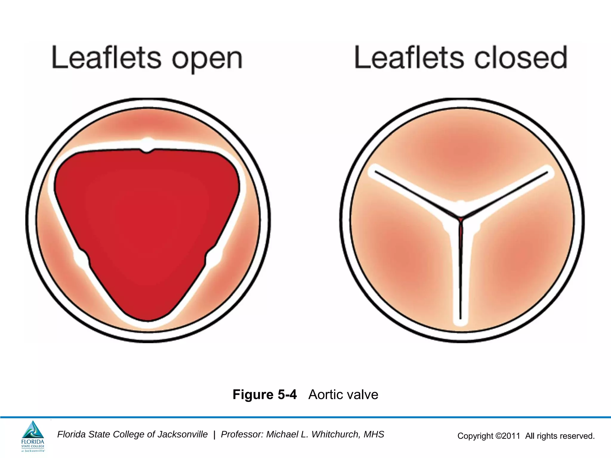
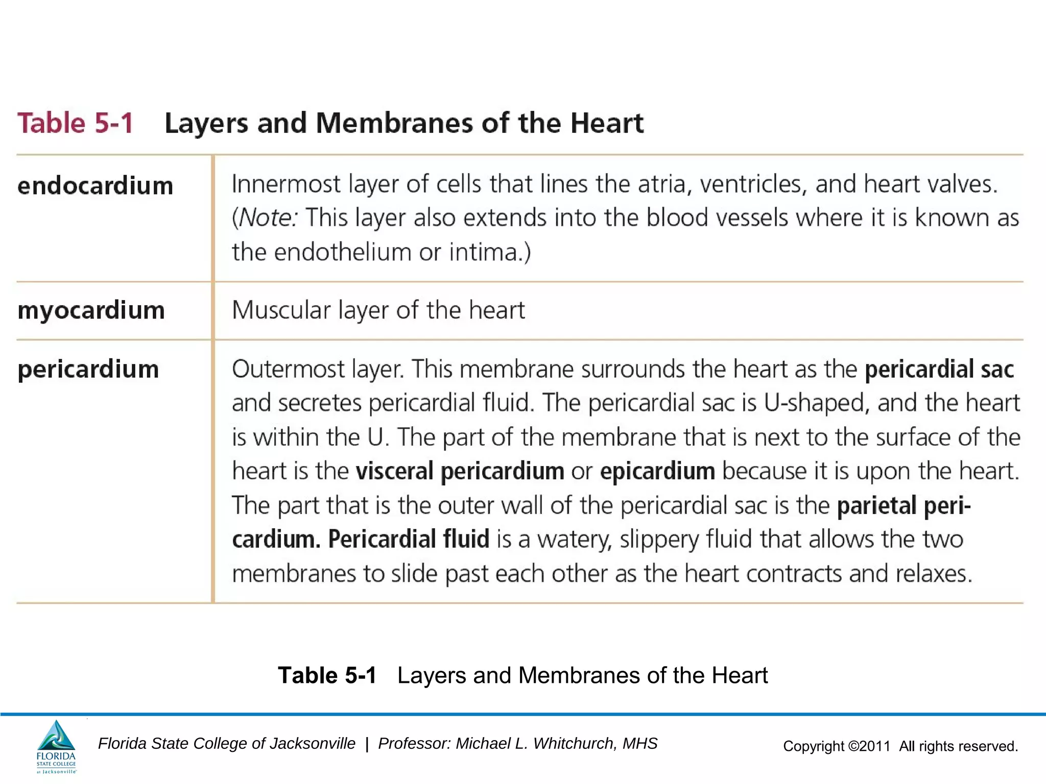
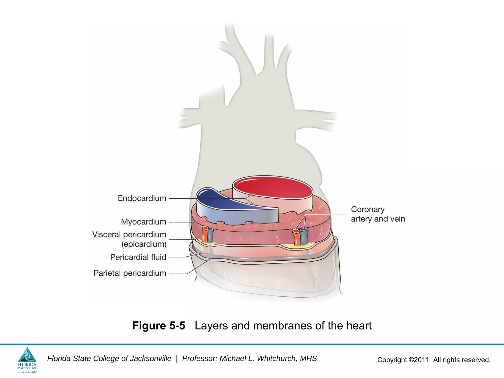
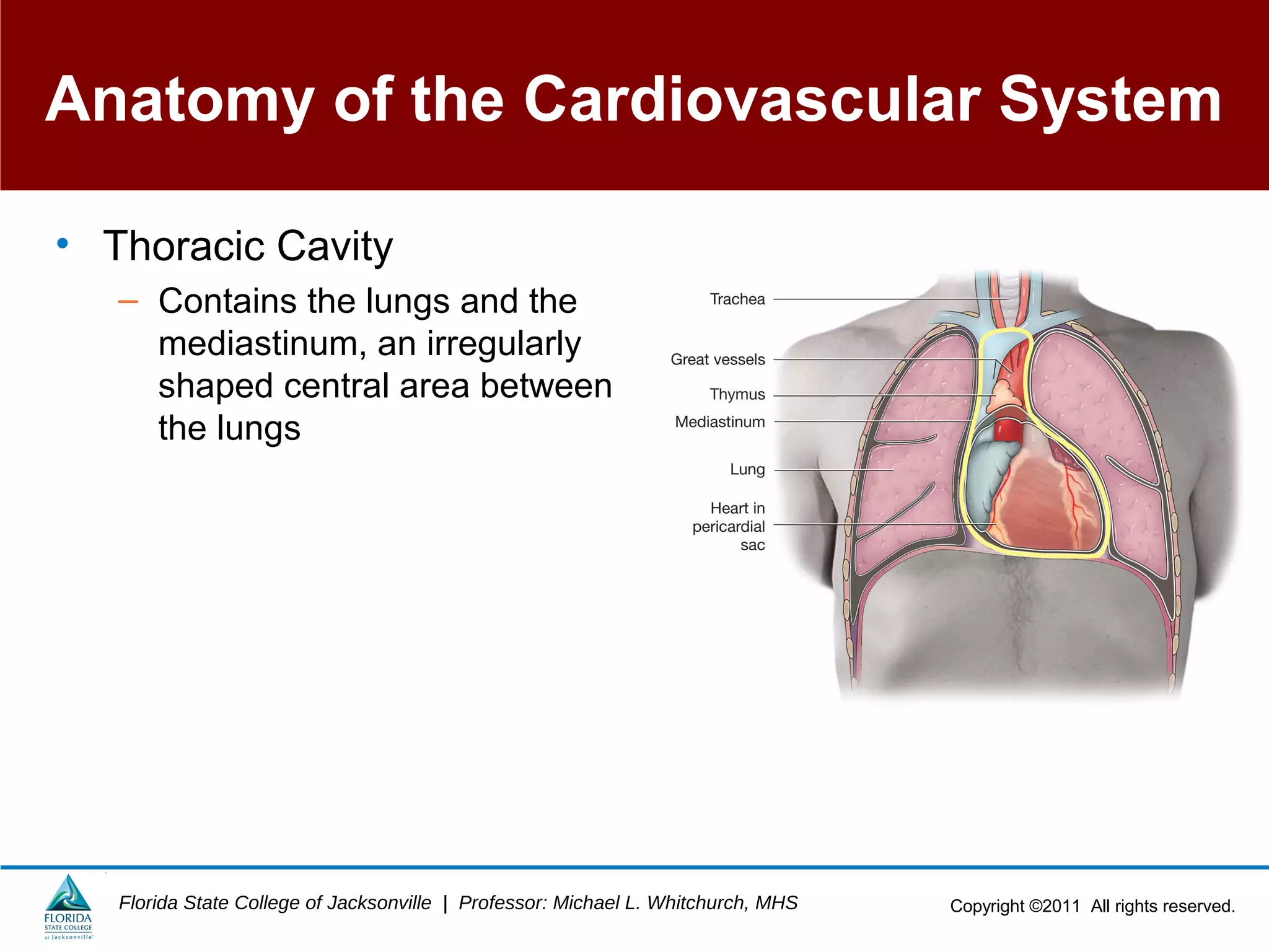
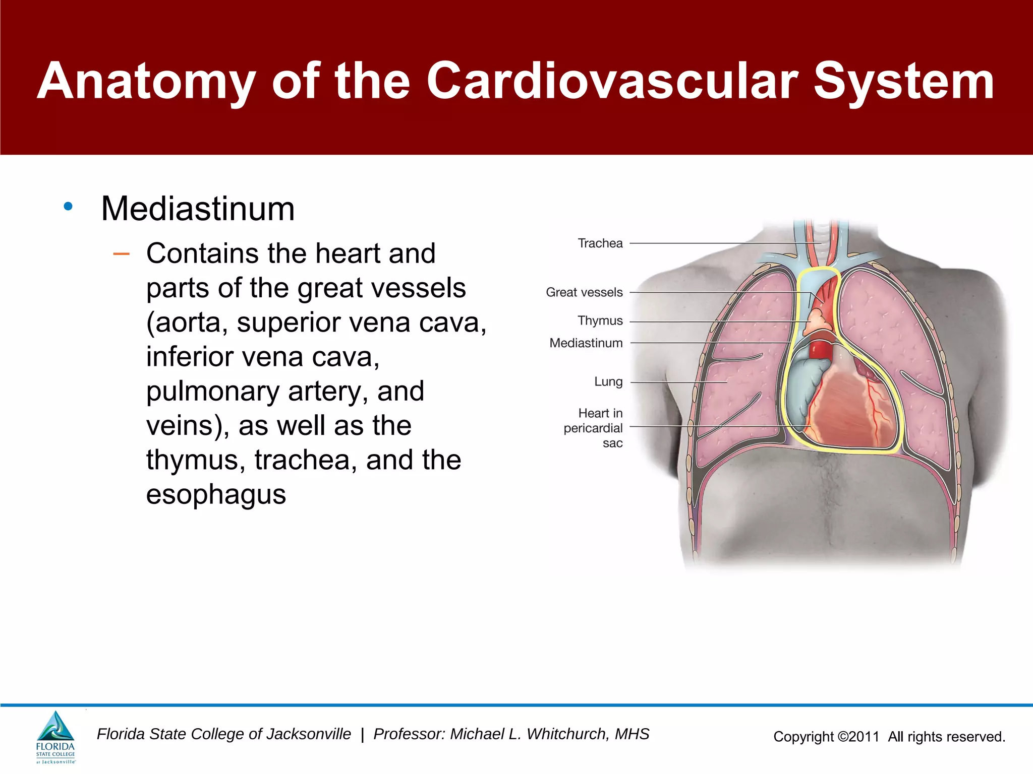
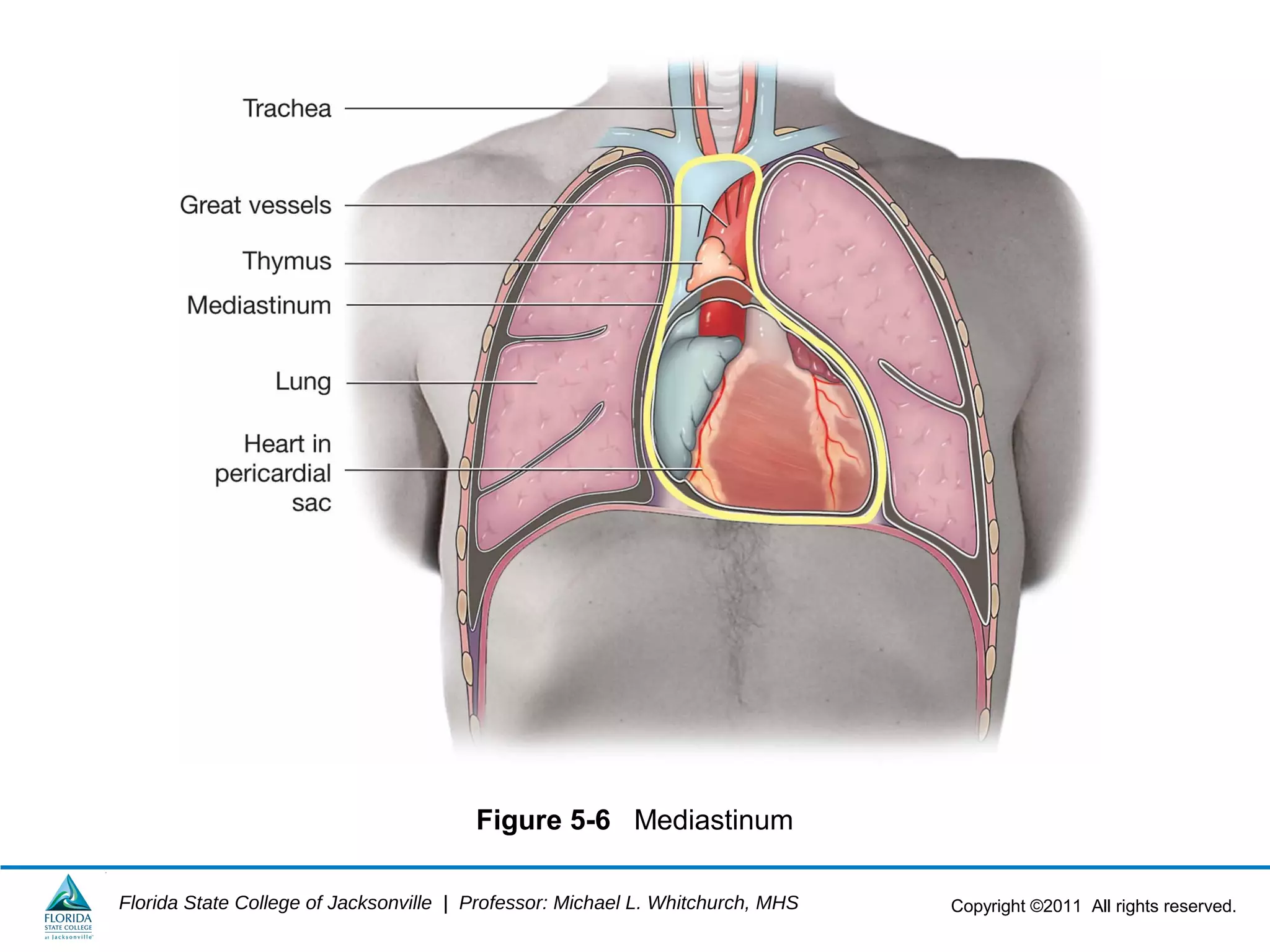
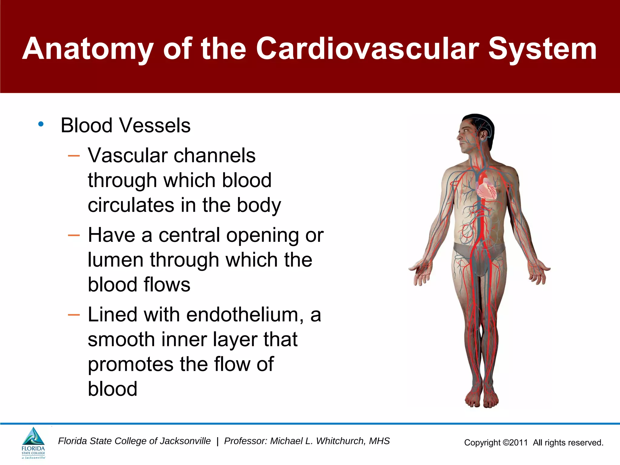
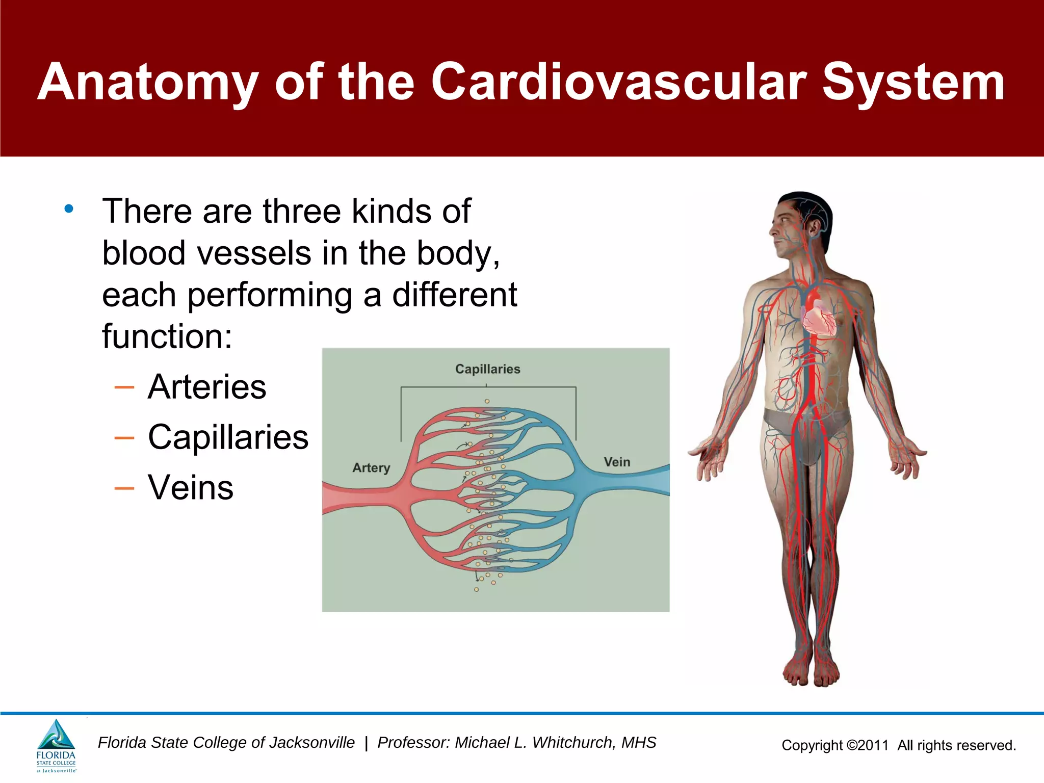
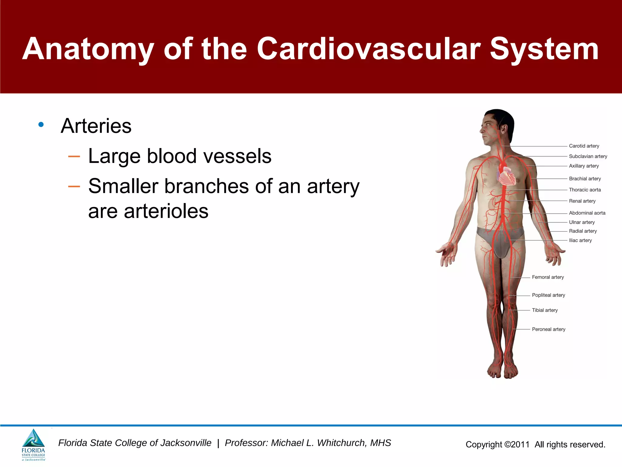
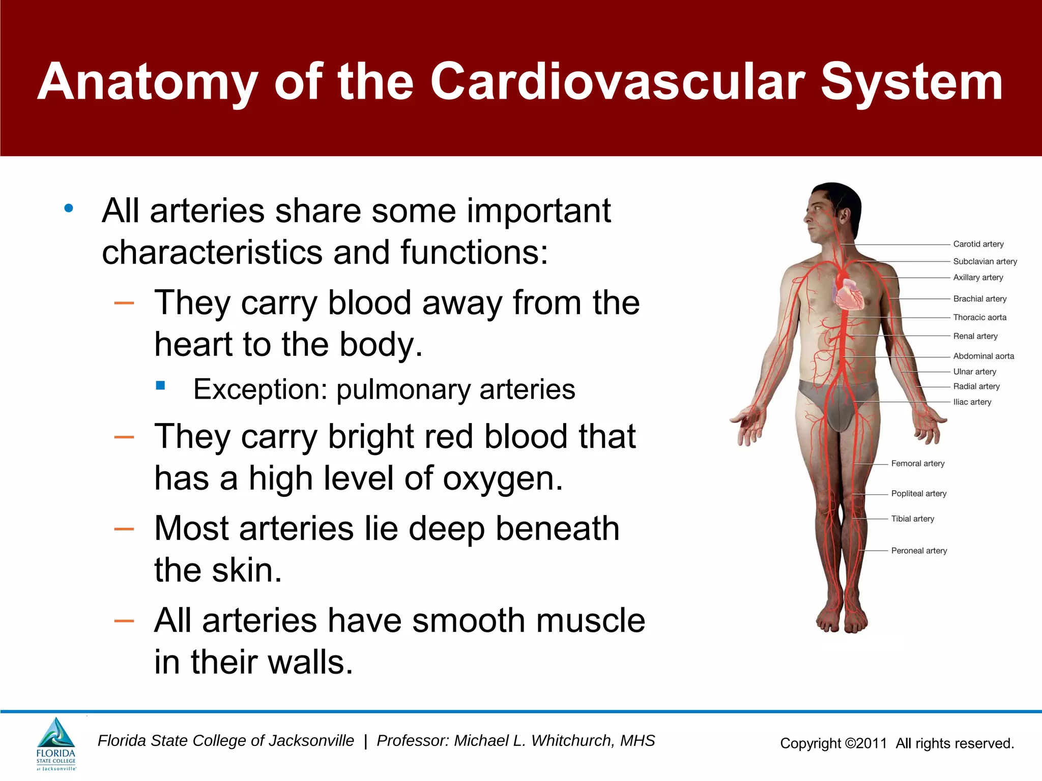
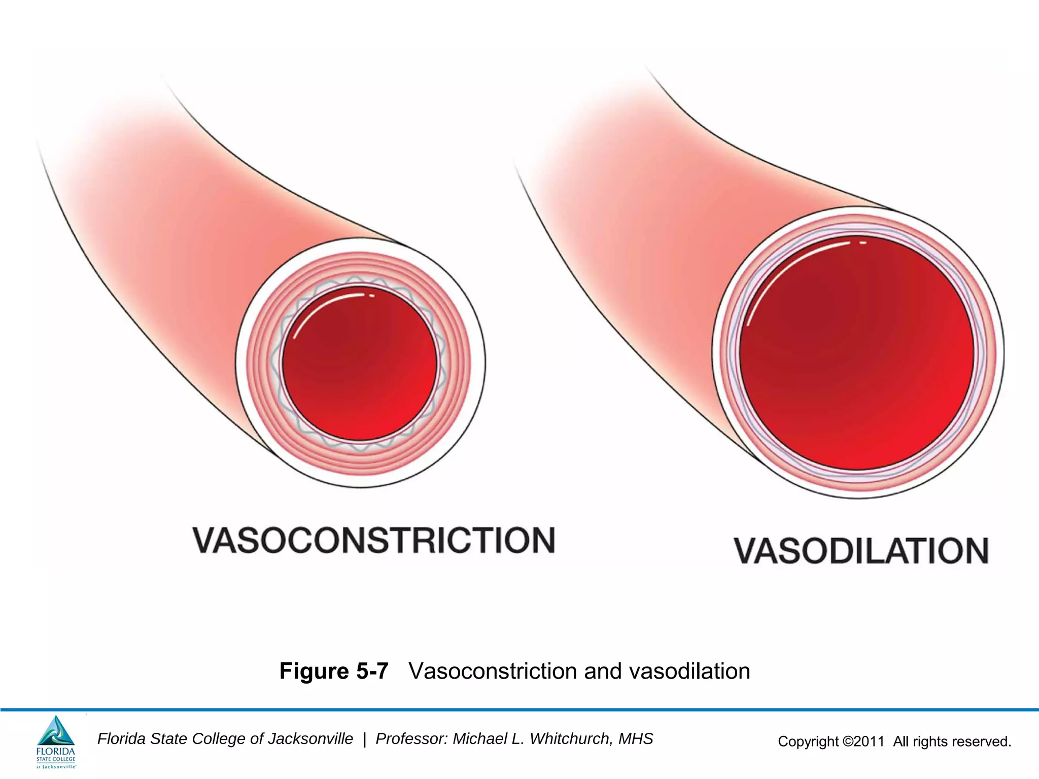
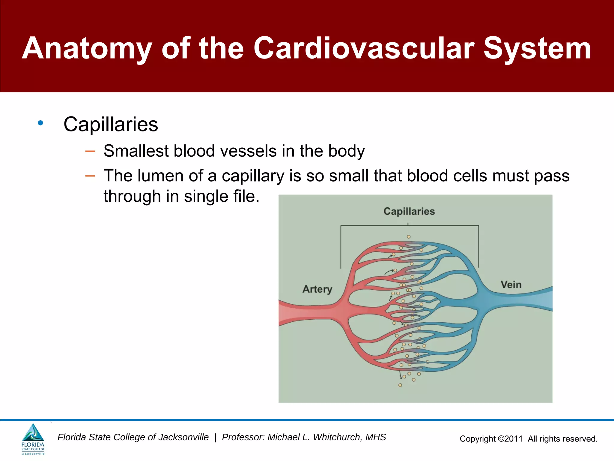
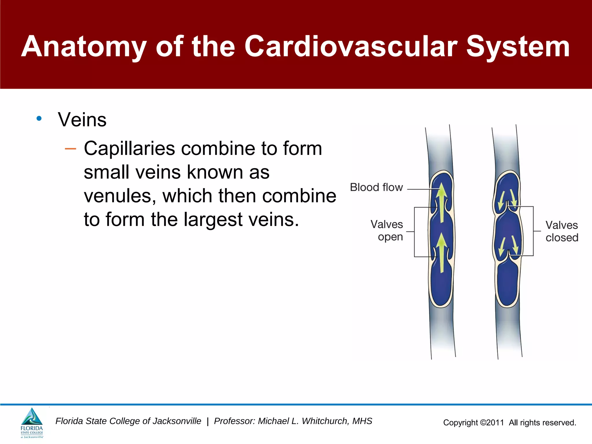
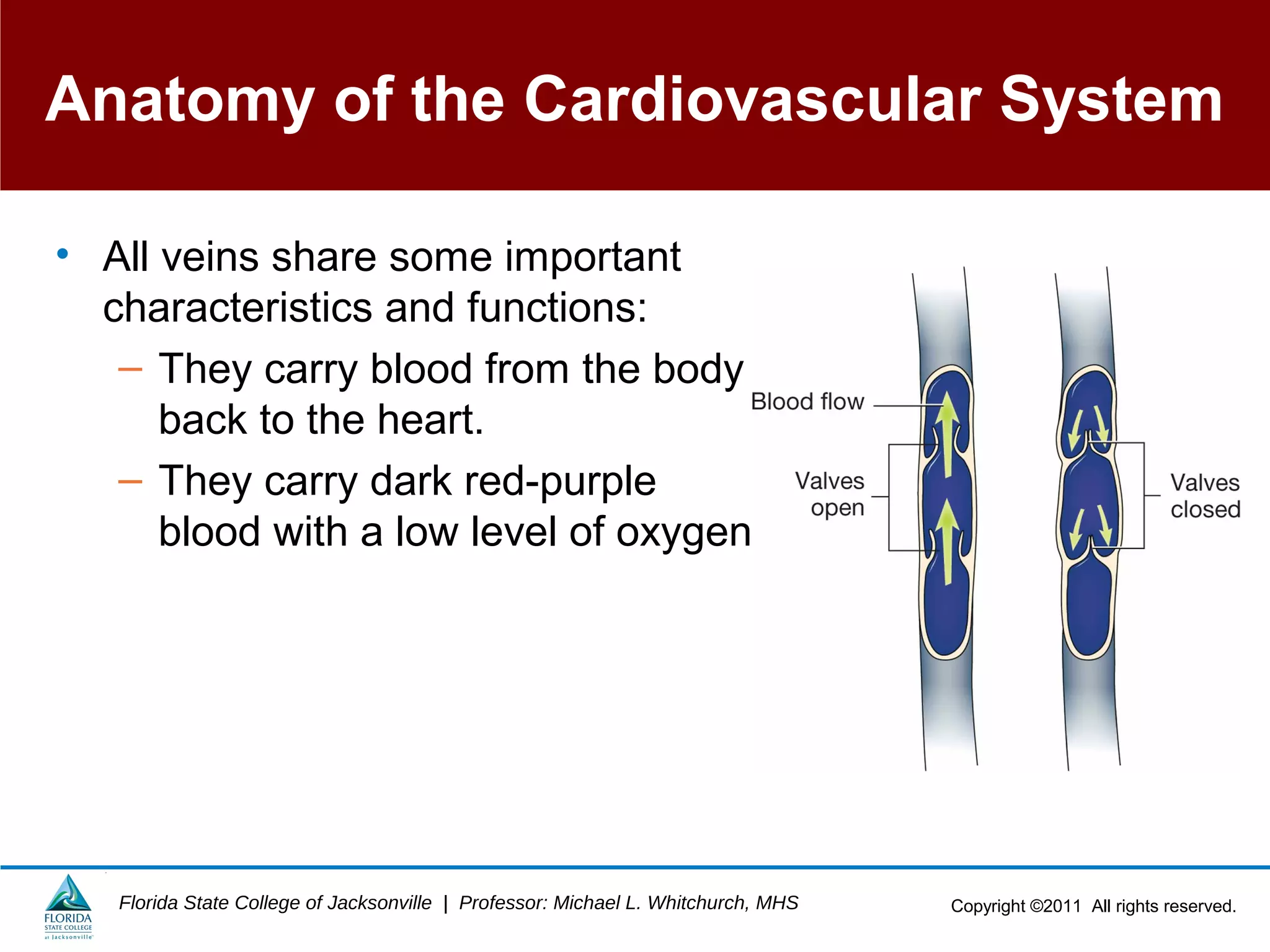
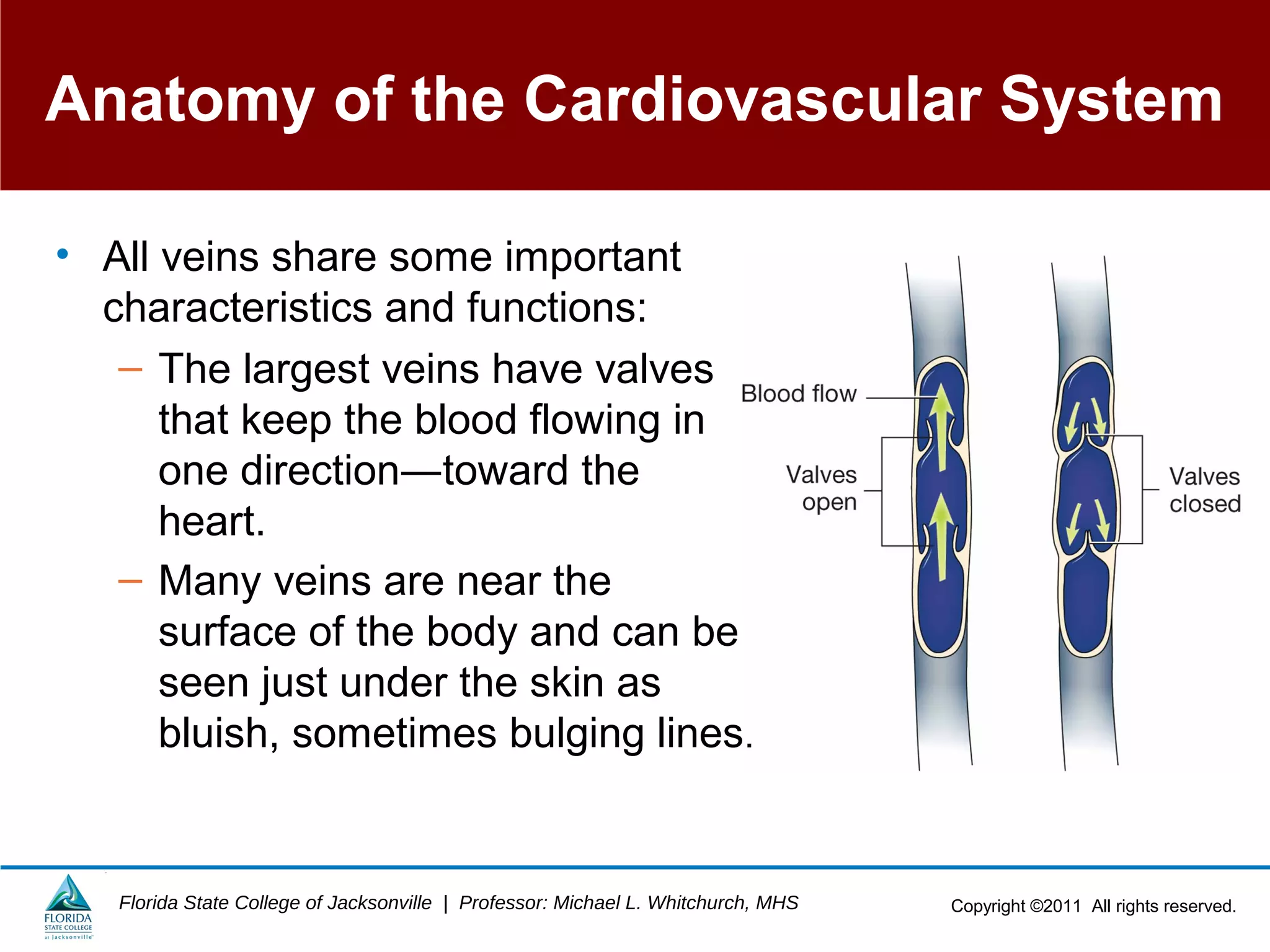
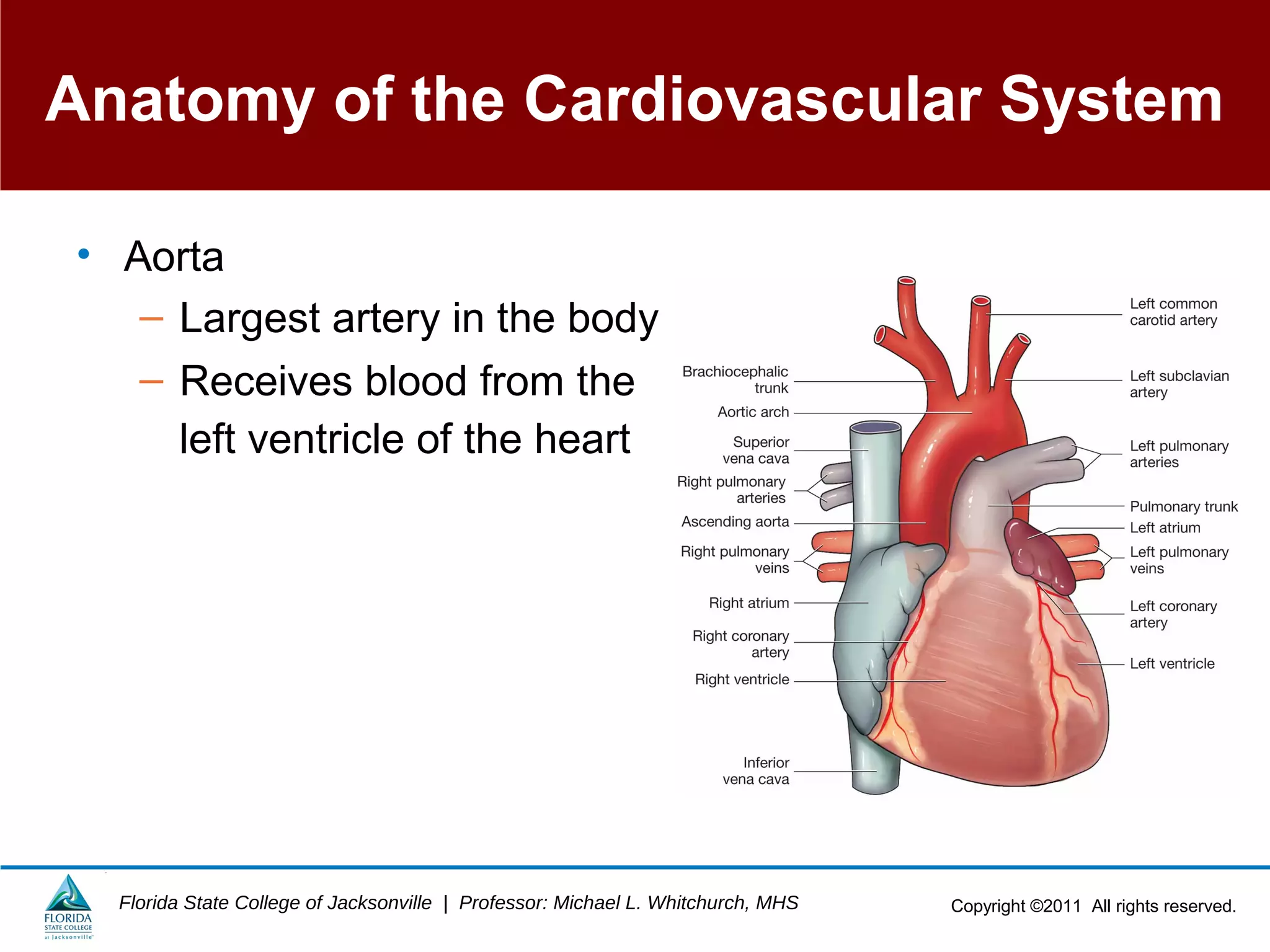
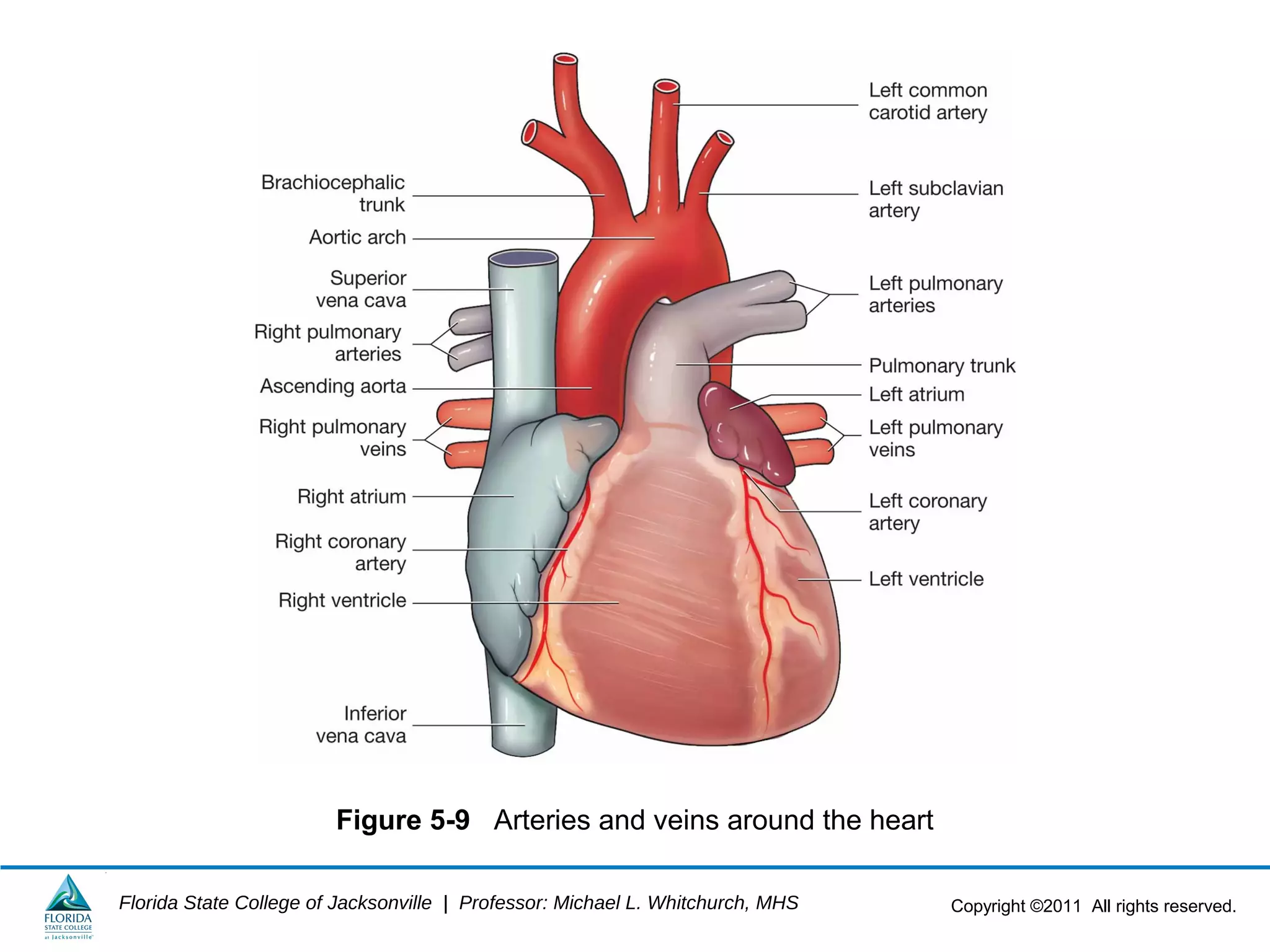
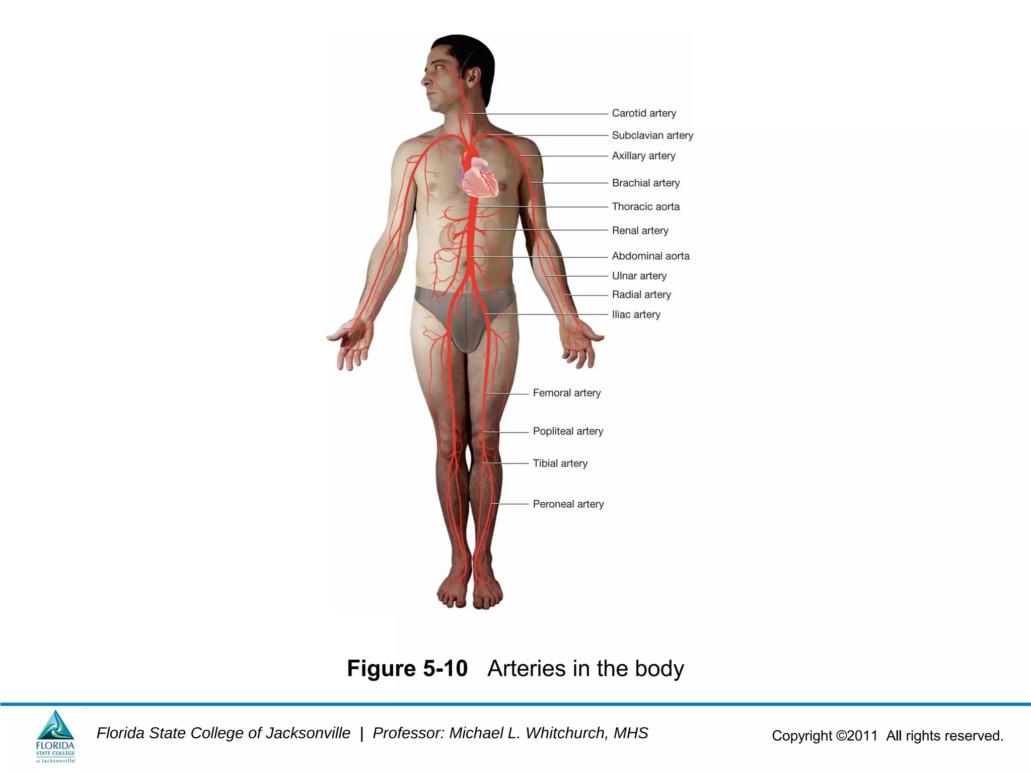
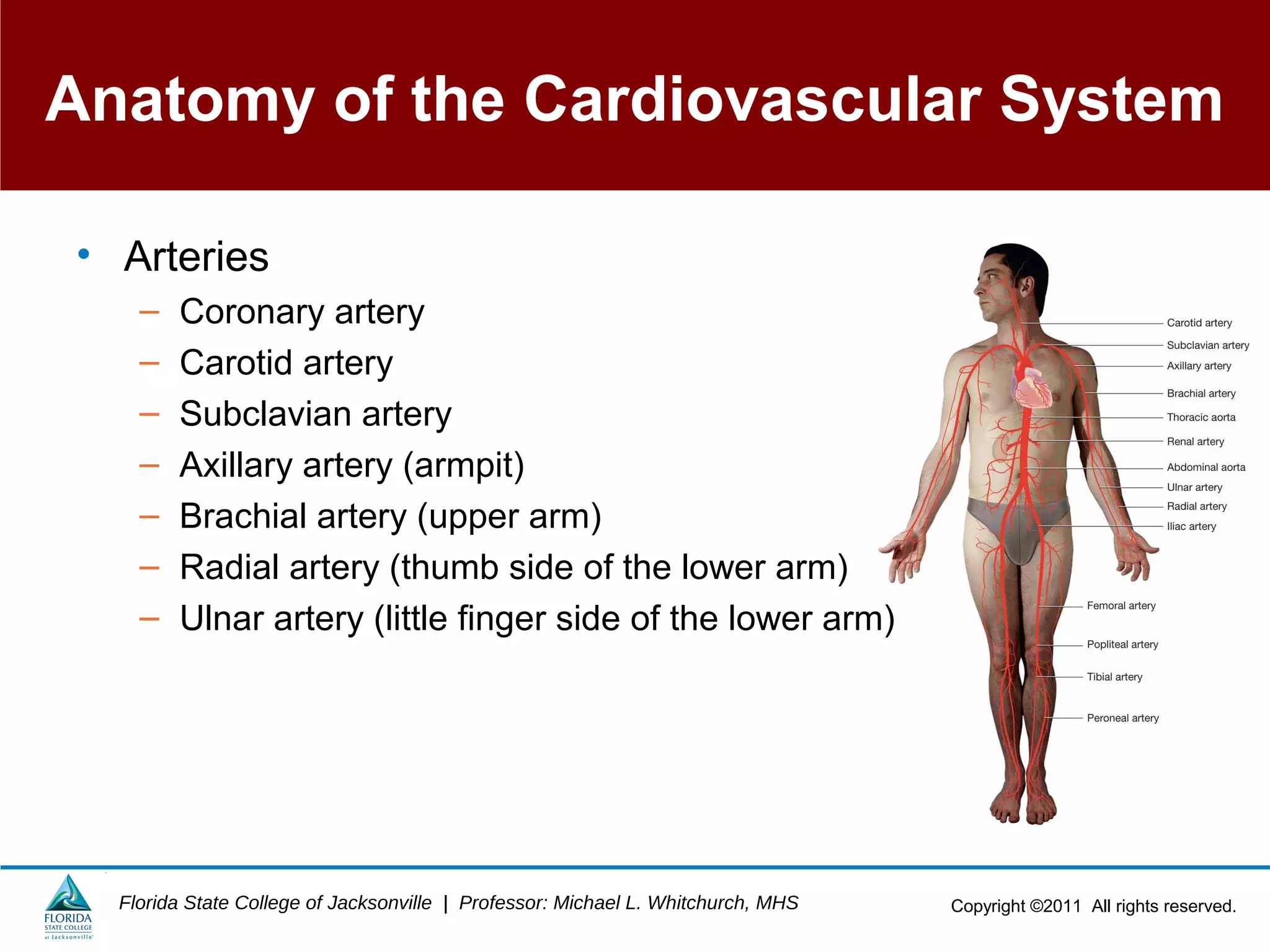
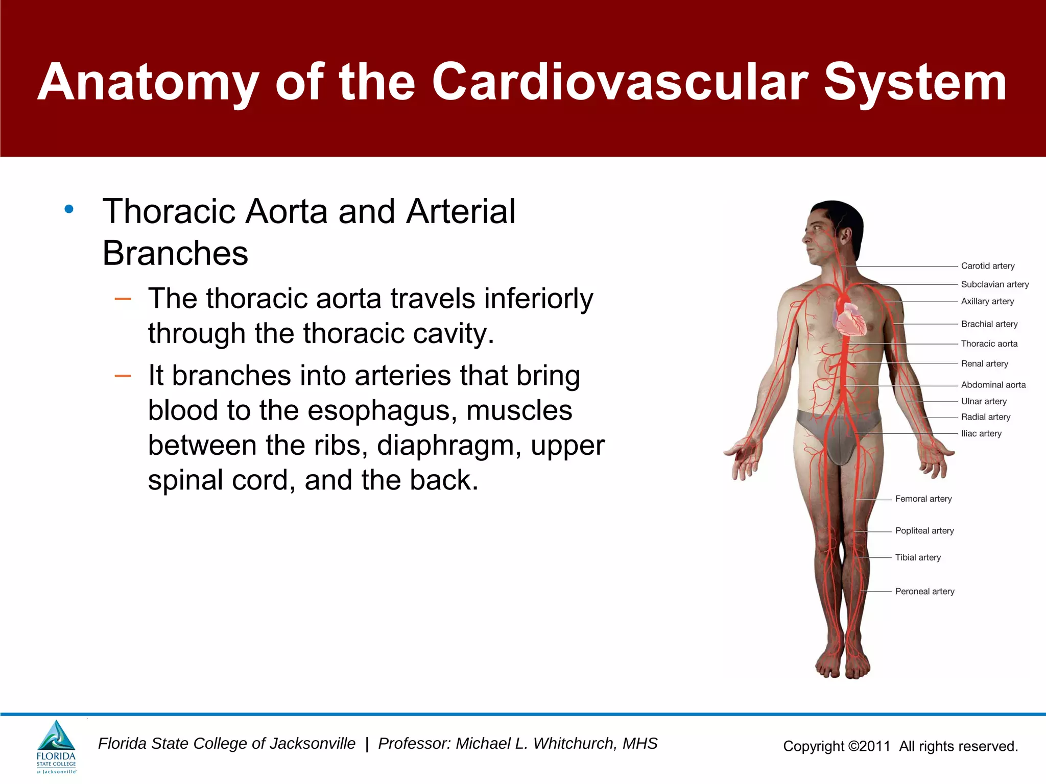
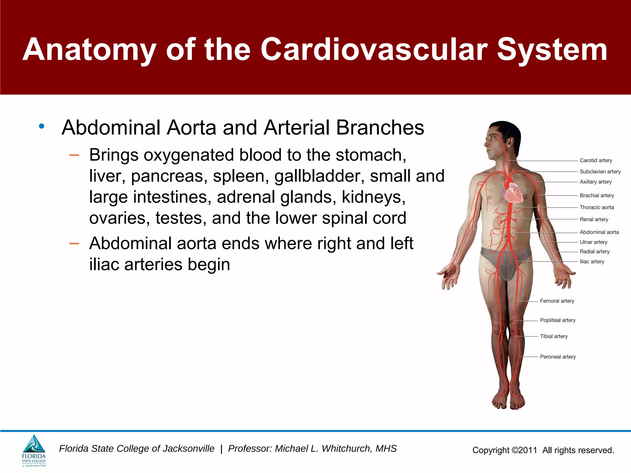
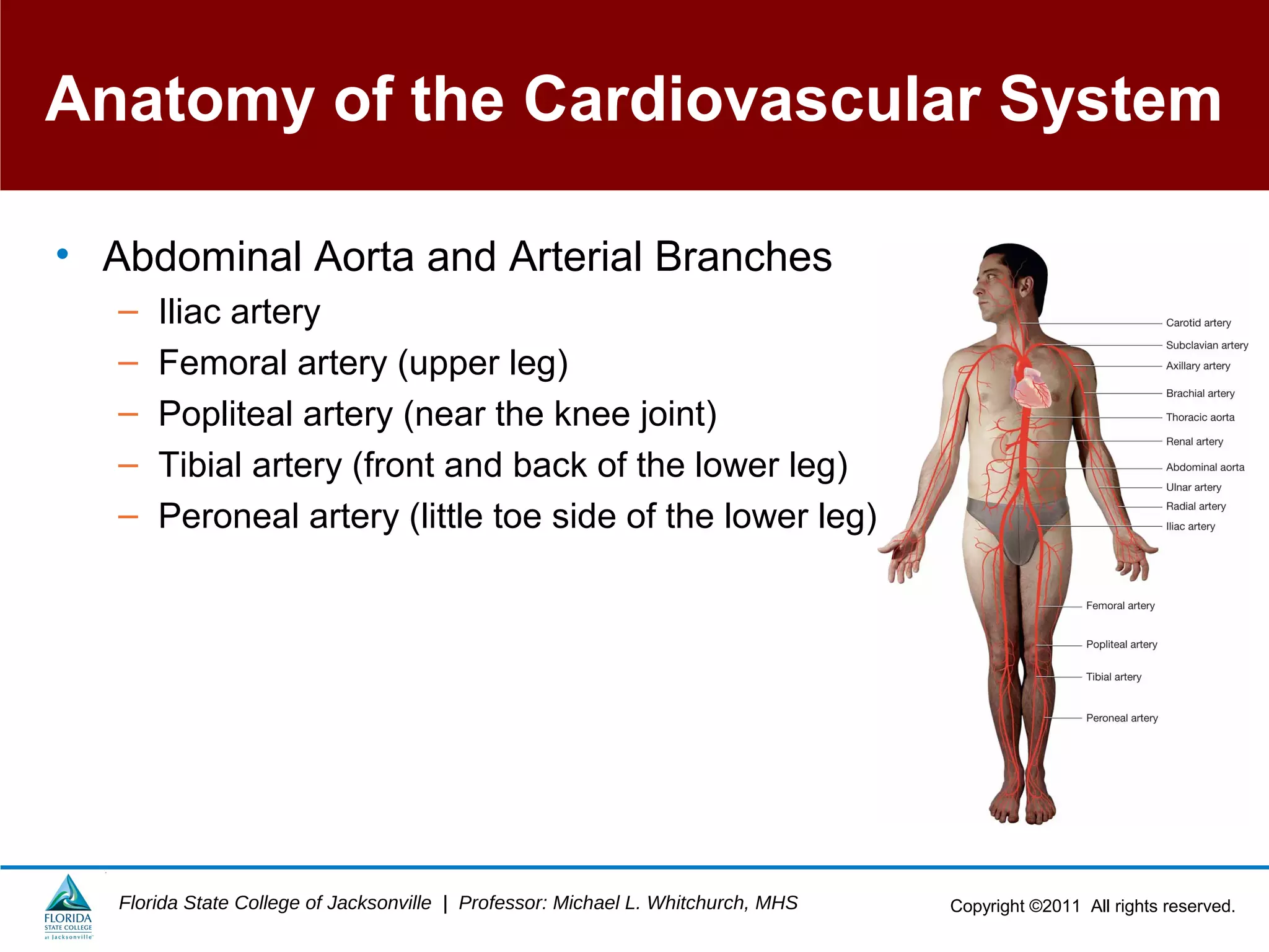
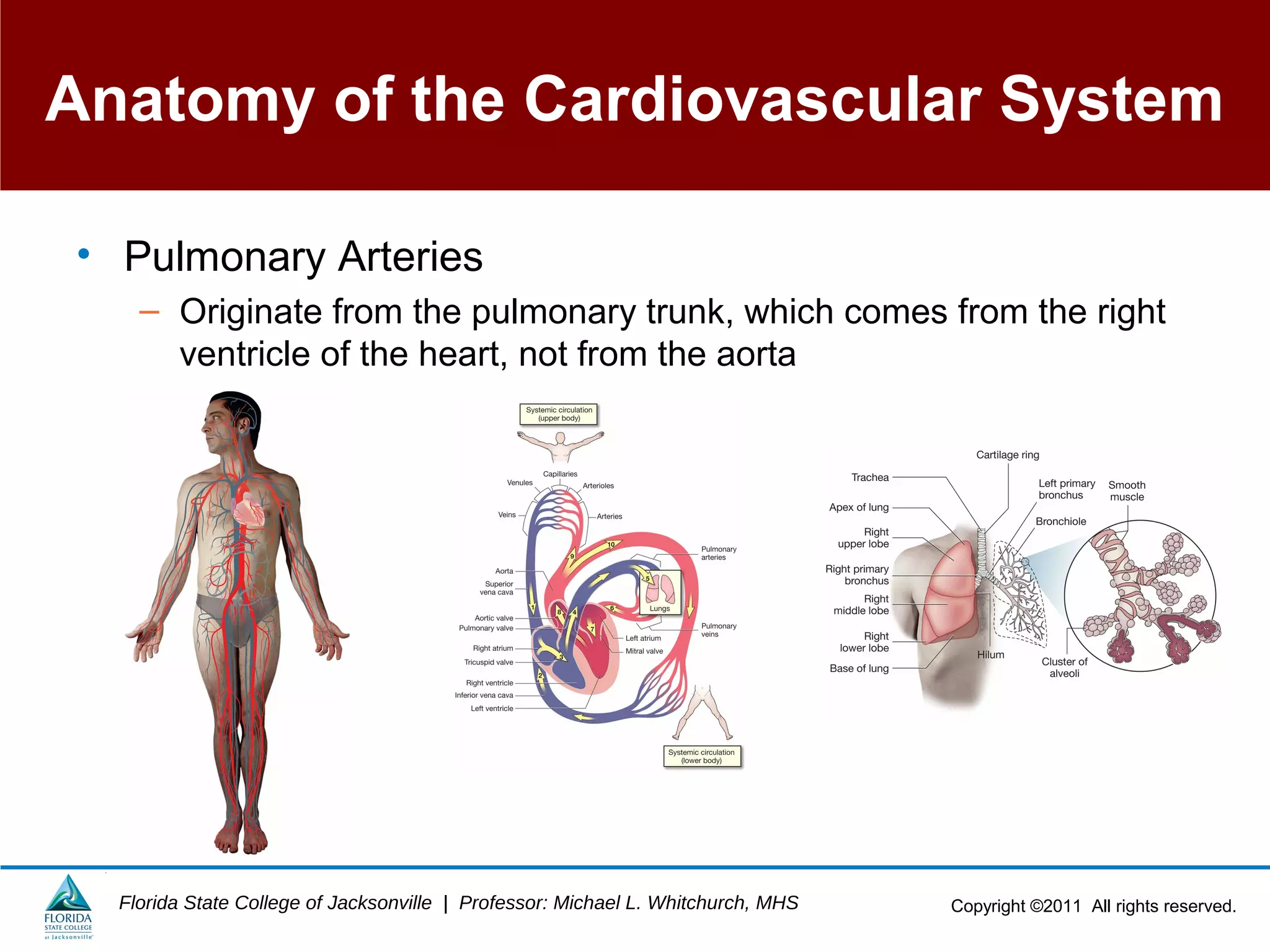
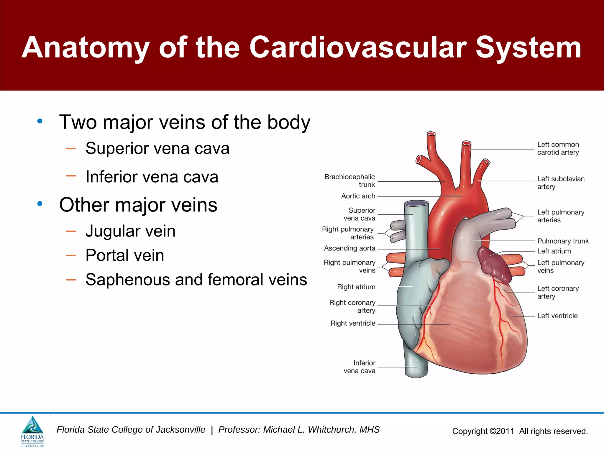
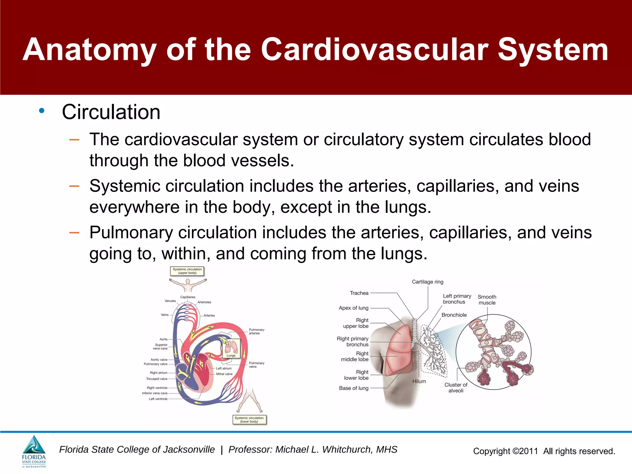
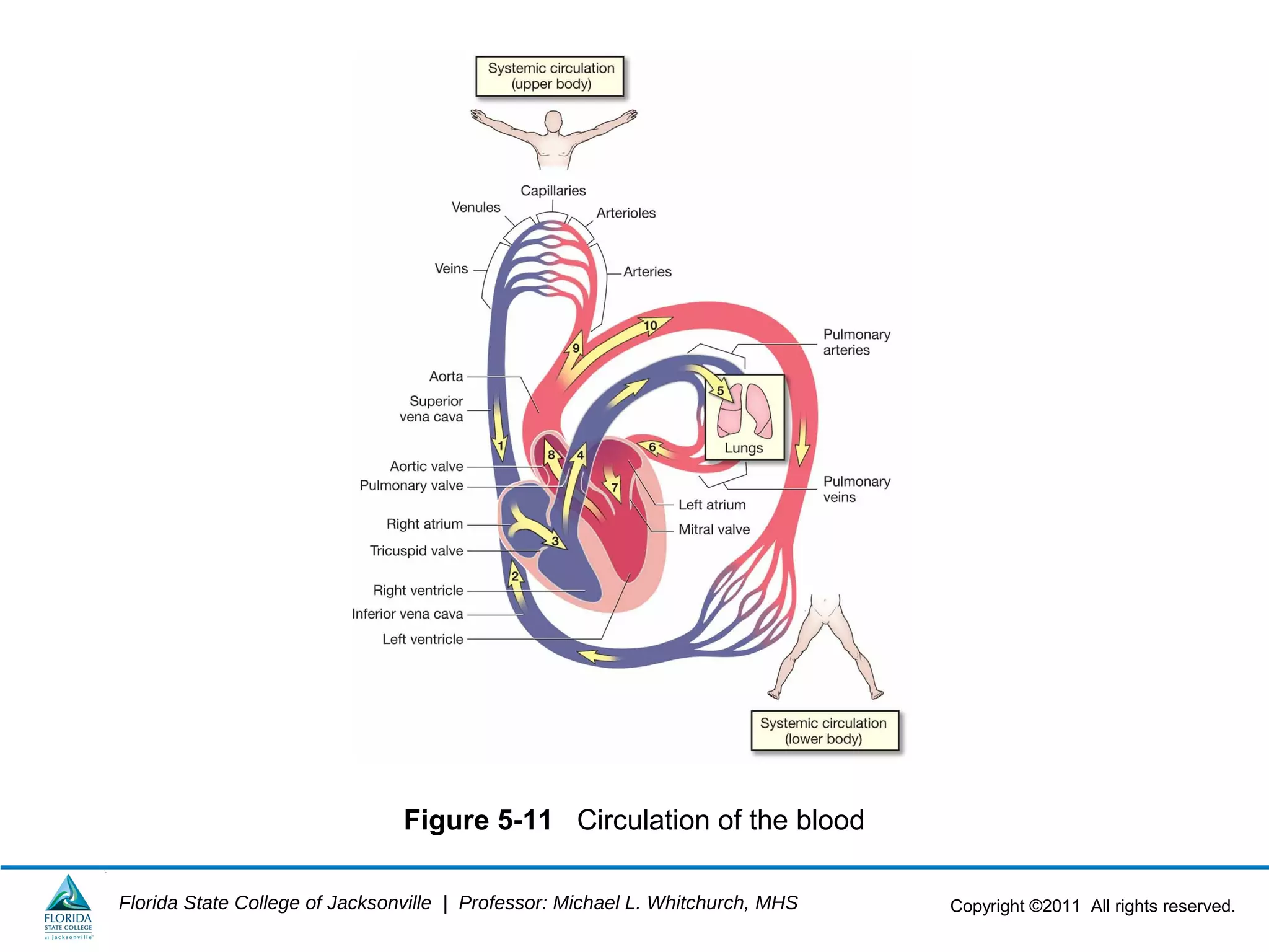
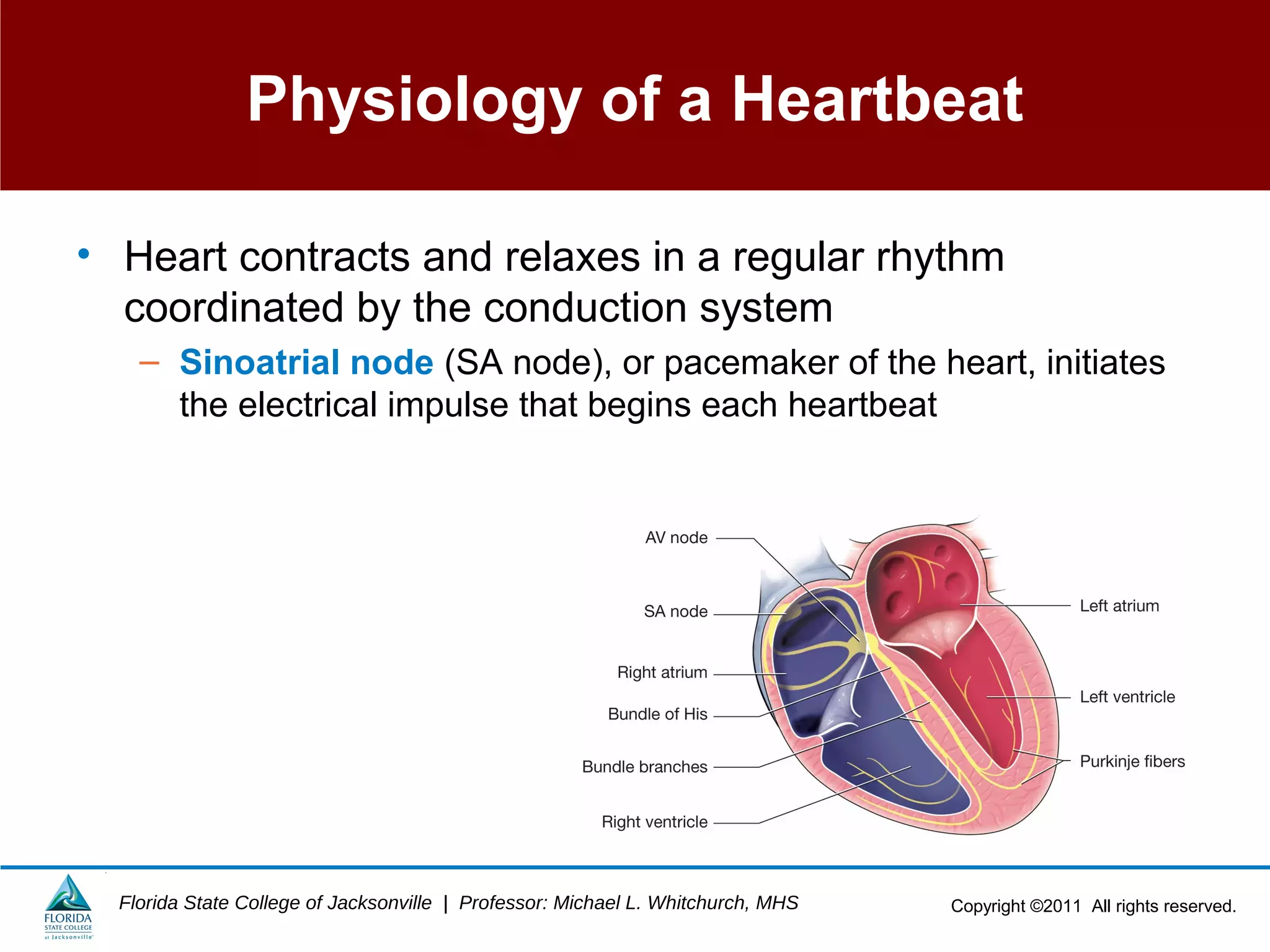
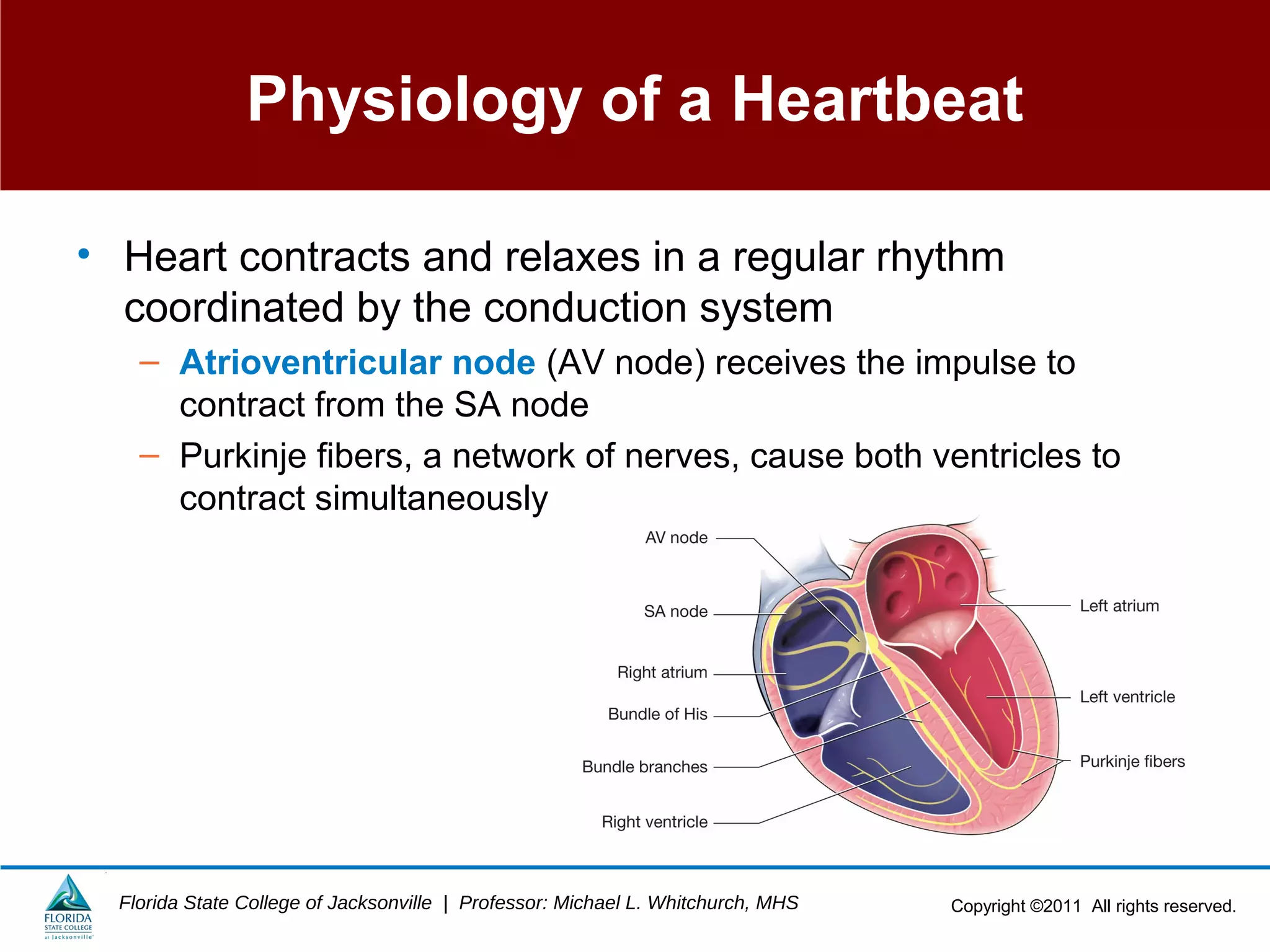
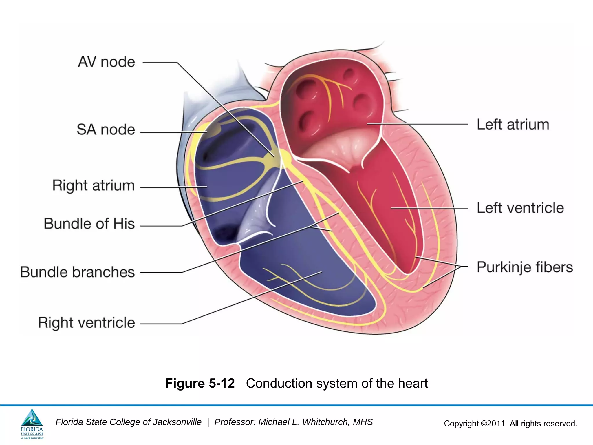
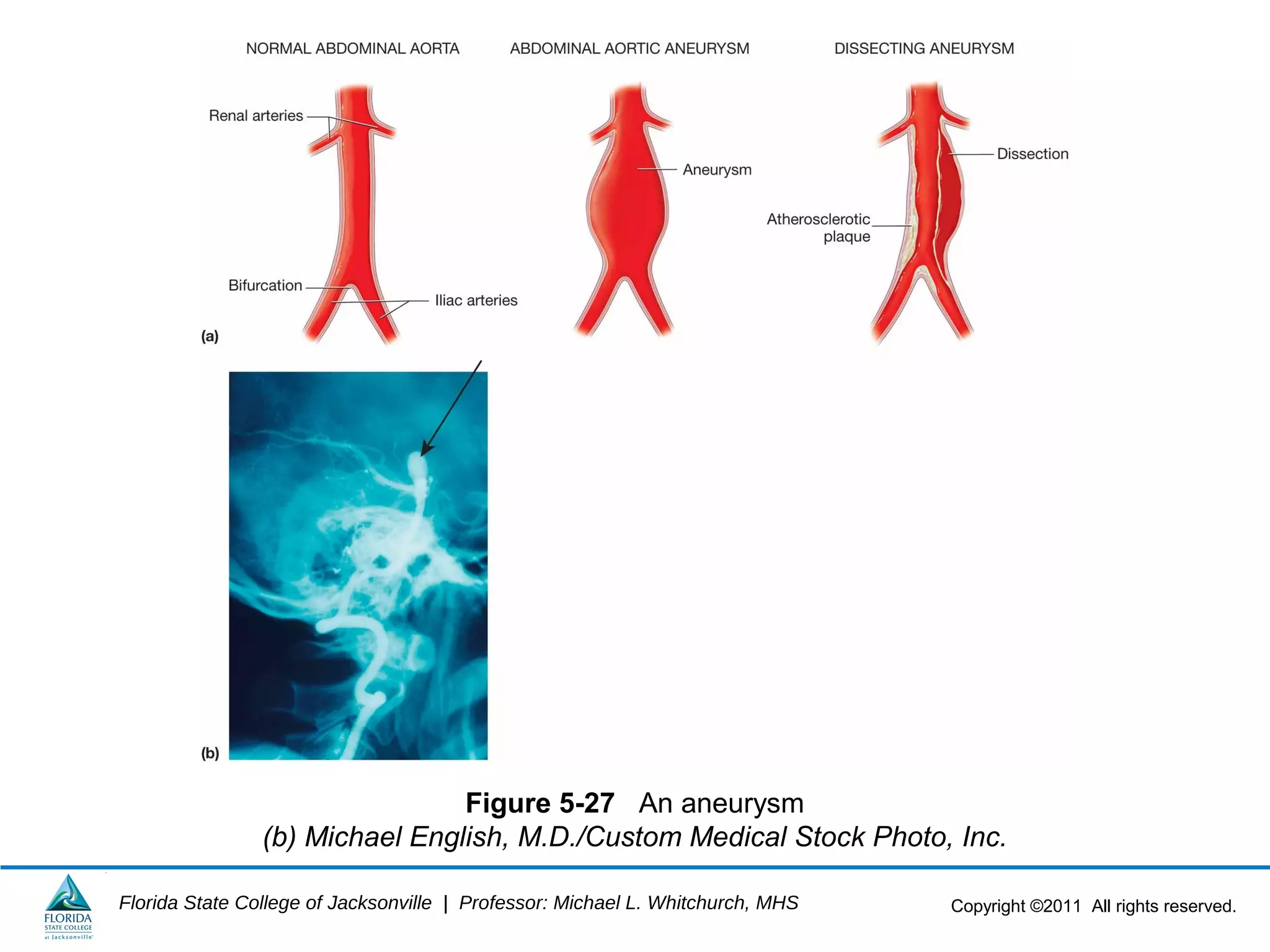
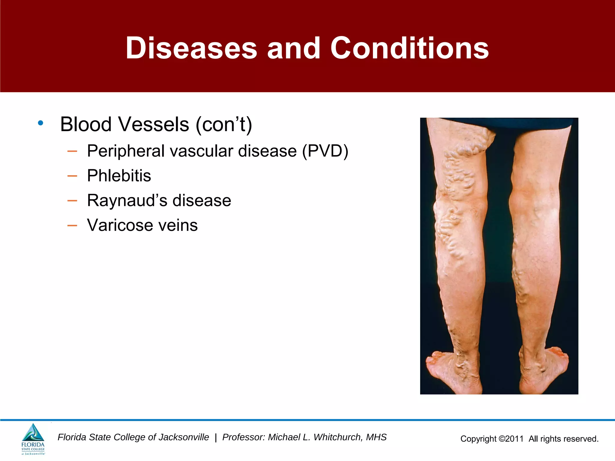
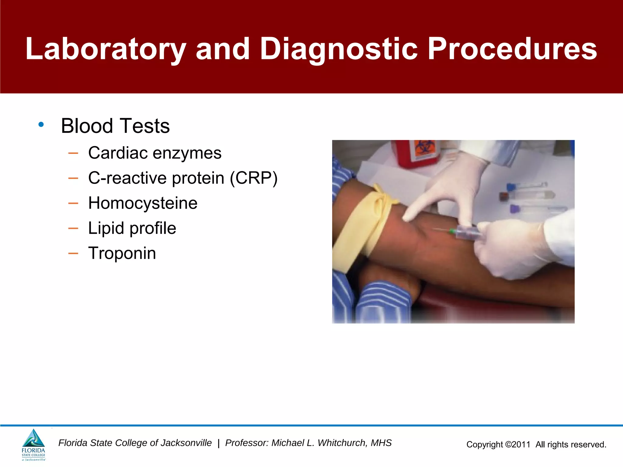
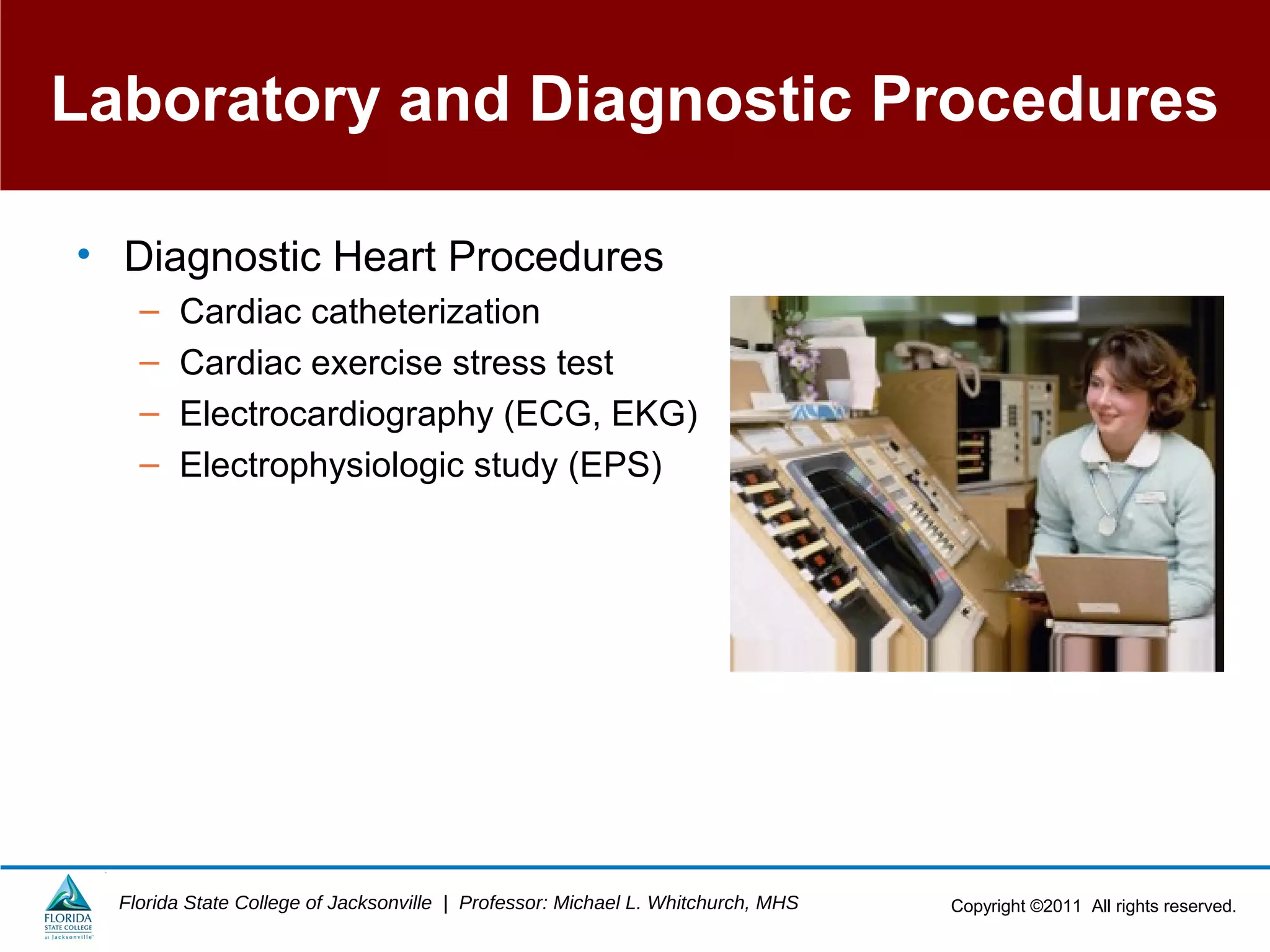
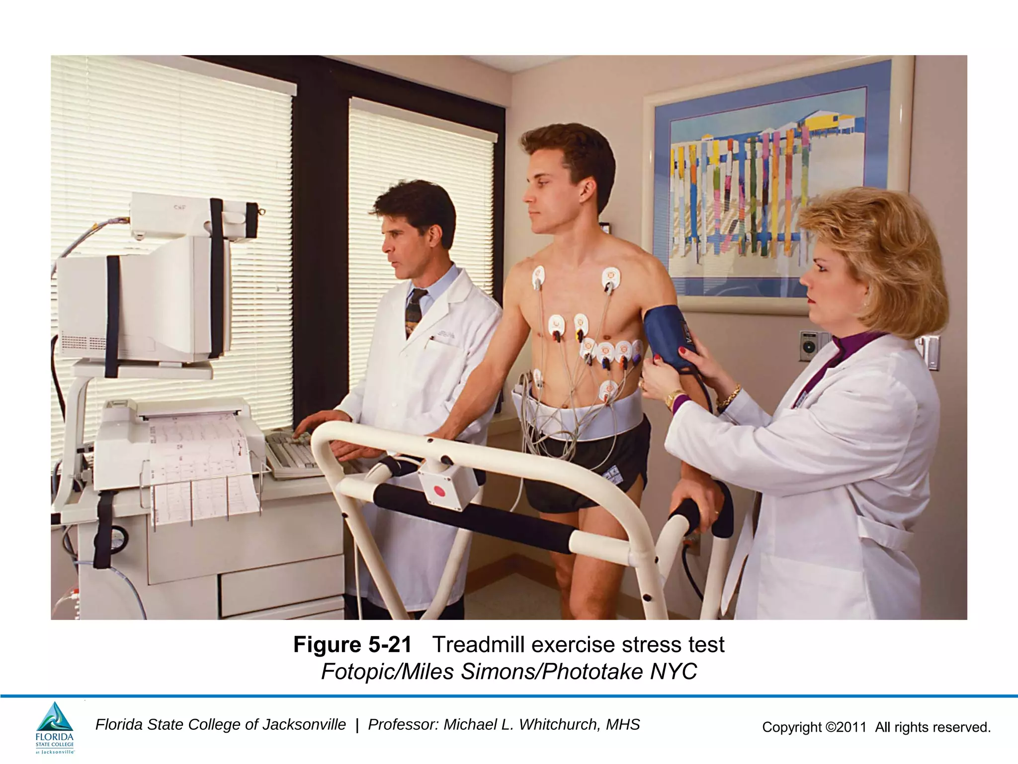
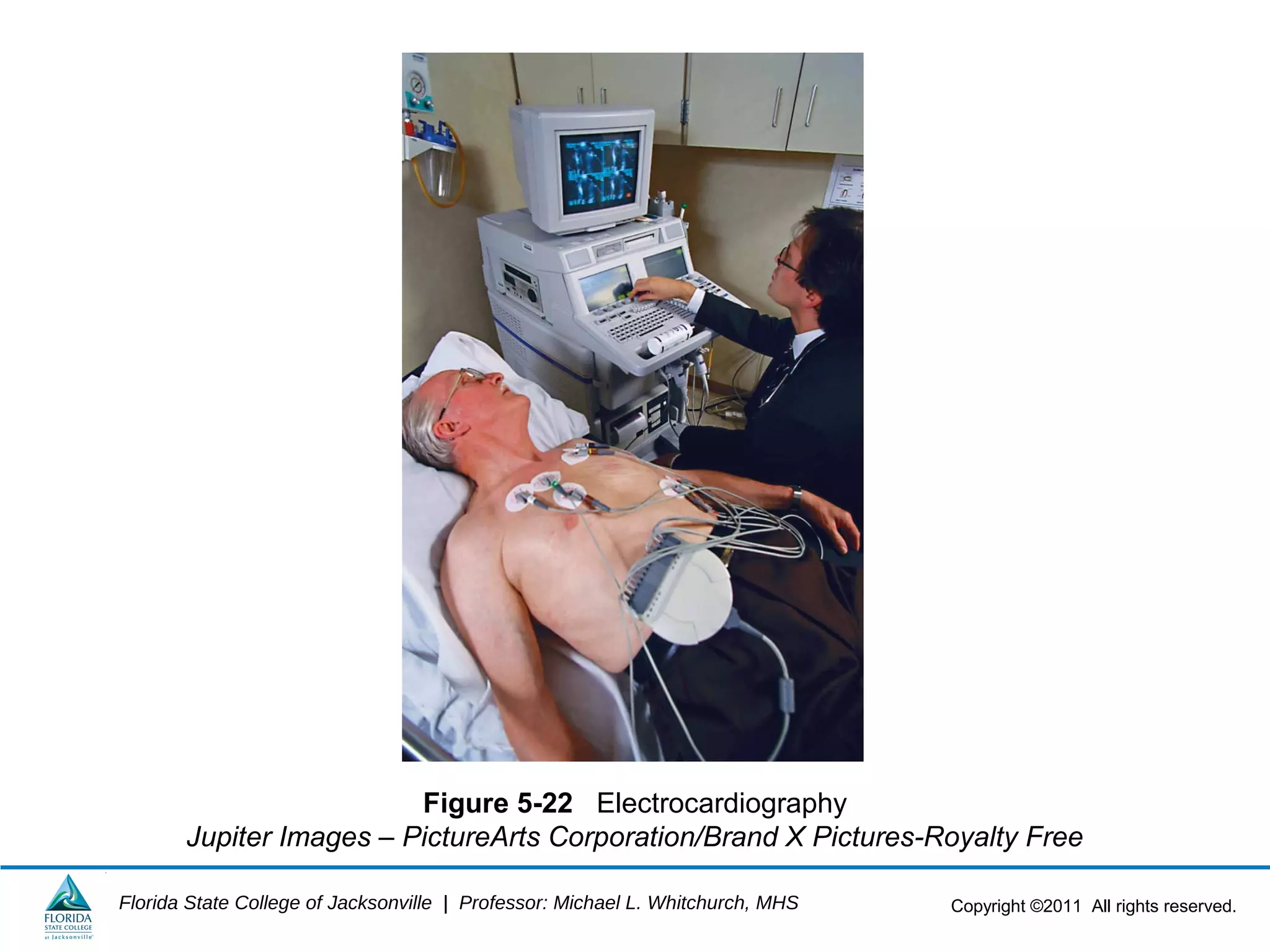
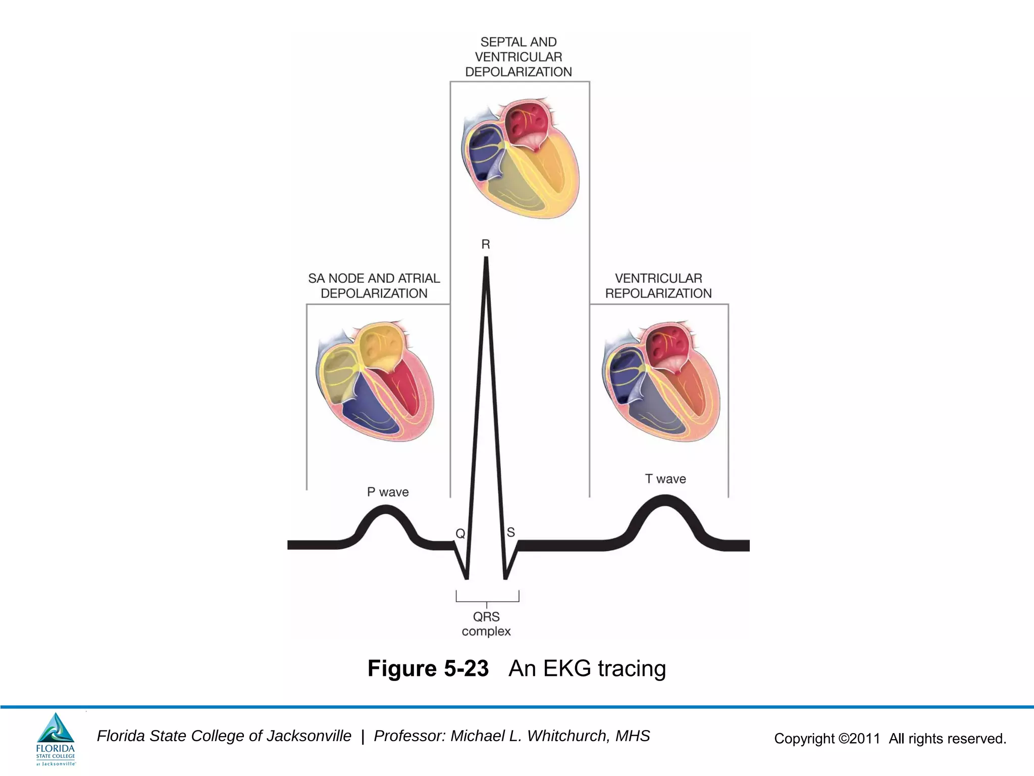
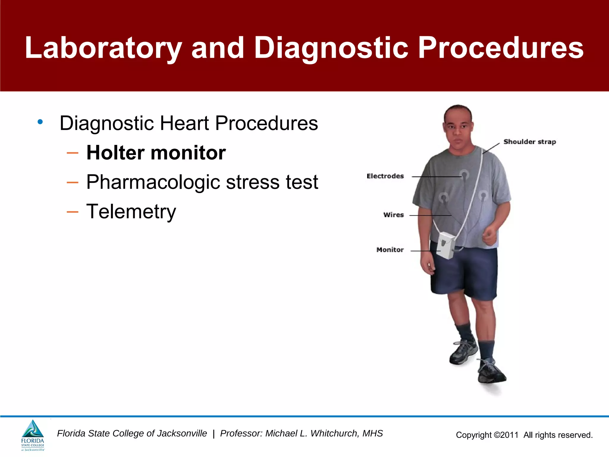
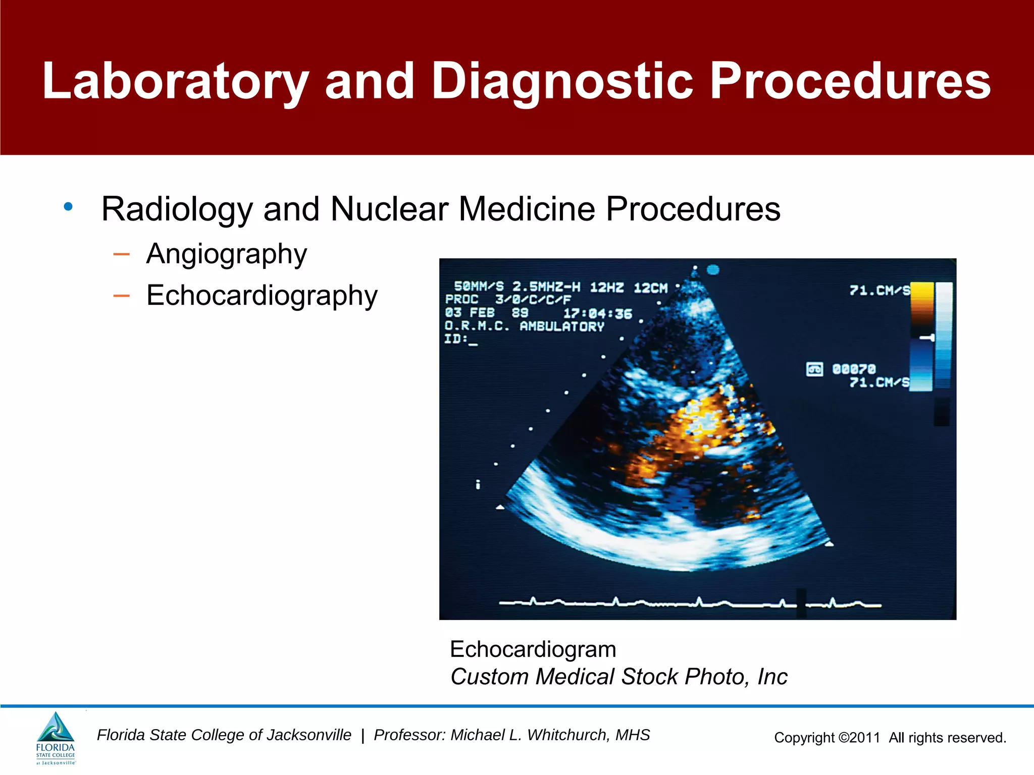
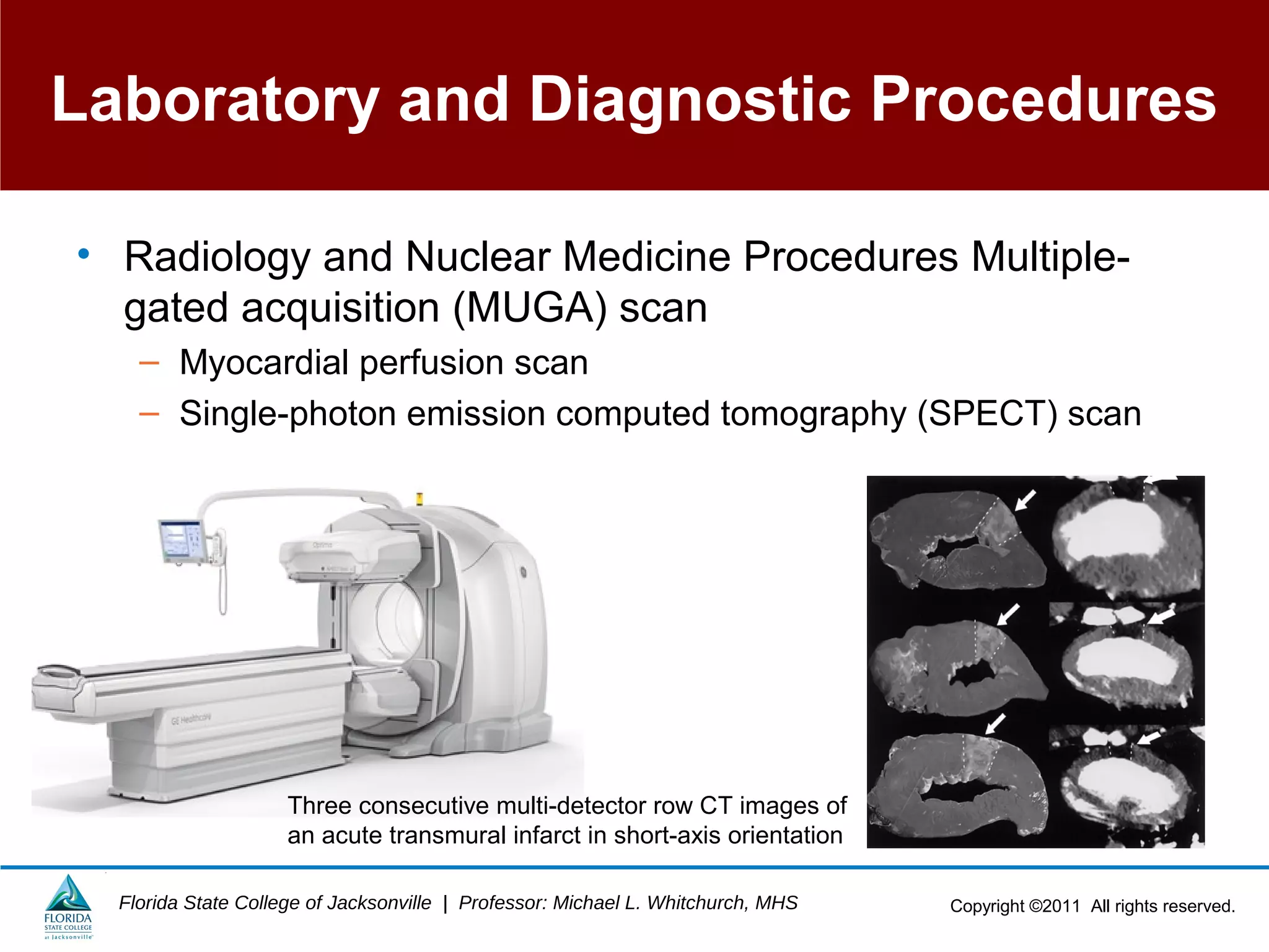
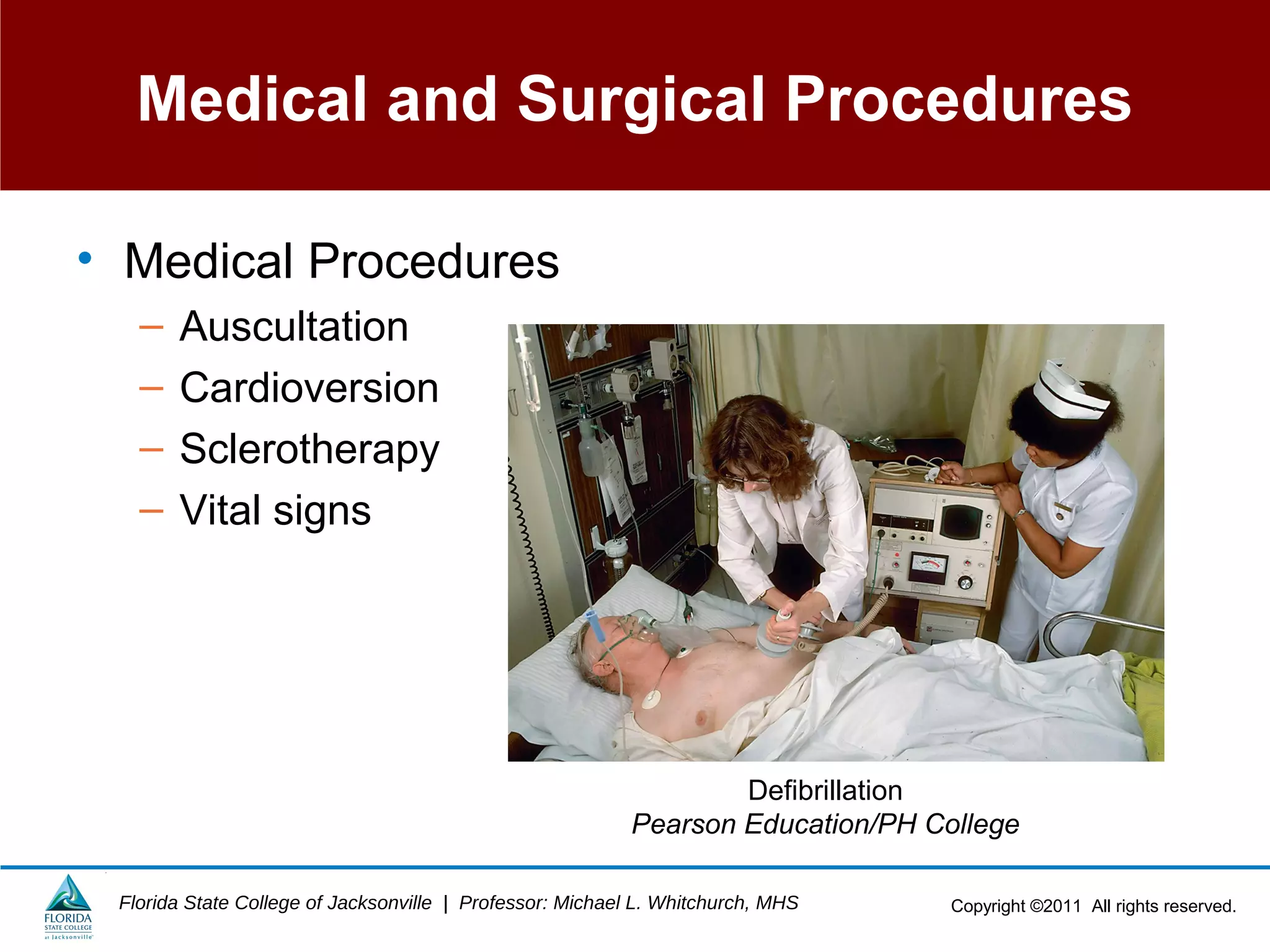
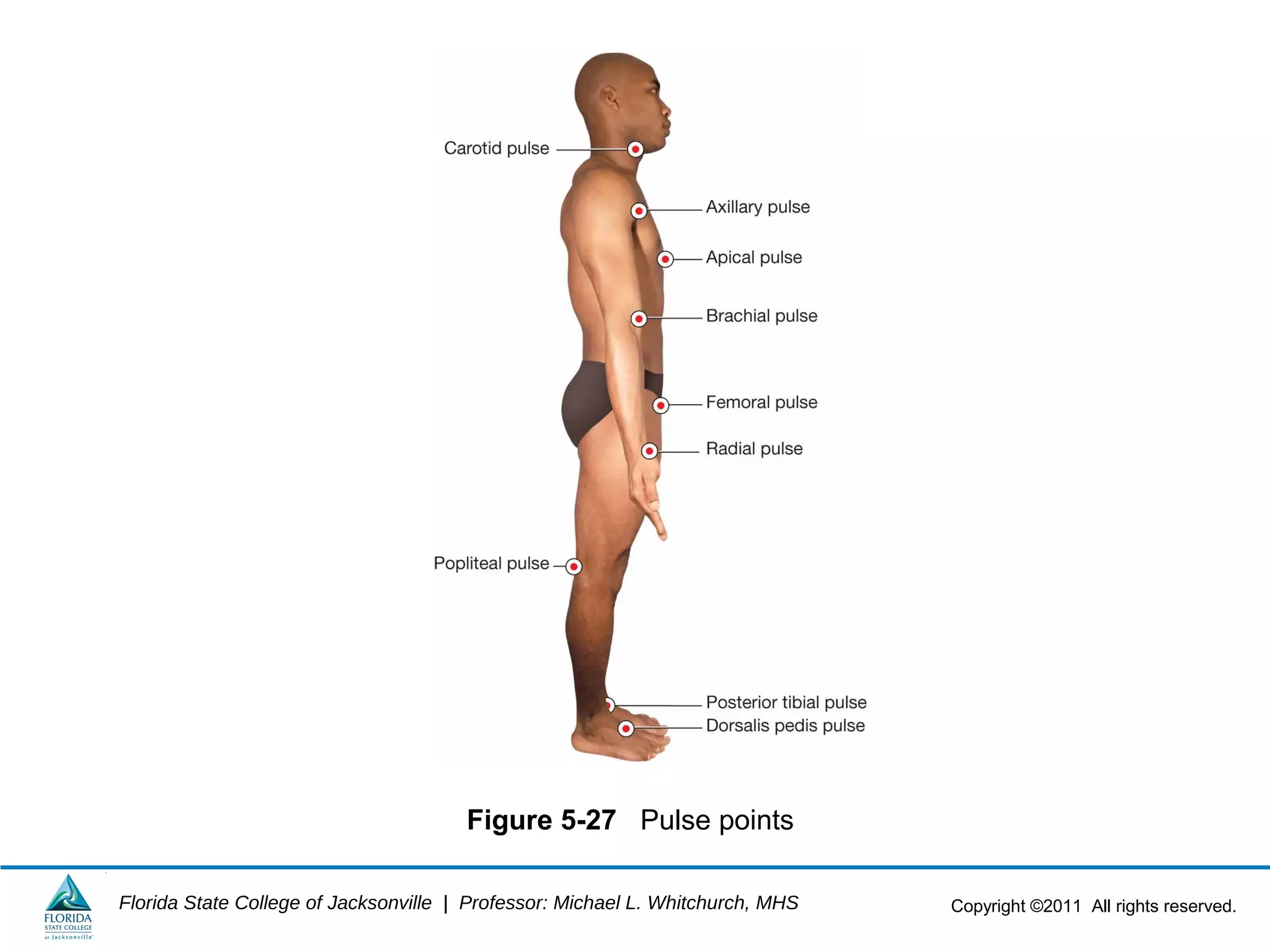
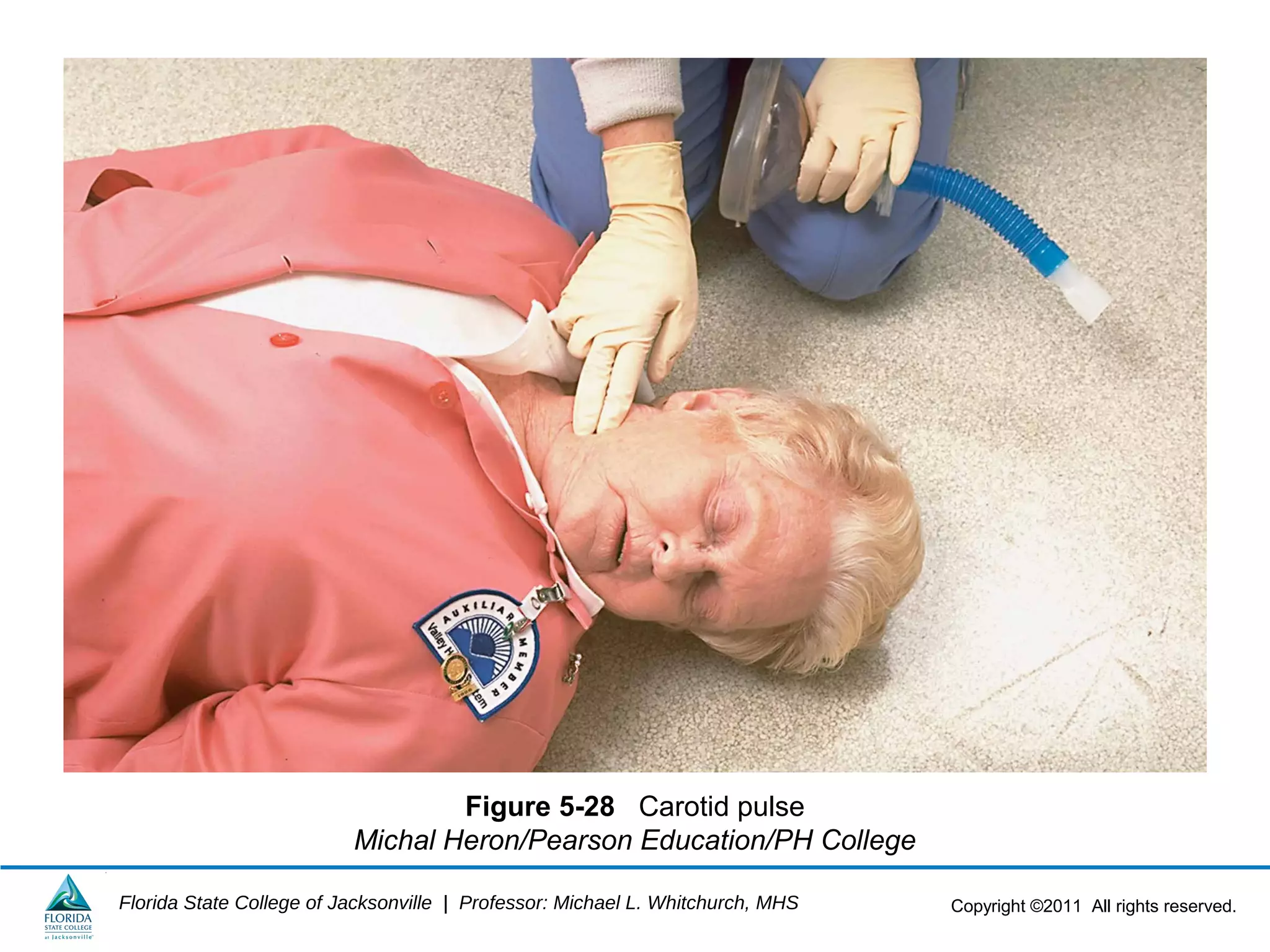
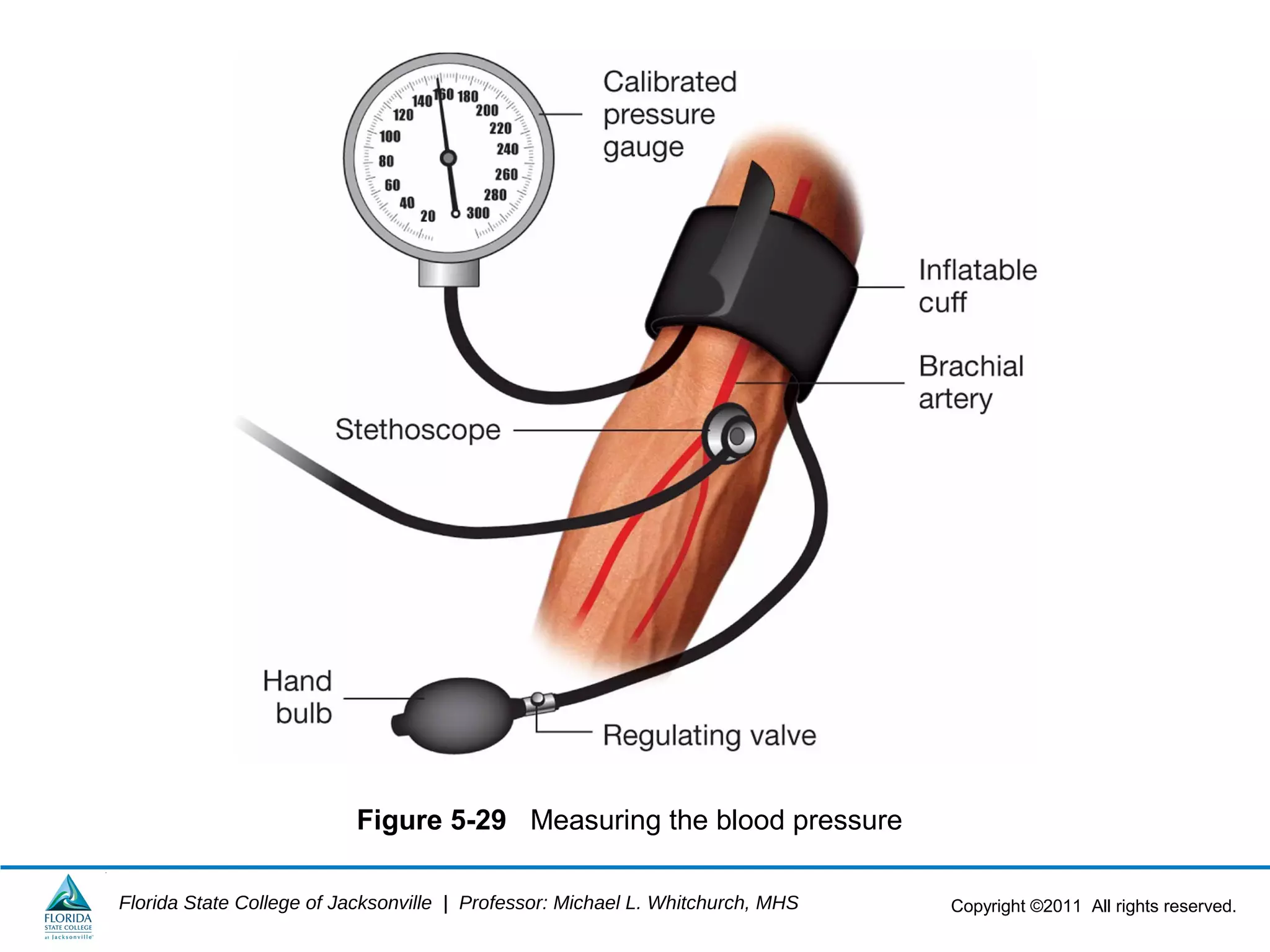
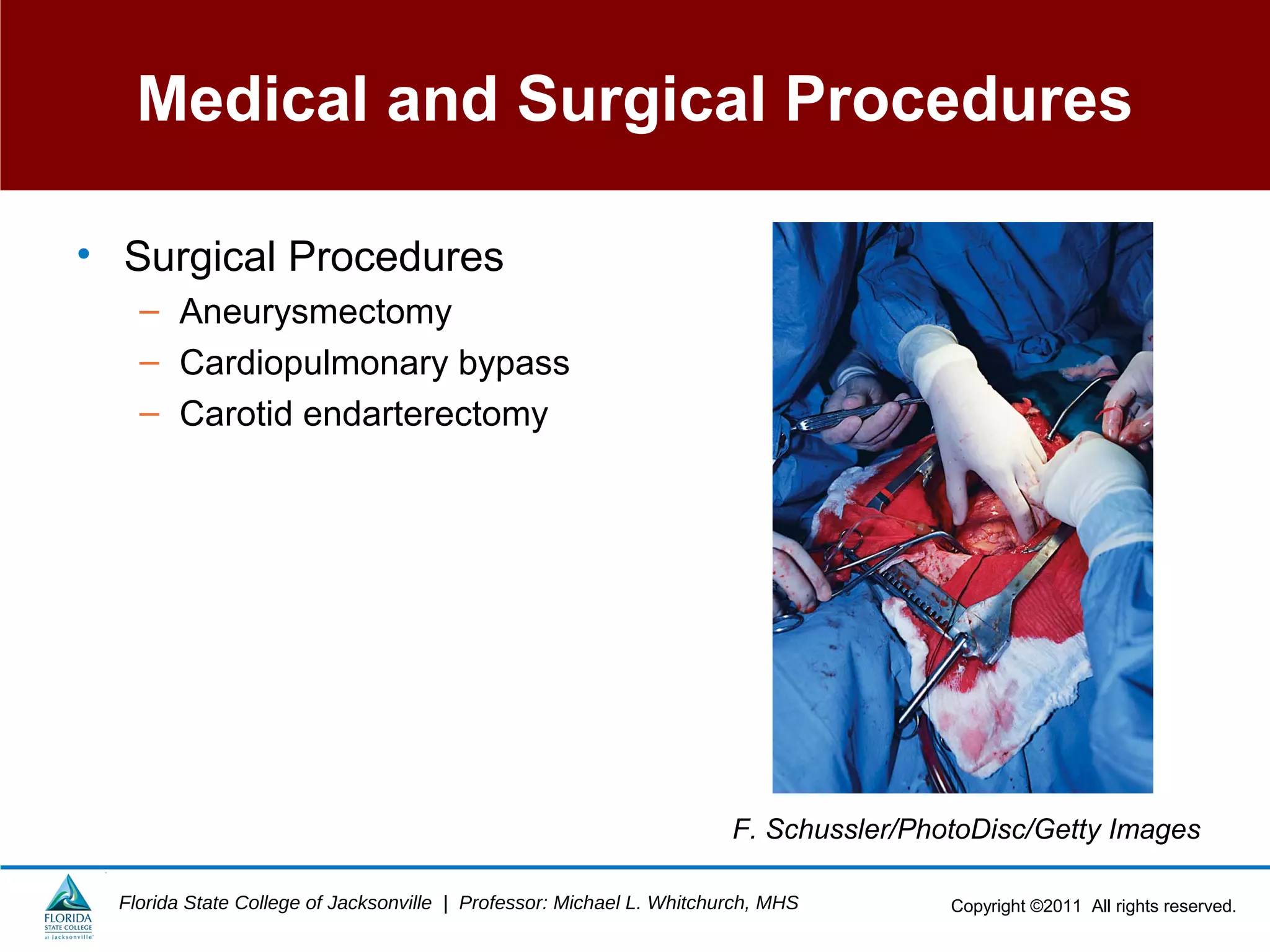
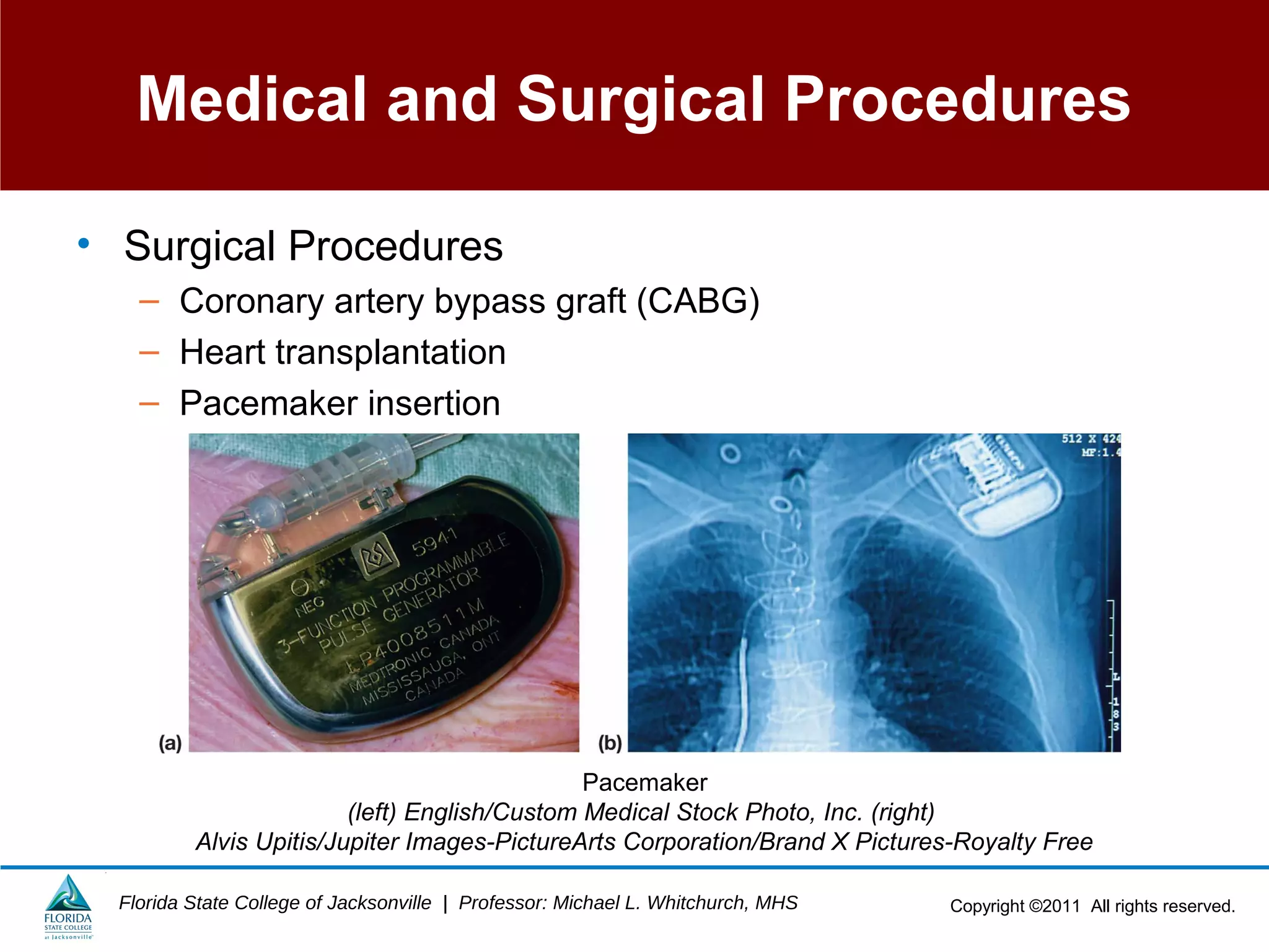
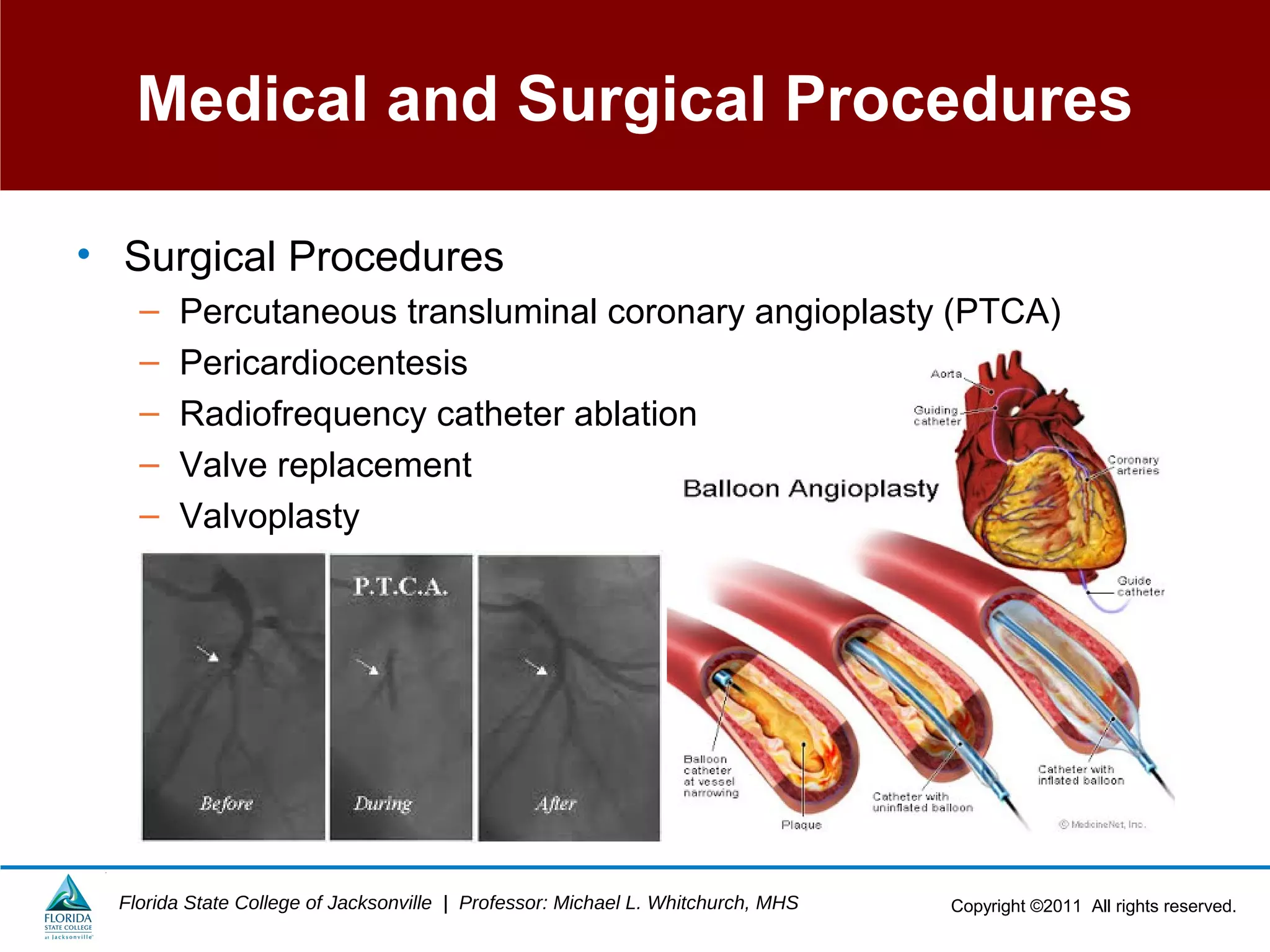
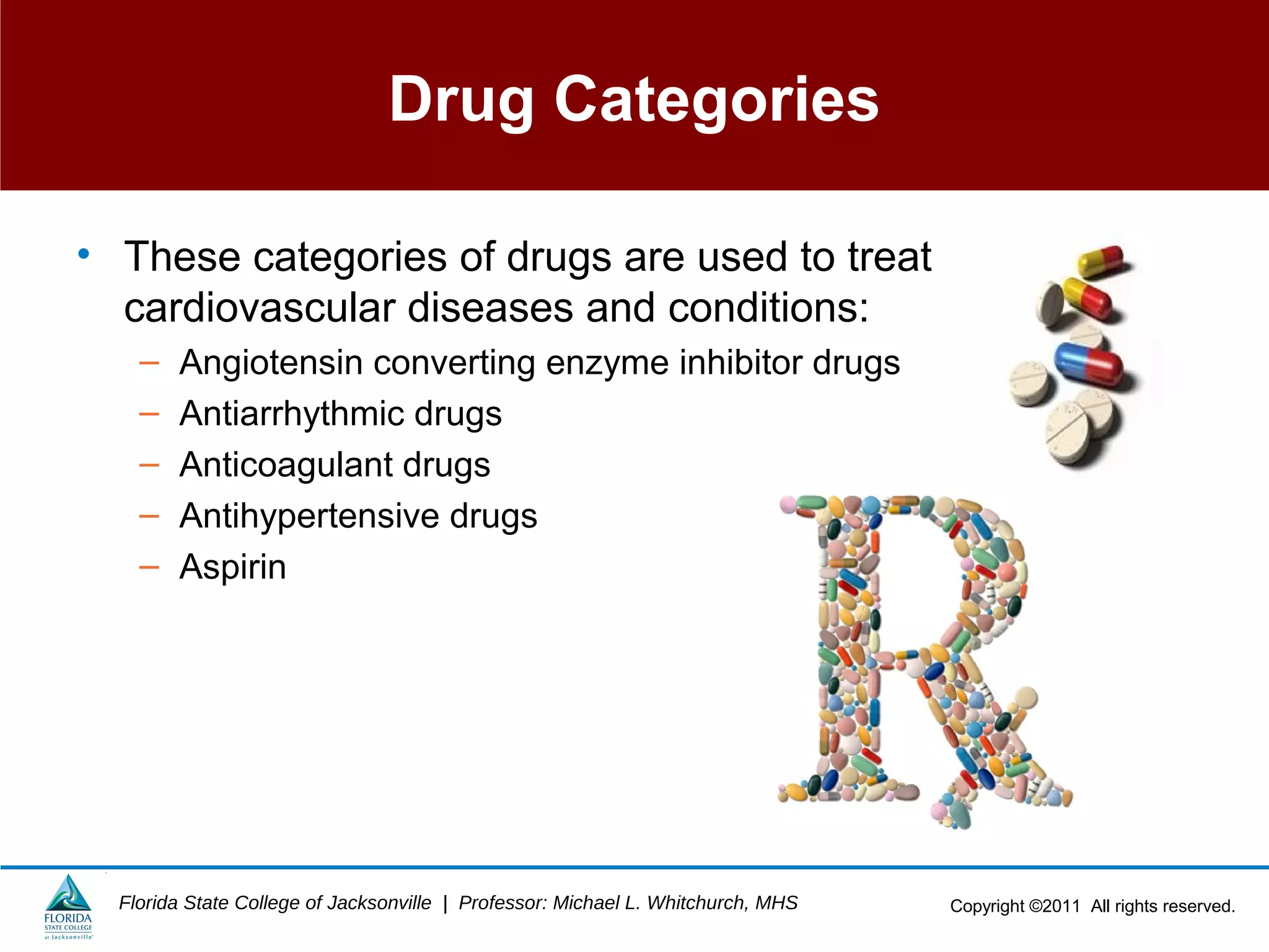
This document provides an overview of the cardiovascular system including its anatomy, physiology, common diseases and conditions, and diagnostic tests. It describes the structures of the heart and blood vessels that make up the cardiovascular system and their functions. Key topics covered include the heart chambers and layers, circulation, conduction system, common cardiovascular diseases like heart attacks and arrhythmias, and laboratory tests used to evaluate cardiovascular health.
The human heart heart length, width, and thickness are 12 cm, 8.5 cm, and 6 cm, respectively. In addition, the mean weight of the heart is 280-340 g in males and 230-280 g in females.
Heart is the center of your cardiovascular system. It is responsible for pumping blood through your body, carrying nutrients in and taking waste out. Having good cardiovascular health is an important factor of optimal wellness.
An adult has a mass of 250-350 grams.
Each day, the average heart beats 100,000 times, pumping about 2,000 gallons (7,571 liters) of blood.
By the end of a long life, a person's heart may have beat (expanded and contracted) more than 3.5 billion times.
Heart has four chambers and four valves present in the heart control the flow of blood in the heart.
The blood vessels are the components of the circulatory system that transport blood throughout the human body. These vessels transport blood cells, nutrients, and oxygen to the tissues of the body. They also take waste and carbon dioxide away from the tissues.
It is a powerpoint presentation that discusses about the lesson or topic: Circulatory System. It also talks about the definition, parts and the concepts about Circulatory System.
The Cardiovascular System: Life's Vital Transport System
The cardiovascular system, comprising the heart, blood vessels, and blood, is a fundamental physiological network in the body.
It facilitates the circulation of oxygen, nutrients, hormones, and immune cells while eliminating waste products.
Essential for maintaining tissue function, energy production, and overall homeostasis.
Defining the Cardiovascular System
The cardiovascular system, also known as the circulatory system, is a complex network responsible for circulating vital substances throughout the body.
Components of the Cardiovascular System
Heart: A muscular organ that pumps blood, generating the force required to propel blood through the blood vessels.
Blood Vessels: A network of tubes that carry blood to and from various body tissues.
Blood: A specialized fluid containing red and white blood cells, platelets, and plasma, essential for nutrient and gas exchange.
overview of heart and its disease
The heart is a powerful muscle that pumps
blood throughout the body by means of a
coordinated contraction.
The contraction is generated by an
electrical activation, which is spread by a
wave of bioelectricity that propagates in a
coordinated manner throughout the
heart.
It is located in thoracic cavity,
posterior to the sternum ,superior
to the diaphragm between the
lungs.
A human heart beats an average of
100,000 times per day. During that
time, it pumps more than 4,300
gallons of blood throughout
the entire body.
EPICARDIUM:
It is the outer layer of the wall of the heart which is composed of
connective tissue covered by epithelium. It is also known as
visceral pericardium.
2) MYOCARDIUM:
It is the muscular middle layer of the wall of the heart. It is
composed of spontaneously contracting cardiac muscle fibers
which allow the heart to contract. It stimulates heart contraction
to pump blood from the ventricles and relaxes the heart to allow
the arteries to receive blood.
ENDOCARDIUM:
It is the inner layer of the heart which consist of epithelial
tissue and connective tissue.
USMLE CVS 005 Blood vessels – Arteries and veins.pdfAHMED ASHOUR
The major blood vessels in the human body form an extensive network that facilitates the transportation of blood, oxygen, and nutrients to various tissues and organs.
Understanding the anatomy and function of major blood vessels is essential for comprehending the circulatory system and diagnosing and treating cardiovascular conditions.
Explore natural remedies for syphilis treatment in Singapore. Discover alternative therapies, herbal remedies, and lifestyle changes that may complement conventional treatments. Learn about holistic approaches to managing syphilis symptoms and supporting overall health.
Title: Sense of Taste
Presenter: Dr. Faiza, Assistant Professor of Physiology
Qualifications:
MBBS (Best Graduate, AIMC Lahore)
FCPS Physiology
ICMT, CHPE, DHPE (STMU)
MPH (GC University, Faisalabad)
MBA (Virtual University of Pakistan)
Learning Objectives:
Describe the structure and function of taste buds.
Describe the relationship between the taste threshold and taste index of common substances.
Explain the chemical basis and signal transduction of taste perception for each type of primary taste sensation.
Recognize different abnormalities of taste perception and their causes.
Key Topics:
Significance of Taste Sensation:
Differentiation between pleasant and harmful food
Influence on behavior
Selection of food based on metabolic needs
Receptors of Taste:
Taste buds on the tongue
Influence of sense of smell, texture of food, and pain stimulation (e.g., by pepper)
Primary and Secondary Taste Sensations:
Primary taste sensations: Sweet, Sour, Salty, Bitter, Umami
Chemical basis and signal transduction mechanisms for each taste
Taste Threshold and Index:
Taste threshold values for Sweet (sucrose), Salty (NaCl), Sour (HCl), and Bitter (Quinine)
Taste index relationship: Inversely proportional to taste threshold
Taste Blindness:
Inability to taste certain substances, particularly thiourea compounds
Example: Phenylthiocarbamide
Structure and Function of Taste Buds:
Composition: Epithelial cells, Sustentacular/Supporting cells, Taste cells, Basal cells
Features: Taste pores, Taste hairs/microvilli, and Taste nerve fibers
Location of Taste Buds:
Found in papillae of the tongue (Fungiform, Circumvallate, Foliate)
Also present on the palate, tonsillar pillars, epiglottis, and proximal esophagus
Mechanism of Taste Stimulation:
Interaction of taste substances with receptors on microvilli
Signal transduction pathways for Umami, Sweet, Bitter, Sour, and Salty tastes
Taste Sensitivity and Adaptation:
Decrease in sensitivity with age
Rapid adaptation of taste sensation
Role of Saliva in Taste:
Dissolution of tastants to reach receptors
Washing away the stimulus
Taste Preferences and Aversions:
Mechanisms behind taste preference and aversion
Influence of receptors and neural pathways
Impact of Sensory Nerve Damage:
Degeneration of taste buds if the sensory nerve fiber is cut
Abnormalities of Taste Detection:
Conditions: Ageusia, Hypogeusia, Dysgeusia (parageusia)
Causes: Nerve damage, neurological disorders, infections, poor oral hygiene, adverse drug effects, deficiencies, aging, tobacco use, altered neurotransmitter levels
Neurotransmitters and Taste Threshold:
Effects of serotonin (5-HT) and norepinephrine (NE) on taste sensitivity
Supertasters:
25% of the population with heightened sensitivity to taste, especially bitterness
Increased number of fungiform papillae
Prix Galien International 2024 Forum ProgramLevi Shapiro
June 20, 2024, Prix Galien International and Jerusalem Ethics Forum in ROME. Detailed agenda including panels:
- ADVANCES IN CARDIOLOGY: A NEW PARADIGM IS COMING
- WOMEN’S HEALTH: FERTILITY PRESERVATION
- WHAT’S NEW IN THE TREATMENT OF INFECTIOUS,
ONCOLOGICAL AND INFLAMMATORY SKIN DISEASES?
- ARTIFICIAL INTELLIGENCE AND ETHICS
- GENE THERAPY
- BEYOND BORDERS: GLOBAL INITIATIVES FOR DEMOCRATIZING LIFE SCIENCE TECHNOLOGIES AND PROMOTING ACCESS TO HEALTHCARE
- ETHICAL CHALLENGES IN LIFE SCIENCES
- Prix Galien International Awards Ceremony
HOT NEW PRODUCT! BIG SALES FAST SHIPPING NOW FROM CHINA!! EU KU DB BK substit...GL Anaacs
Contact us if you are interested:
Email / Skype : kefaya1771@gmail.com
Threema: PXHY5PDH
New BATCH Ku !!! MUCH IN DEMAND FAST SALE EVERY BATCH HAPPY GOOD EFFECT BIG BATCH !
Contact me on Threema or skype to start big business!!
Hot-sale products:
NEW HOT EUTYLONE WHITE CRYSTAL!!
5cl-adba precursor (semi finished )
5cl-adba raw materials
ADBB precursor (semi finished )
ADBB raw materials
APVP powder
5fadb/4f-adb
Jwh018 / Jwh210
Eutylone crystal
Protonitazene (hydrochloride) CAS: 119276-01-6
Flubrotizolam CAS: 57801-95-3
Metonitazene CAS: 14680-51-4
Payment terms: Western Union,MoneyGram,Bitcoin or USDT.
Deliver Time: Usually 7-15days
Shipping method: FedEx, TNT, DHL,UPS etc.Our deliveries are 100% safe, fast, reliable and discreet.
Samples will be sent for your evaluation!If you are interested in, please contact me, let's talk details.
We specializes in exporting high quality Research chemical, medical intermediate, Pharmaceutical chemicals and so on. Products are exported to USA, Canada, France, Korea, Japan,Russia, Southeast Asia and other countries.
The prostate is an exocrine gland of the male mammalian reproductive system
It is a walnut-sized gland that forms part of the male reproductive system and is located in front of the rectum and just below the urinary bladder
Function is to store and secrete a clear, slightly alkaline fluid that constitutes 10-30% of the volume of the seminal fluid that along with the spermatozoa, constitutes semen
A healthy human prostate measures (4cm-vertical, by 3cm-horizontal, 2cm ant-post ).
It surrounds the urethra just below the urinary bladder. It has anterior, median, posterior and two lateral lobes
It’s work is regulated by androgens which are responsible for male sex characteristics
Generalised disease of the prostate due to hormonal derangement which leads to non malignant enlargement of the gland (increase in the number of epithelial cells and stromal tissue)to cause compression of the urethra leading to symptoms (LUTS
New Drug Discovery and Development .....NEHA GUPTA
The "New Drug Discovery and Development" process involves the identification, design, testing, and manufacturing of novel pharmaceutical compounds with the aim of introducing new and improved treatments for various medical conditions. This comprehensive endeavor encompasses various stages, including target identification, preclinical studies, clinical trials, regulatory approval, and post-market surveillance. It involves multidisciplinary collaboration among scientists, researchers, clinicians, regulatory experts, and pharmaceutical companies to bring innovative therapies to market and address unmet medical needs.
Title: Sense of Smell
Presenter: Dr. Faiza, Assistant Professor of Physiology
Qualifications:
MBBS (Best Graduate, AIMC Lahore)
FCPS Physiology
ICMT, CHPE, DHPE (STMU)
MPH (GC University, Faisalabad)
MBA (Virtual University of Pakistan)
Learning Objectives:
Describe the primary categories of smells and the concept of odor blindness.
Explain the structure and location of the olfactory membrane and mucosa, including the types and roles of cells involved in olfaction.
Describe the pathway and mechanisms of olfactory signal transmission from the olfactory receptors to the brain.
Illustrate the biochemical cascade triggered by odorant binding to olfactory receptors, including the role of G-proteins and second messengers in generating an action potential.
Identify different types of olfactory disorders such as anosmia, hyposmia, hyperosmia, and dysosmia, including their potential causes.
Key Topics:
Olfactory Genes:
3% of the human genome accounts for olfactory genes.
400 genes for odorant receptors.
Olfactory Membrane:
Located in the superior part of the nasal cavity.
Medially: Folds downward along the superior septum.
Laterally: Folds over the superior turbinate and upper surface of the middle turbinate.
Total surface area: 5-10 square centimeters.
Olfactory Mucosa:
Olfactory Cells: Bipolar nerve cells derived from the CNS (100 million), with 4-25 olfactory cilia per cell.
Sustentacular Cells: Produce mucus and maintain ionic and molecular environment.
Basal Cells: Replace worn-out olfactory cells with an average lifespan of 1-2 months.
Bowman’s Gland: Secretes mucus.
Stimulation of Olfactory Cells:
Odorant dissolves in mucus and attaches to receptors on olfactory cilia.
Involves a cascade effect through G-proteins and second messengers, leading to depolarization and action potential generation in the olfactory nerve.
Quality of a Good Odorant:
Small (3-20 Carbon atoms), volatile, water-soluble, and lipid-soluble.
Facilitated by odorant-binding proteins in mucus.
Membrane Potential and Action Potential:
Resting membrane potential: -55mV.
Action potential frequency in the olfactory nerve increases with odorant strength.
Adaptation Towards the Sense of Smell:
Rapid adaptation within the first second, with further slow adaptation.
Psychological adaptation greater than receptor adaptation, involving feedback inhibition from the central nervous system.
Primary Sensations of Smell:
Camphoraceous, Musky, Floral, Pepperminty, Ethereal, Pungent, Putrid.
Odor Detection Threshold:
Examples: Hydrogen sulfide (0.0005 ppm), Methyl-mercaptan (0.002 ppm).
Some toxic substances are odorless at lethal concentrations.
Characteristics of Smell:
Odor blindness for single substances due to lack of appropriate receptor protein.
Behavioral and emotional influences of smell.
Transmission of Olfactory Signals:
From olfactory cells to glomeruli in the olfactory bulb, involving lateral inhibition.
Primitive, less old, and new olfactory systems with different path
NVBDCP.pptx Nation vector borne disease control programSapna Thakur
NVBDCP was launched in 2003-2004 . Vector-Borne Disease: Disease that results from an infection transmitted to humans and other animals by blood-feeding arthropods, such as mosquitoes, ticks, and fleas. Examples of vector-borne diseases include Dengue fever, West Nile Virus, Lyme disease, and malaria.
Flu Vaccine Alert in Bangalore Karnatakaaddon Scans
As flu season approaches, health officials in Bangalore, Karnataka, are urging residents to get their flu vaccinations. The seasonal flu, while common, can lead to severe health complications, particularly for vulnerable populations such as young children, the elderly, and those with underlying health conditions.
Dr. Vidisha Kumari, a leading epidemiologist in Bangalore, emphasizes the importance of getting vaccinated. "The flu vaccine is our best defense against the influenza virus. It not only protects individuals but also helps prevent the spread of the virus in our communities," he says.
This year, the flu season is expected to coincide with a potential increase in other respiratory illnesses. The Karnataka Health Department has launched an awareness campaign highlighting the significance of flu vaccinations. They have set up multiple vaccination centers across Bangalore, making it convenient for residents to receive their shots.
To encourage widespread vaccination, the government is also collaborating with local schools, workplaces, and community centers to facilitate vaccination drives. Special attention is being given to ensuring that the vaccine is accessible to all, including marginalized communities who may have limited access to healthcare.
Residents are reminded that the flu vaccine is safe and effective. Common side effects are mild and may include soreness at the injection site, mild fever, or muscle aches. These side effects are generally short-lived and far less severe than the flu itself.
Healthcare providers are also stressing the importance of continuing COVID-19 precautions. Wearing masks, practicing good hand hygiene, and maintaining social distancing are still crucial, especially in crowded places.
Protect yourself and your loved ones by getting vaccinated. Together, we can help keep Bangalore healthy and safe this flu season. For more information on vaccination centers and schedules, residents can visit the Karnataka Health Department’s official website or follow their social media pages.
Stay informed, stay safe, and get your flu shot today!
Tom Selleck Health: A Comprehensive Look at the Iconic Actor’s Wellness Journeygreendigital
Tom Selleck, an enduring figure in Hollywood. has captivated audiences for decades with his rugged charm, iconic moustache. and memorable roles in television and film. From his breakout role as Thomas Magnum in Magnum P.I. to his current portrayal of Frank Reagan in Blue Bloods. Selleck's career has spanned over 50 years. But beyond his professional achievements. fans have often been curious about Tom Selleck Health. especially as he has aged in the public eye.
Follow us on: Pinterest
Introduction
Many have been interested in Tom Selleck health. not only because of his enduring presence on screen but also because of the challenges. and lifestyle choices he has faced and made over the years. This article delves into the various aspects of Tom Selleck health. exploring his fitness regimen, diet, mental health. and the challenges he has encountered as he ages. We'll look at how he maintains his well-being. the health issues he has faced, and his approach to ageing .
Early Life and Career
Childhood and Athletic Beginnings
Tom Selleck was born on January 29, 1945, in Detroit, Michigan, and grew up in Sherman Oaks, California. From an early age, he was involved in sports, particularly basketball. which played a significant role in his physical development. His athletic pursuits continued into college. where he attended the University of Southern California (USC) on a basketball scholarship. This early involvement in sports laid a strong foundation for his physical health and disciplined lifestyle.
Transition to Acting
Selleck's transition from an athlete to an actor came with its physical demands. His first significant role in "Magnum P.I." required him to perform various stunts and maintain a fit appearance. This role, which he played from 1980 to 1988. necessitated a rigorous fitness routine to meet the show's demands. setting the stage for his long-term commitment to health and wellness.
Fitness Regimen
Workout Routine
Tom Selleck health and fitness regimen has evolved. adapting to his changing roles and age. During his "Magnum, P.I." days. Selleck's workouts were intense and focused on building and maintaining muscle mass. His routine included weightlifting, cardiovascular exercises. and specific training for the stunts he performed on the show.
Selleck adjusted his fitness routine as he aged to suit his body's needs. Today, his workouts focus on maintaining flexibility, strength, and cardiovascular health. He incorporates low-impact exercises such as swimming, walking, and light weightlifting. This balanced approach helps him stay fit without putting undue strain on his joints and muscles.
Importance of Flexibility and Mobility
In recent years, Selleck has emphasized the importance of flexibility and mobility in his fitness regimen. Understanding the natural decline in muscle mass and joint flexibility with age. he includes stretching and yoga in his routine. These practices help prevent injuries, improve posture, and maintain mobilit
TEST BANK for Operations Management, 14th Edition by William J. Stevenson, Ve...kevinkariuki227
TEST BANK for Operations Management, 14th Edition by William J. Stevenson, Verified Chapters 1 - 19, Complete Newest Version.pdf
The cardiovascular system, also called the circular system, is one of the busiest systems in the body and works constantly to transport nutrients, oxygen, and waste in and out of the body. Your heart never takes a break. To understand the complexities of the circulatory system, you need to understand the structures that compose the heart, other structures that are part of the system, and how they are all related. You must become familiar with the types of tests and procedures that are available to diagnose and treat cardiovascular problems, which type of cardiovascular specialists can treat the variety of problems that occur within this body system, and the specialized terms and abbreviations associated with the cardiovascular system. You'll see the combining form cardio-throughout this lesson. Several other commonly used prefixes are found in the lesson, such as myo-, endo-, peri-,and hyper-. Many vessels have names that are the same as bones, so you will already know some of these terms.
The heart is a complicated organ. Despite being roughly the size of a clenched fist, your heart averages 70 beats per minute and more than 30 million beats per year to push blood throughout your body. This blood carries nutrients to every cell and then carries the waste products away for elimination. Without nutrients, cells cannot survive. Learning the anatomy of the cardiovascular system will help with your pursuit of a career in health or medicine.
The heart is like a duplex apartment-divided into two parts with a definite wall in between the parts, with each part or apartment containing two rooms. Instead of apartment A and B, we have the right and left heart. Remember when looking at the heart, that you are looking at the patient, not at yourself. So the right side of the patient is on the left side of the illustration.In the heart, the rooms are called chambers, which is actually an old word for room. Two chambers are on the top of the heart, and two are on the bottom. Each top chamber is an atrium (atria is the plural). The two lower chambers are each a ventricle. The wall dividing the heart into the two apartments is the septum. You had this word in the Pulmonology chapter. Do you remember that it divided the nasal cavity into two parts?And the inferior point of the heart is the apex. Just as apartment rooms are separated by walls with doorways and doors, the chambers of the heart have openings and valves to make sure the blood flows only in one direction. If blood flowed in both directions, it would be like people trying to get on and off a bus through the same door at the same time. Instead, it's better if people get on using the front door, and get off through the back door. It's faster and more people can move where they need to go.The heart has a doorway or valve between the two chambers and at the exit of the lower chambers, like a front door. In the upper chambers, the "back doors" are always open. There are no valves there where blood enters the heart.
Let's start on the right side of the heart. The valve between the right atrium and right ventricle is the tricuspid valve. It has three small cusps or leaflets of tissue that can open and close. The cusps allow the blood to flow from the atrium into the ventricle, but not in reverse.After blood has delivered its oxygen and nutrients to the body tissues, it picks up waste and carbon dioxide and brings it to the heart. This blood enters the right atrium, goes through the tricuspid valve into the right ventricle, and then leaves the ventricle to go to the lungs. However, the blood must pass through that "front door" the pulmonary valve. The name of the valve tells you where the blood is going-to the lungs to get rid of carbon dioxide and pick up fresh oxygen.
After blood goes to the lungs, it returns to the heart with a fresh load of oxygen, and much less carbon dioxide. This fresh blood reenters the heart on the left side now.Blood enters the open "back door" of the left atrium, and goes through the mitral valve (or bicuspid valve) into the left ventricle. As you can tell from the word biscupid, this valve only has two cusps. Do you remember how many lobes there were in the right and left lungs? It's the same number as the number of cusps in the valves between the atria and ventricles. On the right side, there are three lobes and three cusps; on the left side, there are two lobes and two cusps in the mitral valve.Blood leaving the left ventricle must go through that front door," the aortic valve. Again, the name of this valve tells you where the blood is going-into the aorta.
Heart Layers and MediastinumThe heart is a pump and it takes muscle tissue to make this pump work. This specialized tissue is called the myocardium which is made of cardiac muscle cells.With so much blood continuously flowing through the heart, it is important that the lining of the heart is very smooth and the endocardium is this smooth layer. Around the outside of the heart is the pericardium, which is composed of two layers, like the pleura of the lungs. And also like the pleura, the two layers of the pericardium create pericardial fluid between them, which prevents friction as the heart beats.The heart and pericardial sac lie between the lungs in the mediastinum. Also in this space are the large blood vessels that enter and leave the heart, the trachea, esophagus and thymus gland.
Table is on page 6 of Ch. 5.
Arteries All blood vessels that leave the heart and carry blood from the heart are arteries. Smaller arteries are arterioles. (Remember the ending –ioles?) Arteries carry bright red blood, rich in oxygen, away from the heart to most of the body, while the pulmonary artery carries darker blood with a higher percentage of carbon dioxide from the right ventricle to the lungs. Arteries have a fairly thick layer of muscle in the artery wall that allows the artery to constrict or relax. These movements are vasoconstriction and vasodilation. When arteries are constricted, blood pressure is increased, and blood pressure lowers when the arteries are dilated, opening the vessel lumen.The heartbeat can be felt in arteries that are close to the surface, such as the pulse in the wrist.
CapillariesCapillaries in some ways are the most important blood vessels, and they are the smallest. They are so small that the walls are only one cell thick, and blood cells must march through in single file.Yet capillaries provide the place where transfer between blood and cells can take place, bringing food and oxygen to cells, and removing carbon dioxide waste.Capillaries are located between the arterioles and venules, completing a loop of circulation between the heart and the tissues of the body.
VeinsVeins are blood vessels that return blood to the heart, and small veins are venules. Most of the "old" blood is returning from the tissues with more carbon dioxide, and it is dark red. Think about the color of the blood when you have a routine blood test done and blood is collected from a vein in your arm.But blood returning to the heart from the lungs, after the blood picks up a fresh load of oxygen, also uses veins. These are the pulmonary veins, and the "fresh" blood is bright red.Veins have only a small amount of muscle in their walls. Also, it is important that blood in your feet return to the heart efficiently, or you will have swollen ankles. In order to do this, veins have valves which prevent blood from flowing backwards toward your feet. It takes muscle action in your legs to move the blood upward.
Textbook page 9 goes into greater detail about the ascending aorta, aortic arch, and arterial branches. I just summarized in slide 37.
CirculationOkay, now let's trace blood in its journey through the body. Let's start at the vena cavae. From the heart to the body and back to the heart is called the systemic circulation. From the heart to the lungs and back to the heart is the pulmonary circulation.
. The Heartbeat: ConductionThe heart is a muscle, and for muscle to contract-or the heart to pump-an electrical stimulus or nerve message must be sent to the muscle tissue. The heart has a special system of nervous tissue, called the conduction system, to make sure the heartbeats are constant.In the right atrium is a small area of nervous tissue that is the sinoatrial node (SA node), or the natural pacemaker. This node is responsible for starting every single heartbeat by itself. When the SA node fires, the stimulus causes the atria to contract, pushing blood through the one-way valves into the ventricles. But the stimulus isn't finished. It continues to the AV node (atrioventricular node). As you can tell by the name, it is located at the junction of the right atrium and ventricle.The electrical stimulus goes down the septum through the bundle of His and then separates to the right and left side, where a nerve ends at a Purkinje fiber. These fibers cause the ventricles to contract, sending blood to the lungs and throughout the body.
Regulation of the HeartbeatWhen the heart is contracting, that period is called systole. Between each heartbeat is a short rest period called diastole, when the atria just passively fills with blood from the vena cavae and pulmonary veins.When not exercising or being anxious, your heart beats about 60–80 beats a minute (bpm). But obviously, if you are moving a lot, your body needs more oxygen, so the nervous system sends a message to increase the rate.Likewise, when you are resting, reading, or sleeping, the heart rate slows.
The heart pumps blood throughout the body like a machine that never stops. As with any machine that is constantly active, many things can go wrong with your heart. It can experience muscle weakness or dysfunction, and circulation might be compromised because of problems with veins, arteries, valves, electrical impulses, or even diseases, such as infections. Congestive Heart Failure Hypertension or coronary heasrt disease can cause the heart to hypertrophy. This enlarged heart, known as cardiomegaly, cannot keep enlarging forever. The muscle will weaken and the heart cannot pump enough blood with each heartbeat. The result is congestive heart failure (CHF), a serious problem that can be life threatening.Peripheral edema is one symptom of CHF if the weakness is on the right side of the heart. Left-sided failure causes pulmonary edema. Myocardial InfarctionOne of the most severe myocardial disorders is myocardial infarction, which results in actual death of some of the muscle tissue. If enough of the tissue is affected, a person can die.Atherosclerosis or a blood clot can completely obstruct a coronary artery, which is known as an infarct. No blood gets through to the muscle tissue at all. Thus, the tissue first reacts with severe chest pain, and then the tissue dies. This is necrosis.While some TV ads say that the person should be quickly given an aspirin to prevent further clotting, first make sure the person is not allergic to aspirin!
Infections of the Heart Layers The lining of the heart and blood vessels is the endocardium. This lining, especially on defective valves, can trap bacteria and become infected, resulting in acute or subacute bacterial endocarditis.Infection or inflammation of the pericardial sac, pericarditis, results in an increased amount of fluid in the sac. This fluid squeezes the heart, making it more difficult to pump efficiently. This problem is cardiac tamponade.Valves that do not close correctly cause an abnormal heart sound called a murmur, kind of like a squeaky door. Rheumatic Heart Disease While many people ignore getting treatment for strep throat, it is a common cause for rheumatic heart disease. The strep bacteria like to cause damage to the mitral and aortic valves, causing growth of vegetations and stenosis. This can be prevented with initial antibiotic therapy for the strep infection.Infection of the mitral valve or a congenital deformity is the cause of mitral valve prolapse. Regurgitation of blood makes an abnormal heart sound as the blood flows backward through the valve.
Conduction Disorders Have you ever had a bad starter on your car? If so, you know that until it's fixed, you probably aren't going anywhere. Your battery can be okay, but if the starter isn't working, it seems like the car is "dead." The heart can have a similar problem. The starter in the heart is the SA node, and if it is not working well, the heart can have a number of problems.Like a "dead" car, a heart that receives no starting stimulus from the SA node for each heartbeat has asystole, also known as cardiac arrest. Perhaps, like a car starter that works intermittently (it always seems to work at the repair shop), the heart's pacemaker works erratically, resulting in bradycardia or tachycardia. Or even more interestingly, the heart can have sick sinus syndrome, in which these two disorders alternate, sometimes with bradycardia and sometimes with tachycardia.An electrical rhythm disorder, such as just described, is known as an arrhythmia or the alternate term, dysrhythmia. Do you know why the first term has two "r"s and the second term only has one? I'll tell you on the next page.
Coronary Artery Disorders Hypertension or coronary heasrt disease can cause the heart to hypertrophy. This enlarged heart, known as cardiomegaly, cannot keep enlarging forever. The muscle will weaken and the heart cannot pump enough blood with each heartbeat. The result is congestive heart failure (CHF), a serious problem that can be life threatening.Peripheral edema is one symptom of CHF if the weakness is on the right side of the heart. Left-sided failure causes pulmonary edema. The coronary arteries are small arteries on the surface of the heart that are responsible for getting food and oxygen to the myocardium. These arteries can become clogged with plaque (atherosclerosis) or can spasm. Either way, less blood reaches the heart muscle and ischemia results.Angina pectoris causes severe, crushing pain in the chest that can spread into the neck or down the left arm. The primary treatment is nitroglycerin, an old ingredient in gunpowder, or another nitrate drug. This drug relaxes and opens the coronary arteries.A general term for any disease of the heart muscle is cardiomyopathy. Can you see the meaning in the word parts?
Renumber as Fig. 5-21.
Blood Tests Blood tests are crucial when diagnosing chest pain. Cardiac enzymes rise when myocardial tissue cells are damaged or die.A lipid profile test determines the levels of HDL and LDL in the blood. HDL is the "happy" or good cholesterol level that should be high, while the LDL is the "lousy" or harmful cholesterol level that should be low to be healthy.A C-reactive protein test reveals the presence of inflammation in the body. Inflammation of blood vessels can result from inflammation anywhere else in the body. Inflamed coronary blood vessel walls become rough, which is a good place for a blood clot to form, possibly leading to a myocardial infarction.
Conduction Monitoring The most common procedure, which you might even have had at some time, is electrocardiography, abbreviated either as ECG or EKG. The "K" in EKG comes from the Greek word for heart, kardia. The printed record of electrical heart activity is an electrocardiogram. The printout is on long pieces of narrow paper, and sometimes a printout is called a rhythm strip.A Holter monitor is used to record a 24-hour electrocardiogram. This allows the physician to note differences in heart rate and rhythm during daily activities. The patient keeps a record of these activities as well as a diary of meals and symptoms.Telemetry is another method of recording heart rate and rhythm in a continuous format on a monitor. Telemetry is usually used in a hospital setting, such as Intensive Care, Coronary Care Unit, or even the Emergency Department.
Diagnostic Heart Procedures The cardiac exercise stress test does indeed place stress on the patient's heart while walking on a treadmill. The heart and blood pressure are monitored during the test. The treadmill angle increases throughout the test, causing the heart to work harder.A more complex procedure is cardiac catheterization. A small plastic tubing, or catheter, is inserted into a vein and threaded through the vein into the heart. Pressure readings are taken and a dye is injected so that the heart chambers can be seen.If the catheter is inserted on the left side, the dye outlines the coronary arteries and their condition can be noted. Sometimes follow-up procedures are necessary if those arteries have any type of blockage.
Medical Procedures The simplest medical procedure is one you have probably experienced-auscultation. The physician can learn a lot about the heart's rhythm, rate, and state of the heart valves just by listening with a stethoscope.Another procedure done for all patients who visit the doctor's office is the taking of vital signs, which includes TPR (temperature, pulse, and respirations) and blood pressure (BP or B/P). Typically, the pulse part of TPR is taken at the radial artery in the wrist, although the pulse can be taken at other pulse points, such as the carotid pulse used at times by paramedics. The apical pulse requires a stethoscope.The systolic (higher number when the heart is pumping) and diastolic (lower number when the heart is resting) blood pressures are measured with a sphygmomanometer.Cardioversion is a much more serious procedure used to restore a normal rhythm for a person with an arrhythmia. Defibrillation is a procedure using a defibrillator to stop the rapid and ineffective pulse of ventricular fibrillation.
Renumber as Fig. 5-30.
.
Arterial Surgical Procedures The term for removing a dangerous aneurysm is aneurysmectomy. Sometimes an artificial artery is used to replace the defective segment.While plaque in the coronary arteries is dangerous, because it could lead to a heart attack, plaque in the carotid arteries restricts blood flow to the brain. Carotid endarterectomy is an effective surgical procedure for enlarging the lumen of these important arteries.Other methods for opening the lumens of plaque-filled arteries are balloon angioplasty and inserting a stent.
Surgical Procedures Do you remember the danger involved with pericarditis-that of cardiac tamponade? The surgical procedure, pericardiocentesis, removes excess fluid from the pericardial sac using a needle. This gives the heart more room to fill with blood and to pump more effectively.Valve replacement is a good solution when a diseased valve does not open sufficiently or close completely, which interferes with proper blood flow. Usually, an artificial valve is placed in the heart.Sometimes, a valve just needs a bit of reconstructive surgery, called valvoplasty.