Download as PDF, PPTX
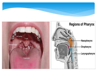
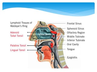
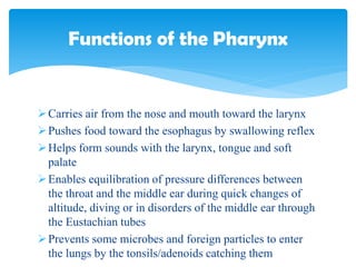
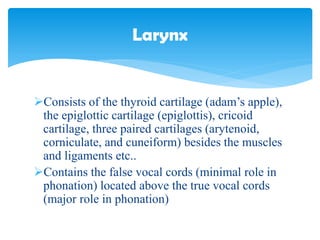
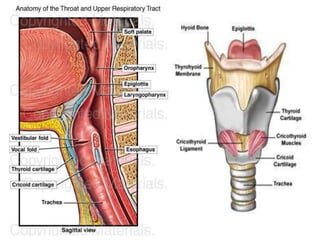
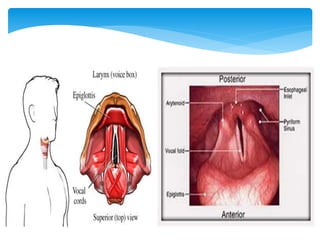
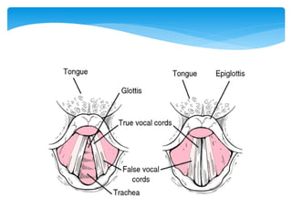
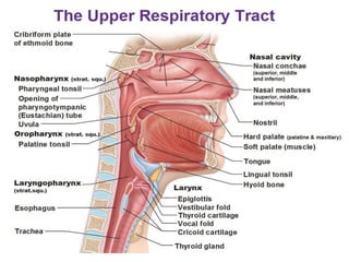
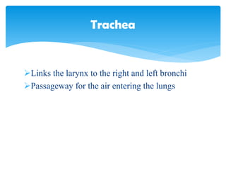
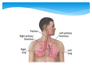
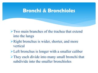

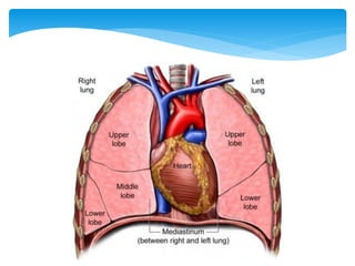
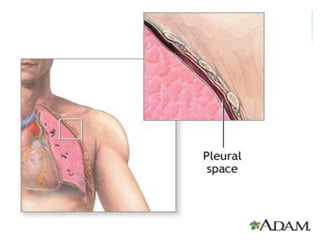
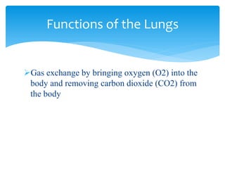
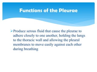

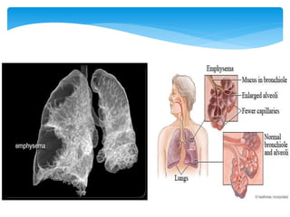

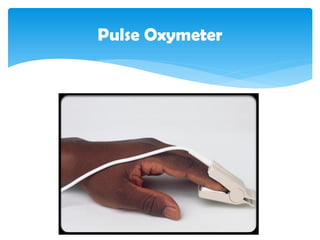
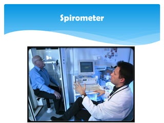


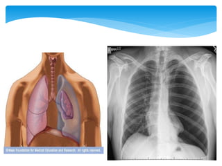


The document provides a detailed overview of the human respiratory system, categorizing its components into the upper and lower respiratory tracts, including structures like the nose, pharynx, larynx, trachea, bronchi, and lungs. It outlines the functions of these structures, such as air passage, sound production, and gas exchange, as well as common disorders affecting the respiratory system. Additionally, it discusses various respiratory volumes, capacities, and tests used to assess respiratory health.


