Pleural effusion (dr. mahesh)
•Download as PPTX, PDF•
111 likes•26,414 views

Pleural Effusion (Radiology)

Recommended
More Related Content
What's hot
What's hot (20)
Viewers also liked
Viewers also liked (20)
Similar to Pleural effusion (dr. mahesh)
Similar to Pleural effusion (dr. mahesh) (20)
More from Bangabandhu Sheikh Mujib Medical University (BSMMU)
More from Bangabandhu Sheikh Mujib Medical University (BSMMU) (10)
Recently uploaded
Recently uploaded (20)
Pleural effusion (dr. mahesh)
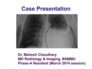
- 1. Dr. Mahesh Chaudhary MD Radiology & Imaging, BSMMU Phase-A Resident (March 2014 session)
- 2. PLEURAL EFFUSIONS DEFINITION- A COLLECTION OF FLUID BETWEEN THE PARIETAL PLEURA AND VISCERAL PLEURA.
- 3. The Right Lung -Three lobes-the superior, middle and inferior, which are separated by the horizontal fissure and the oblique fissure. -10 bronchopulmonary segments The Left Lung -Two lobes which are separated by the oblique fissure. -10 bronchopulmonary segments ANATOMY IN A HEALTHY LUNG
- 5. The main anatomy affected by pleural effusions are the layers in the Lung There are two layers-the parietal pleura and the visceral pleura. • At the Hilum, the parietal pleura folds back on itself to become the visceral pleura. The pleural fluid contains – -contains about 5-15ml of fluid at one time -about 100-200ml of fluid circulates though the pleural space within a 24-hour period -has an alkaline pH of about 7.60 - 7.64 Protein content less than 2% (1-2 g/dL) Glucose content similar to that of plasma Mesothelial cells Macrophages Lymphocytes (few) Sodium, potassium and calcium concentrations similar to that of interstitial fluid. Lactate Dehydrogenase concentration of less than 50% of that of plasma
- 6. ANATOMY OF A HEALTHY LUNG A pleural effusion is an accumulation of fluid between the parietal pleura and the visceral pleura. Chest X-ray frontal view: 100-200ml pleural fluid ANATOMY OF A LUNG WITH A PLEURAL EFFUSION
- 8. ANATOMY & PHYSIOLOGY OF A LUNG WITH A PLEURAL EFFUSION • The fluid accumulates due to the over production of pleural fluid by the mesothelial cells and separates the visceral and parietal pleura. • This fluid can not be drained by the lymphatic system, and so therefore continues to accumulate, resulting in a pleural effusion. • The accumulation of fluid may also be due to changes in hydrostatic pressure or oncotic pressure. The lung has the natural tendency to collapse towards the hilum and this is opposed by forces of similar magnitude in the chest wall tending to expand outward. Thus the parietal and visceral pleura are kept in close apposition. If increase fluid or air collect in the pleural space ,the effect of outward forces on the underlying lung is diminished, and the lung tend to retract toward its hilum.
- 9. Aetiology
- 10. There are 4 different fluids which can accumulate in the pleural space. • Blood HAEMOTHORAX • Pus EMPYEMA • Chyle CHYLOTHORAX • Serous fluid HYDROTHORAX • They can further be classified into TRANSUDATES and EXUDATES depending on – Chemical composition – Mechanism of fluid formation Light’s criteria: Transudate vs. Exudate • Pleural fluid protein / serum protein > 0.5 Pleural fluid LDH / serum LDH > 0.6 Pleural fluid LDH > 2/3 ULN serum LDH
- 11. Pathophysiology Hydrostatic Pressure Oncotic pressure Increased peritoneal fluid
- 12. Mechanisms for pleural fluid accumulation: • Increased hydrostatic pressure (Eg. CCF) • Reduced plasma oncotic pressure (Eg. Hypoproteinaemia) • Increased capillary permeability (Eg.TB, Tumour ) • Reduced lymphatic drainage from pleural space (Obstrustioin by tumour, TB, radiation) • Transdiaphragmatic passage of fluid (Eg. Liver disease, Acute pancreatitis) .
- 13. Transudates • Clear, pale yellow, watery substance • Increase hydrostatic pressure, • Decrease oncotic pressure • Common causes: Congestive heart failure Cirrhosis of the Liver Nephrotic syndrome Hypoproteinaemia Hypothyroidism Acute rheumatic fever
- 14. Exudates • Pale yellow and cloudy substance, has a low pH • Influenced by local factors where fluid absorption is altered (inflammation, infection, cancer) • Rich in white blood cells. • Common causes: Pulmonary TB Pneumonia Bronchial carcinoma Pulmonary infarction Collagen disease (SLE, RA) Lymphoma Meig’s syndrome (Right pleural effusion, Ascites, Ovarian fibroma)
- 15. Blood stained fluid Tends to loculate early CT scan shows higher density measurement Common causes: -Chest injury -Bronchial carcinoma -Pulmonary infarction -Lymphoma Haemothorax
- 16. Chylothorax • Milky fluid due to lymph and fats • Chyle leaks from the thoracic duct due to -damage to the lymphatic vessels. -lymphatic obstruction (tumor) or trauma • High triglyceride levels found in fluid analysis • Common causes: • Traumatic (thoracic surgery), trauma to thoracic duct • Neoplastic ( Bronchial carcinoma, metastasis) • Infective (TB) • Lymphoma (involving thoracic duct)
- 17. Empyema • Pus in pleural space • Yellow, cloudy, and foul odor • Has a pH > 7.2 • Common causes: Pneumonia Rupture of lung abscess, Rupture of sub-phrenic abscess Tuberculosis Infected chest wounds Secondary infection during aspiration of pleural fluid
- 18. Diagnosis of Pleural Effusions • Medical history • Physical examination • Plain film chest x-ray – first line imaging • CT • Ultrasound imaging Diagnosing Pleural Effusions through Imaging
- 19. Characteristics on a supine chest radiograph • Fluid accumulates posteriorly • Affected hemi-thorax appears whiter or paler grey • Apparent thickening of the pleura • Approx 200 mls of fluid present before abnormal pale grey appearance is produced
- 20. First line imaging – Chest x-ray Clear right side hemi-diaphragm and sharp costophrenic angle Area of homogenous Whiteness, with loss of hemi- diaphragm Meniscus shaped upper border Features on a PA or AP erect radiograph
- 21. A large right side pleural effusion The heart has been pushed towards the left side by the fluid Entire white-out of right hemi-thorax
- 22. Lateral decubitus chest radiograph Free layering pleural effusion At least 100ml pleural fluid is necessary
- 26. Loculated fluid
- 28. Loculated effusion (elliptical, pointed margins) in left major fissure CT Scan
- 29. Pleural Effusion Diagnosis through CT Imaging
- 33. CT signs: Pleural effusion vs ascites.
- 34. 4 signs 1.Displaced crus sign: Pleural fluid may collect posterior to the diaphragmatic crux and therefore displace the crus anteriorly, whereas ascites collects anterior to the crus and may cause posterior displacement. 2.Diaphragm sign: As an extension of the displaced crus sign, Any fluid that is on the exterior of the dome of the diaphragms in the pleura, whereas any that is within the dome is ascites 3.Interface sign: The interface between the liver or spleen & pleural fluid is said to be less sharp than that between the liver or spleen and ascites 4.Bare area sign: The peritoneal coronary ligament prevents ascitic fluid from extending over the entire posterior surface of the liver, whereas in a free pleural space, pleural fluid may extend or over the entire posterior costophrenic recess behind the liver
- 35. Ultrasound • No radiation, • Small effusions missed on CXR • Even 20-25 ml of fluid can be detected • Transudate-Anechoic, Exudative- Reflectative +/- • Identify pleural thickening and masses • Used to guide thoracocentisis
- 36. Patient position • Patient seated, arms folded, leaning forward • Unwell patient imaged semi-supine
- 37. MRI • Not used to image pleural effusion • Incidental finding
- 38. Treatment • Needed if patient becomes breathless • Small effusions are left and ‘observed’ • Usually directed at underlying cause (antibiotics for pneumonia) • Underlying cause treated effusion will go away for good • If not it will return within few weeks
- 39. Thoracocentisis • Invasive procedure • Removes fluid from pleural space • Allows lung to expand, making breathing easier • Guided using ultrasound
- 40. Pleurodesis • Chemical inserted into pleural space • Parietal and visceral layers become irritated • Closes space • Painful Pleuroperitoneal Shunt • Internal shunt • Fluid drains from chest into abdominal cavity Pleurectomy • Operation to remove the pleura • Most severe cases
- 42. Have a nice day
Editor's Notes
- Firstly I am going to review the anatomy and physiology of a healthy lung, and then we will consider the anatomy of a lung with a pleural effusion. The right lung accountable for 56% of the total lung volume, and is divided up into 3 lobes-the superior, middle and inferior lobe. This diagram nicely demonstrates these lobes, and the fissures which separate them. (point on the oblique and horizontal fissure)The left lung is slightly smaller as it makes up 44% of the total lung volume, due to the asymmetrical position of the heart. The left lung only has two lobes which are separated by an oblique fissure.
- The lungs have two membranes-the parietal pleura and the visceral pleura. The parietal pleura is the outside membrane which lines the thoracic cavity and the visceral pleura is the inside membrane which lines the lungs. The parietal pleura receives its blood supply from the systematic circulation and contains sensory nerve endings. The visceral pleura receives its blood supply from the low pressure pulmonary circulation and has no sensory nerve endings.The parietal pleura folds back on itself at the hilum to become the visceral pleura, making it a continuous membrane.The mesothelium plays a huge part in pleural effusions. It is a membrane that forms the lining of the pleura, and includes the pleura, the pericardium (the lining around the heart) and the peritoneum (the lining around the abdominal cavity). The main function of the mesothelium is to a produce serous fluid which acts a lubricate that is secreted between the parietal pleura and the visceral pleura to reduce the friction and provide a non-adhesive, protective surface to help facilitate movement.The volume of this pleural fluid in health is around 10ml and has a pH of 7.60- 7.64. there is an oncotoic pressure of 25cm H2O.The contents and characteristics of pleural fluid can alter when a pleural effusion occurs, but typically when the lung is healthy the pleural fluid contains glucose content similar to that of plasma, mesothelial cells, macrophages and lymphocytes. It also contains sodium, potassium and calcium concentrations similar to that of interstitial fluid, and Lactate Dehydrogenase which lizzie will explore later.
- This diagram nicely demonstrates the difference in appearance between the anatomy of a healthy lung and that of a lung with a pleural effusion. As you can see there is an accumulation of fluid between the parietal pleura and the visceral pleura.
- Now we will look at how this anatomy alters when a pleural effusion occurs.The fluid which accumulates tends to be pleural fluid, although it can also be blood or chyle, which is a milky fluid consisting of lymph and fat.The pleural fluid accumulates due to the over production of pleural fluid by the mesothelial cells, and as it can not be drained by the lymphatic system, it continues to collect between the parietal pleura and the visceral pleura resulting in a pleural effusion.Changes in hydrostatic pressure or oncotic pressure may also be responsible for a pleural effusion.Other changes to the anatomy may include pleural thickening and mediastinal pleural thickening, which can be spotted easily using CT, which sadie will cover later in the presentation. Another typical change to anatomy is the appearance of pleural nodules which can range from 2mm to 4cm. These appear as small, round shadows which can appear anywhere in the lung field. I have found this x-ray which nicely demonstrates them.The accumulation of fluid may also cause an increase in pressure, which results in a typical symptom of a pleural effusion of chest pain, although this tends to only occur once there is 500ml of accumulated fluid.
- Filling both CP angle and parallelly extending up the lateral chest wall Sign of increased left arterial pressure or lymphatic spread
- Contour of diaphragm is altered & apex being more lateral than usual Left- increased distance between the gastric bubble and lung baseMove with change of postureDD- Retrocardiac mass, Subphrenic abscess/collection, Phrenic nr palsy, Basal pneumonitis
- Requires less fluid to cause blunting of Posterior CP angle than Lateral CP angleDisplaces highest point of affected hemi diaphragm laterally
- Often associated with free other pleural fluid and may extend into the fissure (fig)…. Rt Para tracheal shadow increased due to enlarged LNComparatively little depth but considerable width, rather like a convex lensExtrapleural opacities tend to have much sharper outlineDiff between pleural mass, thickening or loculated fluid is difficult in CXR ,,, need USG or CT Pseudo or Vanishing tumour
- Occurs secondary to pleural disease or adhesionsNo change In position with change in posture
- CT scan of the chest which demonstrates loculated pleural effusion in the left major fissure (arrow) in a patient after coronary bypass. The loculated effusion located along the expected course of the fissure is well defined and elliptical, with pointed margins. The Effusion is similar to water in its attenuation therefore its appearance is that of fluid.
- Axial CT image of the lower chest in patient with acute lymphoblastic leukemia. A small dependent, crescent-shaped area with attenuation similar to that of water in the right hemithorax is consistent with a tiny right pleural effusion (arrow).
- CT scan of the chest demonstrates a mass in the right upper lobe eating into the pleura (*). Irregular soft-tissue thickening of the pleural surface (arrow) and pleural effusion (E) are present. These findings are most consistent with primary lung neoplasm with pleural metastasis and malignant pleural effusion (adenocarcinoma).
- CT image at the level of the lower chest in a patient with both ascites and right-sided effusion. The ascites is on the right side by the blue color on the image. The effusion is yellow, and a small portion of the right lung is marked pink. The diaphragm is indicated by a red arrow. The interface between the effusion and the liver (yellow arrows) is less defined than the interface between the liver and ascites (blue arrows). The effusion, unlike ascites, extends posterior to the area of the liver.