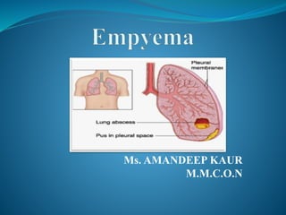
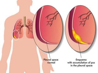
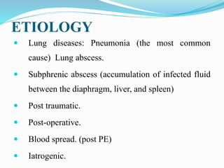
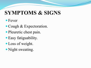
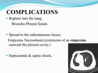
This document defines empyema as the accumulation of pus in the pleural cavity. It is most commonly caused by pneumonia or other lung infections. Key symptoms include fever, chest pain, and cough. Diagnosis involves chest x-ray and thoracentesis. Treatment involves antibiotics, chest tube placement to drain pus, and sometimes fibrinolytics or surgery. Goals are to treat the infection, drain the pleural space, and allow lung re-expansion to prevent chronic issues. Physiotherapy after treatment is important for full recovery.
Lung abscess is a type of liquefactive necrosis of the lung tissue and formation of cavities (more than 2 cm) containing necrotic debris or fluid caused by microbial infection.
Pleural effusion, sometimes referred to as “water on the lungs,” is the build-up of excess fluid between the layers of the pleura outside the lungs. The pleura are thin membranes that line the lungs and the inside of the chest cavity and act to lubricate and facilitate breathing.
Pneumonia is an inflammation of the lung parenchyma caused by various microorganisms, including bacteria, mycobacteria, fungi, and viruses.
Pneumonitis is a more general term that describes the inflammatory process in the lung tissue that may predispose and Pneumonia is an inflammation of the lung parenchyma that is caused by a microbial agent.
place the patient at risk for microbial invasion.
Pneumonia is classified into four: community-acquired pneumonia (CAP) and hospital-acquired pneumonia (HAP), pneumonia in the immunocompromised host, and aspiration pneumonia.
Normally, the pleural space contains a small amount of fluid (5 to 15 mL), which acts as a lubricant that allows the pleural surfaces to move without friction.
But if fluid builds up from either increased production or inadequate removal pleural effusion results.
Pleural effusion B/L or unilateral (parapneumonic process)
Refers to any significant collection of fluid within pleural space.
Any imbalance in formation, absorption lead accumulation of pleural fluid. Common condition:
CHF
Bacterial pneumonia
Malignancy(chest tumor)
Pulmonary embolism
Pleura effusion is a condition refers to a collection of fluid in the pleural space. It is almost secondary to other conditions.
Bronchiectasis is a chronic, irreversible dilation of the bronchi and bronchioles. Or •Bronchiectasis is characterized by permanent, abnormal dilation of one or more large bronchBronchiectasis.
Leading the Way in Nephrology: Dr. David Greene's Work with Stem Cells for Ki...Dr. David Greene Arizona
As we watch Dr. Greene's continued efforts and research in Arizona, it's clear that stem cell therapy holds a promising key to unlocking new doors in the treatment of kidney disease. With each study and trial, we step closer to a world where kidney disease is no longer a life sentence but a treatable condition, thanks to pioneers like Dr. David Greene.
CHAPTER 1 SEMESTER V - ROLE OF PEADIATRIC NURSE.pdfSachin Sharma
Pediatric nurses play a vital role in the health and well-being of children. Their responsibilities are wide-ranging, and their objectives can be categorized into several key areas:
1. Direct Patient Care:
Objective: Provide comprehensive and compassionate care to infants, children, and adolescents in various healthcare settings (hospitals, clinics, etc.).
This includes tasks like:
Monitoring vital signs and physical condition.
Administering medications and treatments.
Performing procedures as directed by doctors.
Assisting with daily living activities (bathing, feeding).
Providing emotional support and pain management.
2. Health Promotion and Education:
Objective: Promote healthy behaviors and educate children, families, and communities about preventive healthcare.
This includes tasks like:
Administering vaccinations.
Providing education on nutrition, hygiene, and development.
Offering breastfeeding and childbirth support.
Counseling families on safety and injury prevention.
3. Collaboration and Advocacy:
Objective: Collaborate effectively with doctors, social workers, therapists, and other healthcare professionals to ensure coordinated care for children.
Objective: Advocate for the rights and best interests of their patients, especially when children cannot speak for themselves.
This includes tasks like:
Communicating effectively with healthcare teams.
Identifying and addressing potential risks to child welfare.
Educating families about their child's condition and treatment options.
4. Professional Development and Research:
Objective: Stay up-to-date on the latest advancements in pediatric healthcare through continuing education and research.
Objective: Contribute to improving the quality of care for children by participating in research initiatives.
This includes tasks like:
Attending workshops and conferences on pediatric nursing.
Participating in clinical trials related to child health.
Implementing evidence-based practices into their daily routines.
By fulfilling these objectives, pediatric nurses play a crucial role in ensuring the optimal health and well-being of children throughout all stages of their development.
One of the most developed cities of India, the city of Chennai is the capital of Tamilnadu and many people from different parts of India come here to earn their bread and butter. Being a metropolitan, the city is filled with towering building and beaches but the sad part as with almost every Indian city
Deep Leg Vein Thrombosis (DVT): Meaning, Causes, Symptoms, Treatment, and Mor...The Lifesciences Magazine
Deep Leg Vein Thrombosis occurs when a blood clot forms in one or more of the deep veins in the legs. These clots can impede blood flow, leading to severe complications.
CHAPTER 1 SEMESTER V PREVENTIVE-PEDIATRICS.pdfSachin Sharma
This content provides an overview of preventive pediatrics. It defines preventive pediatrics as preventing disease and promoting children's physical, mental, and social well-being to achieve positive health. It discusses antenatal, postnatal, and social preventive pediatrics. It also covers various child health programs like immunization, breastfeeding, ICDS, and the roles of organizations like WHO, UNICEF, and nurses in preventive pediatrics.
Antibiotic Stewardship by Anushri Srivastava.pptxAnushriSrivastav
Stewardship is the act of taking good care of something.
Antimicrobial stewardship is a coordinated program that promotes the appropriate use of antimicrobials (including antibiotics), improves patient outcomes, reduces microbial resistance, and decreases the spread of infections caused by multidrug-resistant organisms.
WHO launched the Global Antimicrobial Resistance and Use Surveillance System (GLASS) in 2015 to fill knowledge gaps and inform strategies at all levels.
ACCORDING TO apic.org,
Antimicrobial stewardship is a coordinated program that promotes the appropriate use of antimicrobials (including antibiotics), improves patient outcomes, reduces microbial resistance, and decreases the spread of infections caused by multidrug-resistant organisms.
ACCORDING TO pewtrusts.org,
Antibiotic stewardship refers to efforts in doctors’ offices, hospitals, long term care facilities, and other health care settings to ensure that antibiotics are used only when necessary and appropriate
According to WHO,
Antimicrobial stewardship is a systematic approach to educate and support health care professionals to follow evidence-based guidelines for prescribing and administering antimicrobials
In 1996, John McGowan and Dale Gerding first applied the term antimicrobial stewardship, where they suggested a causal association between antimicrobial agent use and resistance. They also focused on the urgency of large-scale controlled trials of antimicrobial-use regulation employing sophisticated epidemiologic methods, molecular typing, and precise resistance mechanism analysis.
Antimicrobial Stewardship(AMS) refers to the optimal selection, dosing, and duration of antimicrobial treatment resulting in the best clinical outcome with minimal side effects to the patients and minimal impact on subsequent resistance.
According to the 2019 report, in the US, more than 2.8 million antibiotic-resistant infections occur each year, and more than 35000 people die. In addition to this, it also mentioned that 223,900 cases of Clostridoides difficile occurred in 2017, of which 12800 people died. The report did not include viruses or parasites
VISION
Being proactive
Supporting optimal animal and human health
Exploring ways to reduce overall use of antimicrobials
Using the drugs that prevent and treat disease by killing microscopic organisms in a responsible way
GOAL
to prevent the generation and spread of antimicrobial resistance (AMR). Doing so will preserve the effectiveness of these drugs in animals and humans for years to come.
being to preserve human and animal health and the effectiveness of antimicrobial medications.
to implement a multidisciplinary approach in assembling a stewardship team to include an infectious disease physician, a clinical pharmacist with infectious diseases training, infection preventionist, and a close collaboration with the staff in the clinical microbiology laboratory
to prevent antimicrobial overuse, misuse and abuse.
to minimize the developme
How many patients does case series should have In comparison to case reports.pdfpubrica101
Pubrica’s team of researchers and writers create scientific and medical research articles, which may be important resources for authors and practitioners. Pubrica medical writers assist you in creating and revising the introduction by alerting the reader to gaps in the chosen study subject. Our professionals understand the order in which the hypothesis topic is followed by the broad subject, the issue, and the backdrop.
https://pubrica.com/academy/case-study-or-series/how-many-patients-does-case-series-should-have-in-comparison-to-case-reports/
3. Definition
Also known as:- Pyothorax Or Purulent
Pleuritis. The word Empyema comes from the
greek word Empyein, which means :pus –
producing (suppurates).
Accumulation of Pus in the Pleural cavity.
5. ORGANISMS
The most common:
Staph. aureus .(90% of causes in infants & children)
Strept. pneuomonie.
H. influenzae
6. ETIOLOGY
Lung diseases: Pneumonia (the most common
cause) Lung abscess.
Subphrenic abscess (accumulation of infected fluid
between the diaphragm, liver, and spleen)
Post traumatic.
Post-operative.
Blood spread. (post PE)
Iatrogenic.
8. Pathophysiology
Presence of Parapneumonic Effusion
Release of inflammatory mediators
↑permeability of the capilliaries
Attracts WBCs to the site Escape of albumin & other protein
from the capillaries
↑ Pleural fluid
Presence of Free-flowing, Protein Rich Pleural Fluid (Stage I)
Inflammation worsens
9. Cont…
Attracts more WBCs to the site.
Extensive purulent exudate production.
Initiation of fibroblastic (collagen and other proteins)
activity (Stage II)
Adherence of the two pleural membranes (Stage III)
Formation of a “Peel”
10. CLINICAL STAGES
Acute stage :
within the first 2 weeks of the onset.
Chronic Stage :
after 2 weeks or with the formation of the thick peel
and loculations.
11. Causes of Chronicity:
Inadequate Tube Drainage.
Chronic pulmonary Disease( T.B. or Fungal Infection)
Immuno-supressed patients.
Presence of Foreign body within the pleural space.
12. SYMPTOMS & SIGNS
Fever
Cough & Expectoration.
Pleuretic chest pain.
Easy fatiguability.
Loss of weight.
Night sweating.
13. COMPLICATIONS
Rupture into the lung;
Broncho-Pleural fistula
Spread to the subcutaneous tissue;
Empyema Necessitasis) (extension of an empyema
outward the pleural cavity.)
Septicaemia & septic shock.
15. Light’s criteria
Pleural fluid protein: serum protein > O.5 (1-2 g/dL)
Pleural fluid : serum LDH >0.6 (Lactate dehydrogenase
less than 50% of plasma)
16. Other minor criteria
Cholesterol > 45mg/dl (< 45 mg/dL).
pH <7.2 (7.60-7.64)
Glucose < 50% serum (similar to that of plasma)
17. Goals of the treatment
Treat the infection
Drain the purulent effusion adequately and
completely
Re-expand the lung to fill the pleural space
Eliminate complications and avoid chronicity
18. Antibiotic treatment
As soon as the bacteriologic sample are recovered
Pneumonia
Amoxicillin, Metronidazole
Amox-clavulanic acid
Nosocomial
Tazobactam or Imipenem
Aminoglycoside or Quinolone
Adapted to the laboratory results
21. Primary treatment options
Antibiotics alone;
Récurrent thoracocentèsis
Insertion of chest drain alone or in combination with
fibrinolytics
VATS(Video-Assisted Thoracic Surgery).
Open decortication:- the operation of removing fibrous
scar tissue that prevents expansion of the lung.
22. Thoracocentesis
Big caliber needle
Mostly diagnosis technique
Therapeutically used if the liquid remains.
Theoretically allows pleural lavage
23. Chest Tube
As soon as the liquid is thick
Localization
free: axillary
loculated: Chest imaging using
ultrasonography and/or computed
tomography
Size: 20 à 24
Bedside
24. Pleural Lavage
Isotonic saline
Modalités
3 way stopcock
Directly through the CT: 250 to 500 ml
Cautiously if suspicion of broncho-pleural fistula
Timing:
Immediately after CT placement
Once a day until the liquid is clear
25. NOXYFLEX (noxytioline)
Local disinfectant (formaldéhyde)
2,5 g diluted in at least 100ml isotonic saline
Maximum: 5g/day
Incompatible with iodine polyvidone,chlorhexidin,
chlorine solution, lactic acid
26. Fibrinolytics
Urokinase: 100 000 or 300 000 IU conditioning
Streptokinase: 250000 IU conditioning
250.000 IU in 10-20 ml isotonic saline
Don’t evacuate before 24 to 48 heures
Constantly associated with fever (38-39°C)
Then evacuate
Pleural lavage
clamp 4h ( Chest 1996)
27. Video-assisted thoracic surgery
Collection<10 cm: unusual
Visual control of the CT position
5 mm introducer, 4 mm optical
Collection>10 cm
10 mm introducer
Two or three ports are made in the chest
One port is utilised for the camera and the others for
grasping instruments
Free fluid is evacuated and loculations drained under
thoracoscopic visualisation.
Fibrinous adhesions are separated and the pleural debris
removed from the pleural lining using endoscopic
grasping forceps or by extensive irrigation and suction.
Following the procedure, one or two chest drains are then
placed in the portholes.
29. Physiotherapy
Key to a correct evolution
After CT removal
Often and for a long time…..
Decrease surgery
Decrease long term pain and functionnal limitations