Pleural Effusion for Undergraduates
•Download as PPTX, PDF•
130 likes•14,236 views

This document provides an overview of pleural effusion, including: - Pleural effusion is abnormal fluid accumulation in the pleural space between the lungs and chest wall. Fluid builds up due to changes in pressure or permeability. - Effusions are classified as transudative or exudative based on their mechanism and composition. Causes include infections, cancers, heart failure, and other conditions. - Symptoms depend on the underlying cause but may include chest pain, difficulty breathing, and cough. Diagnosis involves physical exam, imaging like x-rays, and analyzing pleural fluid obtained via thoracentesis. - Management consists of treating the underlying condition medically or surgically with drainage

More Related Content
What's hot
What's hot (20)
Similar to Pleural Effusion for Undergraduates
Similar to Pleural Effusion for Undergraduates (20)
More from Sesha Sai
Recently uploaded
Recently uploaded (20)
Pleural Effusion for Undergraduates
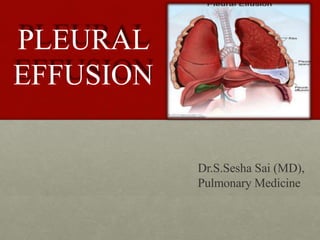
- 1. PLEURAL EFFUSION Dr.S.Sesha Sai (MD), Pulmonary Medicine
- 2. Overview • Introduction • Classification • Pathogenesis • Etiology • Clinical features • Investigations • Management
- 3. • Pleural effusion is defined as abnormal accumulation of fluid in the pleural space, i.e., the space between parietal and visceral pleura • The pleural space contains normally 0.3ml/kg body weight of pleural fluid1. There is a continuous circulation of this fluid and the lymphatic vessels can cope with several millilitres of extra fluid per 24hours • Fluid accumulates in the pleural cavity due to either altered hydrostatic and oncotic pressures or altered permeability of the pleura Introduction
- 4. More Definitions ? • Parapneumonic Effusion : pleural effusion associated with bacterial pneumonia, bronchiectasis, or lung abscess . • Loculated Effusion : Fluid anatomically confined and not freely flowing in the pleural space when there are adhesions between the visceral and the parietal pleura . • Sub-Pulmonic Effusion:accumulation of fluid between the lung & the diaphragm which gives the false impression of an elevated hemi-diaphragm
- 5. Pleural Effusion Parapneumonic effusion Loculated Effusion Subpulmonic Effusion
- 6. Composition of pleural fluid • Clear ultra filtrate of plasma • Volume • Cells/ mm3 0.3 mL/kg 1000 – 5000 60% 30% 5% 5% 1-2 g/dL <50% plasma level(105-333IU/L) plasma level(90-120) ≥ plasma level(7.6-7.64) • Mesothelial cells • Monocytes • Lymphocytes • PMN’s • Protein • LDH • Glucose • pH
- 7. •Can be unilateral or bilateral and classified A)Based on site Apical Interlobar Sub-pulmonic Mediastinal B)Based on mechanism and type of pleural fluid Transudative (alteration in hydrostatic and oncotic pressure) Exudative (alteration in pleural permeability) Classification
- 8. c) Based on mechanism and type of pleural fluid formed Pyogenic Chylous Haemothorax Pseudochylous Hydrothorax Urinothorax
- 9. Transudative pleural effusions Alteration of hydrostatic and oncotic factors that increase the formation or decrease the absorption of pleural fluid (e.g., increased mean capillary pressure [heart failure] or decreased oncotic pressure [cirrhosis or nephrotic syndrome]).
- 10. Exudative pleural effusions Damage or disruption of the normal pleural membranes or vasculature (e.g., tumor involvement of the pleural space, infection, inflammatory conditions, or trauma) leads to increased capillary permeability or decreased lymphatic drainage.
- 11. Pathogenesis • Increased vascular permeability allows migration of inflammatory cells (neutrophils, lymphocytes, and eosinophils) into the pleural space. • The process is mediated by a number of cytokines such as interleukin IL-1, IL-6, IL-8, tumour necrosis factor (TNF)-alpha and platelet activating factor released by mesothelial cells lining the pleural space. The result is the exudative stage of a pleural effusion. This progresses to the fibro-purulent stage due to increased fluid accumulation and bacterial invasion across the damaged epithelium. • Neutrophil migration occurs as well as activation of the coagulation cascade leading to pro-coagulant activity and decreased fibrinolysis. Deposition of fibrin in the pleural space then leads to septation or loculation. The pleural fluid pH and glucose level falls while LDH levels increase.
- 12. Etiology • EXUDATIVE Infective: Pneumonia, Bronchiectasis, Pancreatitis, TB, Lung abscess Collagen vascular disease: SLE, Rheumatoid arthritis, Polyarteritis Neoplastic: leukemias and lymphomas Uremia Drugs: Bromocriptine, amiodarone, nitofurantoin, dantrolene, INH, PAS Postradiation Traumatic
- 13. • TRANSUDATIVE: Renal cause: Nephrotic syndrome Cardiac cause: Congestive cardiac failure Hepatic cause: Hepatic failure Nutritional: Protein energy malnutrition Hypothyroidism
- 14. • PYOGENIC: Lung abscess Septicemia Chest wall injuries Rupture of oesophagus Rupture of subphrenic abscess Rupture of liver abscess
- 15. • CHYLOUS: Trauma to thoracic duct Tumour (mediastinal lymphoma) Tuberculosis Lymphatic obstruction
- 16. • HEMOTHORAX: Chest wall injuries Bleeding disorders Neoplasms-leukemias, lymphoma, mesothelioma Drugs-anticoagulants Pulmonary infarction
- 17. • PSEUDOCHYLOUS: Rheumatoid pleuritis Tuberculosis or paragonimiasis(lung fluke infection) • HYDROTHORAX: Congestive heart failure Hepatic & Renal failure
- 18. Clinical Presentation The underlying cause of the effusion usually dictates the symptoms, although patients may be asymptomatic. Pleural inflammation, abnormal pulmonary mechanics, and worsened alveolar gas exchange produce symptoms and signs of disease.
- 19. Symptomsand signs Inflammation of the parietal pleura leads to pain in local (intercostal) involved areas or referred (phrenic) distributions (shoulder). Dyspnea is frequent and may be present and out of proportion to the size of the effusion. Cough can occur.
- 20. Physical examination Inspection: Absent or diminished movements of affected side Fullness of chest with bulging intercostal spaces Palpation: Diminished breath sounds over the site of the effusion Decreased or absent tactile fremitus Percussion: Stony dullness to percussion Auscultation: Absence of breath sounds over the effusion Vocal resonance absent Signs of pneumonia like bronchial breathing, crackles etc.
- 21. Investigations Total and differential leucocyte counts • Acute phase reactants-white cell count, total neutrophil count, CRP, ESR, pro-calcitonin distinguish bacterial from viral causes Radiological examination • X-ray chest PA view done in erect position-a total of 300mL of fluid is needed to diagnose pleural effusion clinically and radiologically • Even 50mL of fluid can be demonstrated radiologically in lateral decubitus
- 22. Findings • Obliteration of cardiophrenic and costophrenic angles • Loculated effusions • Subpulmonic effusion-collection of fluid below the diaphragm will lead to elevation of diaphragm, confirmed by X-ray in lateral decubitus • Lateral decubitus on side of effusion will show a shift in the fluid level • Tracheal and mediastinal shifts are seen in massive effusion
- 24. Ultrasonogram Useful in differentiating between loculated pleural effusion and tumour CT Scan Helpful if the effusion is minimal or loculated Pleural fluid aspiration (Thoracocentesis) Diagnostic: Helps to differentiate between exudates and transudates Therapeutic: Massive collection or rapid collection of pleural fluid Severe respiratory distress Suspected empyema Massive mediastinal shift
- 25. Gross appearance • Straw-coloured • Blood stained • Purulent • Chylous
- 26. Transudate & Exudate Features Transudates Exudates Appearance Clear/Straw coloured Cloudy, purulent, opalascent Protein < 3g/100mL >3g/100mL pH >7.2 <7.2 Glucose >40mg/dL <40mg/dL LDH Low, <200IU/L High,>200IU/L Cells <1000/mm3 >1000/mm3
- 27. LIGHT’S CRITERIA: • Atleast one of the following criteria should be satisfied to identify exudates: Pleural fluid to serum total protein ratio- more than 0.5 Pleural fluid to serum LDH ratio- more than 0.6 Pleural fluid LDH- more than two-third of serum LDH None of these criteria should be satisfied in a transudative effusion
- 28. Pleural fluid appearance • Most transudates are clear, straw colored, nonviscid, and without odor • Red-tinged pleural effusions indicate the presence of blood.
- 29. Bloody pleural fluid • If the blood is due to thoracentesis, the degree of discoloration should clear during the aspiration. • Bloody pleural fluid usually indicates the presence of malignancy, pulmonary embolism (PE), or trauma.
- 30. Hemothorax • The presence of gross blood should lead to the measurement of a pleural fluid hematocrit. • Hemothorax is defined as a pleural fluid to blood hematocrit ratio of >0.5, and chest tube drainage should be considered.
- 31. Eosinophilia (>10% of total nucleated cell count) is suggestive of air or blood in the pleural space. If air or blood is not present in the pleural space, consideration should be given to fungal and parasitic infection, drug- induced disease, PE, asbestos-related disease, and Churg-Strauss syndrome.
- 32. • Lymphocytosis (>50% of the total nucleated cell count) is suggestive of malignancy or tuberculosis. • Mesothelial cells argues against the diagnosis of tuberculosis. • Plasma cells suggest a diagnosis of multiple myeloma.
- 33. Glucose concentration A glucose concentration of <60 mg/dL is probably due to tuberculosis, malignancy, rheumatoid arthritis, or parapneumonic effusion. For parapneumonic pleural effusions with a glucose of <60 mg/dL, tube thoracostomy should be considered.
- 34. Pleural fluid with a low pH A pH of <7.3 is seen with • empyema, • tuberculosis, • malignancy, • collagen vascular disease, or • esophageal rupture.
- 35. Amylase • An elevation of amylase suggests that the patient has pancreatic disease, malignancy, or esophageal rupture. • Malignancy and esophageal rupture have salivary amylase elevations and not pancreatic amylase elevations.
- 36. Turbid or milky fluid After it is centrifuged. • If the supernatant clears, the cloudiness is likely due to cells and debris. • If the supernatant remains turbid, pleural lipids should be measured. Elevation of triglycerides (>110 mg/dL) suggests that a chylothorax is present, usually due to disruption of the thoracic duct from trauma, surgery, or malignancy (i.e., lymphoma).
- 37. Cytology Cytology is positive in approximately 60% of malignant effusions. The volume of pleural fluid analyzed does not impact the yield of cytologic diagnosis. Repeat thoracentesis increases the diagnostic yield.
- 38. Urinothorax Due to obstructive uropathy Urine arrives in the pleural space either retroperitoneally under the posterior diaphragm, or via the retroperitoneal lymphatics Pleural fluid smells of urine. Pleural fluid Creatinine ≈ Serum Creatinine
- 39. Other investigations • Suspected TB • Adenosine deaminase (> 50 IU/L) • Beta2 - microglobulin • Lysozyme III (> 20mcg/mL) • PCR (Sens 100%, Spec 95%) • AFB (smear 10-20%; cx 25- 50%) • Suspected Rheumatoid • Pleural RF • Low glucose • Suspected SLE • Serum Complement • Pleural ANA • LE cells • Suspected Pneumonia • pH • Suspected Pancreatitis • Pleural Amylase
- 40. Pleural Biopsy • Can be done at maximum dullness on percussion or at a maximum thickening of pleura. Abram’s pleural biopsy needle is used for biopsy • Most helpful in evaluating for TB • Limited utility for CA (40-50% positive) Repeat cytology x 3 • Sarcoid, fungal: might be helpful
- 41. Management SUPPORTIVE TREATMENT • Oxygen is necessary if SpO2 <92% • Fluid therapy if child dehydrated or unable/unwilling in drinking water • Initiate IV antibiotics • Analgesics and antipyretics • Chest radiography & U/S
- 42. Medical • Treat the cause Pneumonia- initial antibiotic treatment A) Following community acquired pneumonia • Cefuroxime • Co-amoxiclav • Penicillin & flucloxacillin • Amoxicillin & flucloxaxillin • Clindamycin B) Hospital acquired pneumonia • Broader spectrum antibiotics that cover aerobic gram negative rods
- 43. • Tuberculosis- Category I treatment 2HRZE+4HRE Prednisolone 1-2mg/kg orally 4-6weeks promotes rapid absorption of the pleural fluid and prevents fibrosis • Congestive cardiac failure- treat with diuretics and other anti-failure medications
- 44. Surgical • Pleural fluid aspiration is done by using a wide bore needle. If the fluid is thick and cannot be drained by a needle, an intercostal drainage(under water seal) at the most dependant part should be done.
- 45. Tube Thoracostomy •Pneumothorax •Pleural fluid loculated •Recurrent Pleural effusion - malignancy •Effusion filling more than half the hemithorax •Air fluid level – Hydro/Pyopneumothorax •Pus in the pleural space - Empyema •Hemothorax/Chylothorax •Para pneumonic effusion oPositive stain for microorganisms oPositive pleural fluid cultures oPleural fluid pH <7.2 oPleural fluid glucose <60 mg/dL
- 46. Underwater seal bag is used as one way valve mechanism. The air or fluid/pus from the pleura enters the underwater drainage bag, but the atmospheric air cannot enter pleura due to under water seal
- 49. Failure to drain with a single small-bore tube should also lead to thoracic surgery consultation to avoid delays in case video assisted thoracoscopy (VATS) becomes necessary. VATS
- 50. Chemical pleurodesis Chemical pleurodesis is an effective therapy for recurrent effusions. This treatment is recommended in patients whose symptoms are relieved with initial drainage but who have rapid reaccumulation of fluid. Talc slurry - Effective and inexpensive. Doxycycline or minocycline can also be instilled into the pleural space via a chest tube. Pain is more prevalent and severe following doxycycline and minocycline than following talc.
- 51. Chronic indwelling pleural catheters Provide good control of effusion-related symptoms via intermittent drainage.
- 52. THANK YOU