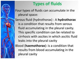
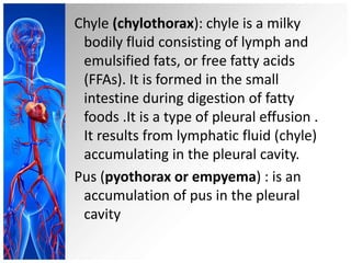
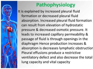
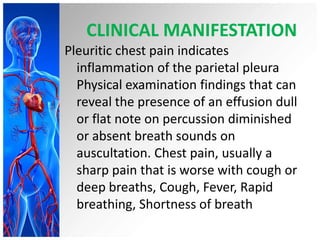
Downloaded 2,015 times

![• G.Burrows CM, Mathews WC, Colt HG. Predicting
survival in patientswith recurrent symptomatic
malignant pleural effusions: an assessment of the
prognostic values of physiologic, morphologic, and
quality of life measures of extent of disease. Chest.
2000;117(1):73-78. [PubMed]
• H.Anderson CB, Philpott GW, Gerguson TB. The
treatment of malignant pleural effusion.
Cancer.1974;33(4):916–922.](https://image.slidesharecdn.com/pleuraleffusion-131227054021-phpapp02/85/Pleural-Effusion-33-320.jpg)


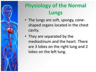
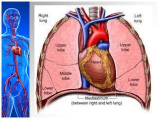
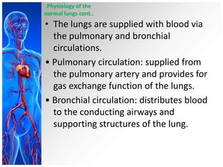
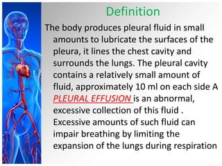
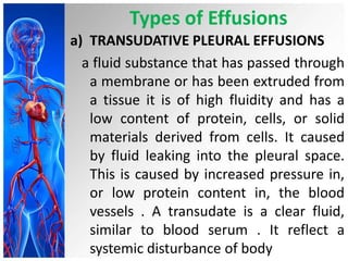
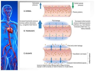
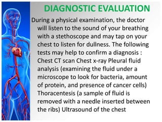
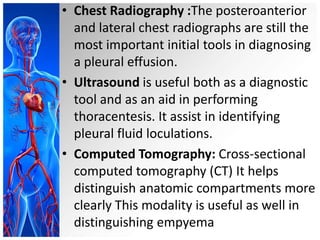
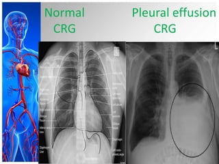
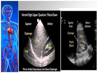
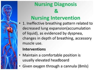
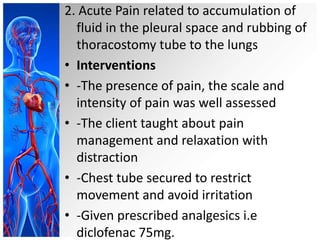
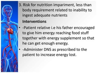
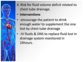
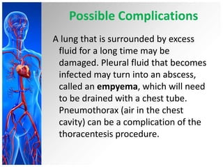
This document provides information on pleural effusions including the physiology of normal lungs, definition of pleural effusion, types of effusions, causes, pathophysiology, clinical manifestations, diagnostic evaluations, treatment, nursing diagnosis and interventions, and possible complications. A pleural effusion is an abnormal collection of fluid in the pleural space between the lungs and chest wall. Effusions can be transudative or exudative depending on the fluid characteristics and underlying cause. Diagnosis involves imaging and fluid analysis. Treatment focuses on removing fluid and treating the underlying cause. Nursing care centers around pain management, breathing exercises, and preventing infections.


