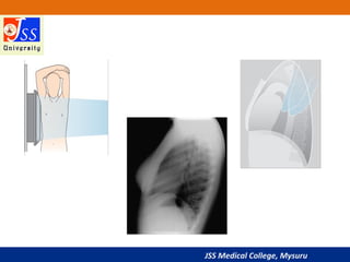
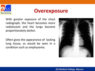
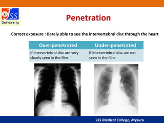
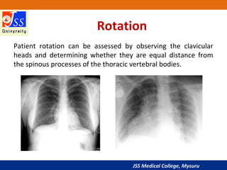
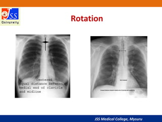
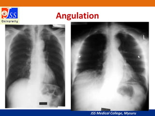
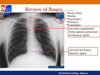
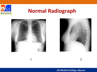
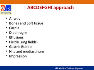
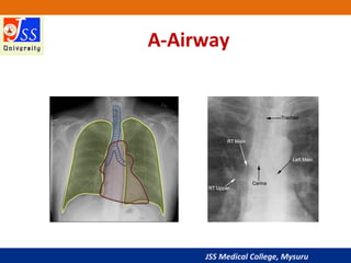
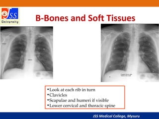
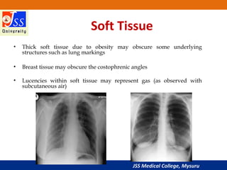
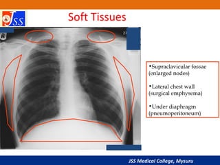
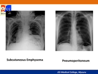
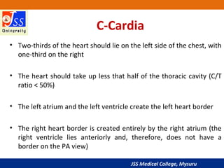
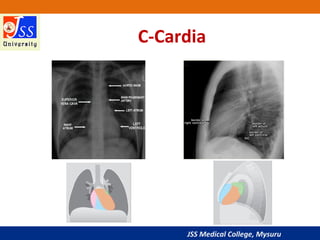
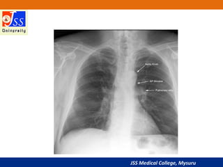
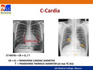
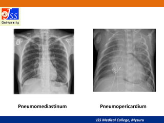
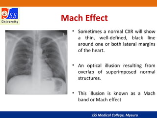
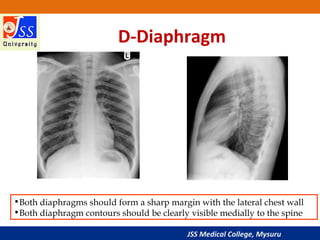
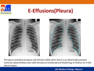
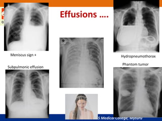
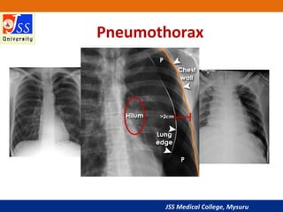
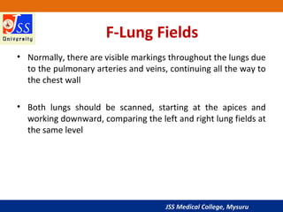
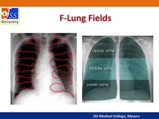
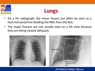
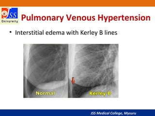
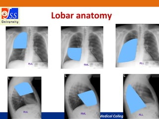
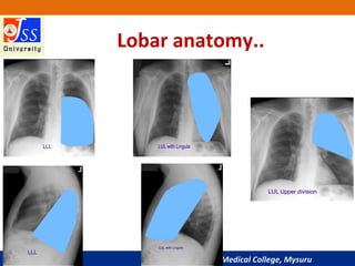
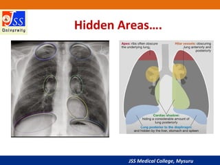
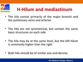
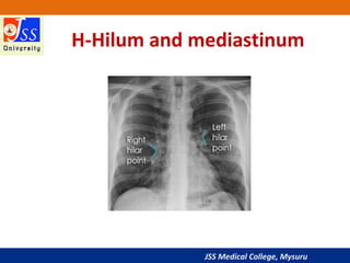
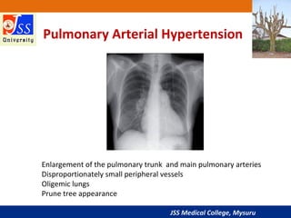
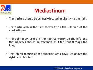
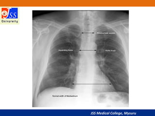
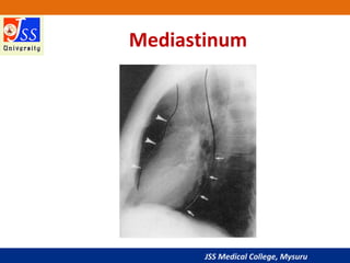
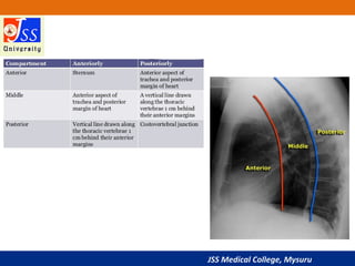
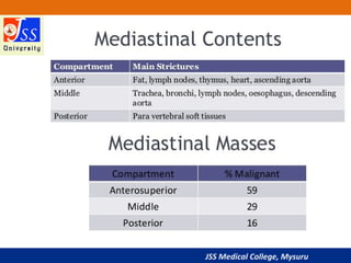
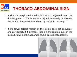
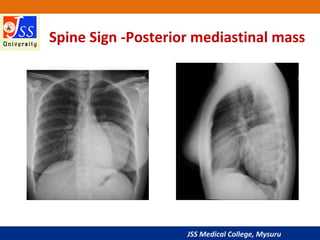
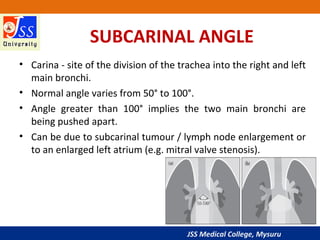
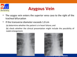
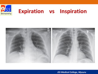
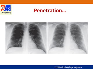
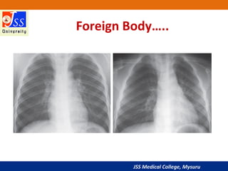
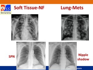
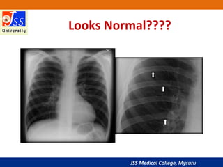
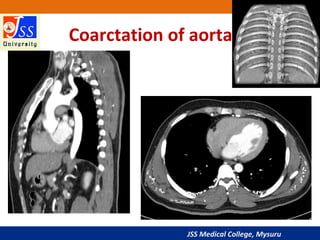
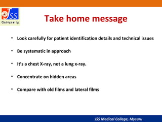
The basics of Chest Radiology explained for the undergraduate students. The technical aspects including the various views, exposure, rotation and breath described.
The inside out approach of interpretation explained. The ABCDEFGH description includes Airway, Bones & soft tissue, Cardiac shadow, Diaphragm, Effusion (pleura), Fields (lungs), Gastric bubble and Hila & mediastinum.
The basic cardiac and lung pathologies discussed.
A triphasic, or triple-phase, CT scan is an enhanced CT technique mostly used to evaluate liver lesions. This technique acquires images at 3 different time points, or phases, following the administration of a contrast.
Interstitial lung disease is a general category that includes many different lung conditions. All interstitial lung diseases affect the interstitium, a part of the lungs' anatomic structure.
Some of the types of interstitial lung disease include:
Interstitial pneumonia: Bacteria, viruses, or fungi may infect the interstitium of the lung. A bacterium called Mycoplasma pneumonia is the most common cause.
Idiopathic pulmonary fibrosis : A chronic, progressive form of fibrosis (scarring) of the interstitium. Its cause is unknown.
Nonspecific interstitial pneumonitis: Interstitial lung disease that's often present with autoimmune conditions (such as rheumatoid arthritis or scleroderma).
Brief Presentation on clinical examination of Respiratory System with Report of Normal case
references:
macleod's clinical examination 13th edition
hutchinson clinical methods
R Alagappan - Manual of Practical Medicine, 4th Edition
Learn Chest X-Ray With Its Normal Positioning & Radio-AnatomyDr.Santosh Atreya
Learn Chest X-Ray With Its Normal Positioning & Radio-Anatomy..For some image description please go through the text book "David Sutton" because i have described these image during my presentation Verbally..There are many animations used inside this presentation so to see all the pictures which are placed layer by layer with the help of animations you simple need to download this presentation first.... Thanx.
in this tutorial i am speaking about chest x-ray quality that include :
1- Inclusion
2- inspiration/lung
3- volume
4- projection
5- penetration
6- Rotation
7- artifact
i try to make it easy and simple for medical students and junior doctors to help them in clinical life.
Artficial Intelligence in Health universities(JSS) Vikram Patil
Artificial intelligence (AI) is a growing phenomenon
Will soon facilitate wide-scale changes in Health education and Healthcare delivery.
Students and Educators need to have the knowledge of AI in relation to learning and research
They need to know extent to which it will impact on education and healthcare delivery
The ppt is useful to understand the role of Universities in the era of AI
These simplified slides by Dr. Sidra Arshad present an overview of the non-respiratory functions of the respiratory tract.
Learning objectives:
1. Enlist the non-respiratory functions of the respiratory tract
2. Briefly explain how these functions are carried out
3. Discuss the significance of dead space
4. Differentiate between minute ventilation and alveolar ventilation
5. Describe the cough and sneeze reflexes
Study Resources:
1. Chapter 39, Guyton and Hall Textbook of Medical Physiology, 14th edition
2. Chapter 34, Ganong’s Review of Medical Physiology, 26th edition
3. Chapter 17, Human Physiology by Lauralee Sherwood, 9th edition
4. Non-respiratory functions of the lungs https://academic.oup.com/bjaed/article/13/3/98/278874
Title: Sense of Smell
Presenter: Dr. Faiza, Assistant Professor of Physiology
Qualifications:
MBBS (Best Graduate, AIMC Lahore)
FCPS Physiology
ICMT, CHPE, DHPE (STMU)
MPH (GC University, Faisalabad)
MBA (Virtual University of Pakistan)
Learning Objectives:
Describe the primary categories of smells and the concept of odor blindness.
Explain the structure and location of the olfactory membrane and mucosa, including the types and roles of cells involved in olfaction.
Describe the pathway and mechanisms of olfactory signal transmission from the olfactory receptors to the brain.
Illustrate the biochemical cascade triggered by odorant binding to olfactory receptors, including the role of G-proteins and second messengers in generating an action potential.
Identify different types of olfactory disorders such as anosmia, hyposmia, hyperosmia, and dysosmia, including their potential causes.
Key Topics:
Olfactory Genes:
3% of the human genome accounts for olfactory genes.
400 genes for odorant receptors.
Olfactory Membrane:
Located in the superior part of the nasal cavity.
Medially: Folds downward along the superior septum.
Laterally: Folds over the superior turbinate and upper surface of the middle turbinate.
Total surface area: 5-10 square centimeters.
Olfactory Mucosa:
Olfactory Cells: Bipolar nerve cells derived from the CNS (100 million), with 4-25 olfactory cilia per cell.
Sustentacular Cells: Produce mucus and maintain ionic and molecular environment.
Basal Cells: Replace worn-out olfactory cells with an average lifespan of 1-2 months.
Bowman’s Gland: Secretes mucus.
Stimulation of Olfactory Cells:
Odorant dissolves in mucus and attaches to receptors on olfactory cilia.
Involves a cascade effect through G-proteins and second messengers, leading to depolarization and action potential generation in the olfactory nerve.
Quality of a Good Odorant:
Small (3-20 Carbon atoms), volatile, water-soluble, and lipid-soluble.
Facilitated by odorant-binding proteins in mucus.
Membrane Potential and Action Potential:
Resting membrane potential: -55mV.
Action potential frequency in the olfactory nerve increases with odorant strength.
Adaptation Towards the Sense of Smell:
Rapid adaptation within the first second, with further slow adaptation.
Psychological adaptation greater than receptor adaptation, involving feedback inhibition from the central nervous system.
Primary Sensations of Smell:
Camphoraceous, Musky, Floral, Pepperminty, Ethereal, Pungent, Putrid.
Odor Detection Threshold:
Examples: Hydrogen sulfide (0.0005 ppm), Methyl-mercaptan (0.002 ppm).
Some toxic substances are odorless at lethal concentrations.
Characteristics of Smell:
Odor blindness for single substances due to lack of appropriate receptor protein.
Behavioral and emotional influences of smell.
Transmission of Olfactory Signals:
From olfactory cells to glomeruli in the olfactory bulb, involving lateral inhibition.
Primitive, less old, and new olfactory systems with different path
Prix Galien International 2024 Forum ProgramLevi Shapiro
June 20, 2024, Prix Galien International and Jerusalem Ethics Forum in ROME. Detailed agenda including panels:
- ADVANCES IN CARDIOLOGY: A NEW PARADIGM IS COMING
- WOMEN’S HEALTH: FERTILITY PRESERVATION
- WHAT’S NEW IN THE TREATMENT OF INFECTIOUS,
ONCOLOGICAL AND INFLAMMATORY SKIN DISEASES?
- ARTIFICIAL INTELLIGENCE AND ETHICS
- GENE THERAPY
- BEYOND BORDERS: GLOBAL INITIATIVES FOR DEMOCRATIZING LIFE SCIENCE TECHNOLOGIES AND PROMOTING ACCESS TO HEALTHCARE
- ETHICAL CHALLENGES IN LIFE SCIENCES
- Prix Galien International Awards Ceremony
Factory Supply Best Quality Pmk Oil CAS 28578–16–7 PMK Powder in Stockrebeccabio
Factory Supply Best Quality Pmk Oil CAS 28578–16–7 PMK Powder in Stock
Telegram: bmksupplier
signal: +85264872720
threema: TUD4A6YC
You can contact me on Telegram or Threema
Communicate promptly and reply
Free of customs clearance, Double Clearance 100% pass delivery to USA, Canada, Spain, Germany, Netherland, Poland, Italy, Sweden, UK, Czech Republic, Australia, Mexico, Russia, Ukraine, Kazakhstan.Door to door service
Hot Selling Organic intermediates
micro teaching on communication m.sc nursing.pdfAnurag Sharma
Microteaching is a unique model of practice teaching. It is a viable instrument for the. desired change in the teaching behavior or the behavior potential which, in specified types of real. classroom situations, tends to facilitate the achievement of specified types of objectives.
Lung Cancer: Artificial Intelligence, Synergetics, Complex System Analysis, S...Oleg Kshivets
RESULTS: Overall life span (LS) was 2252.1±1742.5 days and cumulative 5-year survival (5YS) reached 73.2%, 10 years – 64.8%, 20 years – 42.5%. 513 LCP lived more than 5 years (LS=3124.6±1525.6 days), 148 LCP – more than 10 years (LS=5054.4±1504.1 days).199 LCP died because of LC (LS=562.7±374.5 days). 5YS of LCP after bi/lobectomies was significantly superior in comparison with LCP after pneumonectomies (78.1% vs.63.7%, P=0.00001 by log-rank test). AT significantly improved 5YS (66.3% vs. 34.8%) (P=0.00000 by log-rank test) only for LCP with N1-2. Cox modeling displayed that 5YS of LCP significantly depended on: phase transition (PT) early-invasive LC in terms of synergetics, PT N0—N12, cell ratio factors (ratio between cancer cells- CC and blood cells subpopulations), G1-3, histology, glucose, AT, blood cell circuit, prothrombin index, heparin tolerance, recalcification time (P=0.000-0.038). Neural networks, genetic algorithm selection and bootstrap simulation revealed relationships between 5YS and PT early-invasive LC (rank=1), PT N0—N12 (rank=2), thrombocytes/CC (3), erythrocytes/CC (4), eosinophils/CC (5), healthy cells/CC (6), lymphocytes/CC (7), segmented neutrophils/CC (8), stick neutrophils/CC (9), monocytes/CC (10); leucocytes/CC (11). Correct prediction of 5YS was 100% by neural networks computing (area under ROC curve=1.0; error=0.0).
CONCLUSIONS: 5YS of LCP after radical procedures significantly depended on: 1) PT early-invasive cancer; 2) PT N0--N12; 3) cell ratio factors; 4) blood cell circuit; 5) biochemical factors; 6) hemostasis system; 7) AT; 8) LC characteristics; 9) LC cell dynamics; 10) surgery type: lobectomy/pneumonectomy; 11) anthropometric data. Optimal diagnosis and treatment strategies for LC are: 1) screening and early detection of LC; 2) availability of experienced thoracic surgeons because of complexity of radical procedures; 3) aggressive en block surgery and adequate lymph node dissection for completeness; 4) precise prediction; 5) adjuvant chemoimmunoradiotherapy for LCP with unfavorable prognosis.
MANAGEMENT OF ATRIOVENTRICULAR CONDUCTION BLOCK.pdfJim Jacob Roy
Cardiac conduction defects can occur due to various causes.
Atrioventricular conduction blocks ( AV blocks ) are classified into 3 types.
This document describes the acute management of AV block.
HOT NEW PRODUCT! BIG SALES FAST SHIPPING NOW FROM CHINA!! EU KU DB BK substit...GL Anaacs
Contact us if you are interested:
Email / Skype : kefaya1771@gmail.com
Threema: PXHY5PDH
New BATCH Ku !!! MUCH IN DEMAND FAST SALE EVERY BATCH HAPPY GOOD EFFECT BIG BATCH !
Contact me on Threema or skype to start big business!!
Hot-sale products:
NEW HOT EUTYLONE WHITE CRYSTAL!!
5cl-adba precursor (semi finished )
5cl-adba raw materials
ADBB precursor (semi finished )
ADBB raw materials
APVP powder
5fadb/4f-adb
Jwh018 / Jwh210
Eutylone crystal
Protonitazene (hydrochloride) CAS: 119276-01-6
Flubrotizolam CAS: 57801-95-3
Metonitazene CAS: 14680-51-4
Payment terms: Western Union,MoneyGram,Bitcoin or USDT.
Deliver Time: Usually 7-15days
Shipping method: FedEx, TNT, DHL,UPS etc.Our deliveries are 100% safe, fast, reliable and discreet.
Samples will be sent for your evaluation!If you are interested in, please contact me, let's talk details.
We specializes in exporting high quality Research chemical, medical intermediate, Pharmaceutical chemicals and so on. Products are exported to USA, Canada, France, Korea, Japan,Russia, Southeast Asia and other countries.
Explore natural remedies for syphilis treatment in Singapore. Discover alternative therapies, herbal remedies, and lifestyle changes that may complement conventional treatments. Learn about holistic approaches to managing syphilis symptoms and supporting overall health.
Phone Us ❤85270-49040❤ #ℂall #gIRLS In Surat By Surat @ℂall @Girls Hotel With...
Approach to Chest X-Ray and Interpretation
1. JSS Medical College, Mysuru
Normal Chest X-Ray &
Approach to Chest X-Ray
Dr.Vikram Patil
Assistant Professor, Radiology
JSS Medical College and Hospital, Mysuru
2. JSS Medical College, Mysuru
Introduction
• Most of the chest x-rays you will see will be
normal
• In order to recognise abnormality, you need
to know what a normal CXR looks like
3. JSS Medical College, Mysuru
General Principles
• Have a systematic approach
• Interpret the CXR in conjunction with the
clinical findings
• Always compare with previous CXR if available
to assess for change
• Ask yourself “Does my interpretation make
sense?”
4. JSS Medical College, Mysuru
Before we start……
Relative Densities
The images seen on a chest radiograph result from the differences in
densities of the materials in the body.
The hierarchy of relative densities from least dense (Black) to most
dense (white) :
•Gas (air in the lungs)
•Fat (fat layer in soft tissue)
•Water (same density as heart and blood vessels)
•Bone (the most dense of the tissues)
•Metal (foreign bodies)
5. JSS Medical College, Mysuru
Before Interpreting the Radiograph …
1. Patient identification details
2. X-Ray view-PA, AP, Lateral….
3. Breath : Inspiration or Expiration
4. X-ray penetration : Under or Over penetrated
5. Rotation
6. JSS Medical College, Mysuru
Four major views of a chest
radiograph:
• Posterior-anterior (PA)
• Lateral
• Anterior-posterior (AP)
• Lateral Decubitus
7. JSS Medical College, Mysuru
Posterior-anterior (PA) Position
• The standard position for obtaining a routine adult chest
radiograph
• Patient stands upright with the anterior wall of chest placed
against the front of the film
• The shoulders are rotated forward enough to touch the film,
ensuring that the scapulae do not obscure a portion of the lung
fields
• Usually taken with the patient in full inspiration
• The PA film is viewed as if the patient is standing in front of you
9. JSS Medical College, Mysuru
Anterior-posterior (AP)
Position
• Used when the patient is debilitated, immobilized, or unable
to cooperate with the PA procedure
• Film is placed behind the patient’s back with the patient in a
supine position
• Heart is at a greater distance from the film hence appear
more magnified than in a PA
• The scapulae are usually visible in the lung fields because they
are not rotated out of the view as they are in a PA
11. JSS Medical College, Mysuru
PA view AP view
Scapula Seen in periphery of thorax Seen over lung fields
Clavicles Project over lung fields Above the apex of lung fields
Ribs Posterior ribs distinct Anterior ribs are distinct
Marker PA AP
View
12. JSS Medical College, Mysuru
Lateral Position
• Patient stands upright with the left side of the chest
against the film and the arms raised over the head
• Allows the viewer to see behind the heart and
diaphragmatic dome
• Typically used in conjunction with a PA view of the
same side of chest to help determine the three-
dimensional position of organs or abnormal densities
14. JSS Medical College, Mysuru
Lateral Decubitus Position
• The patient lies on either the right or left side rather than in
the standing position as with a regular lateral radiograph
• The radiograph is labeled according to the side that is placed
down (a left lateral decubitus radiograph would have the
patient’s left side down against the film)
• Often useful in revealing a pleural effusion that cannot be
easily observed in an upright view, since the effusion will
collect in the dependent position
16. JSS Medical College, Mysuru
Three Main Factors Determine the
Technical Quality of the
Radiograph
• Inspiration
• Penetration
• Rotation
17. JSS Medical College, Mysuru
Inspiration
The chest radiograph should be obtained with the
patient in full inspiration to help assess intrapulmonary
abnormalities.
At full inspiration, the diaphragm should be observed
at about the level of the 8th
to 10th
rib posteriorly, or the
5th
to 6th
rib anteriorly.
18. JSS Medical College, Mysuru
Inspiration Expiration
Good Inspiration:
•6 anterior ribs visible
•10 posterior ribs visible
19. JSS Medical College, Mysuru
Penetration
On a properly exposed chest radiograph:
•The lower thoracic vertebrae should be visible through
the heart
•The bronchovascular structures behind the heart
(trachea, aortic arch, pulmonary arteries, etc.) should
be seen
20. JSS Medical College, Mysuru
Underexposure
In an underexposed chest
radiograph, the cardiac shadow is
opaque, with little or no visibility of
the thoracic vertebrae.
The lungs may appear much denser
and whiter, gives appearance of
infiltrates.
21. JSS Medical College, Mysuru
Overexposure
With greater exposure of the chest
radiograph, the heart becomes more
radiolucent and the lungs become
proportionately darker.
Often gives the appearance of lacking
lung tissue, as would be seen in a
condition such as emphysema.
22. JSS Medical College, Mysuru
Penetration
Over-penetrated Under-penetrated
If intervertebral disc are very
clearly seen in the film
If intervertebral disc are not
seen in the film
Correct exposure : Barely able to see the intervertebral disc through the heart
23. JSS Medical College, Mysuru
Rotation
Patient rotation can be assessed by observing the clavicular
heads and determining whether they are equal distance from
the spinous processes of the thoracic vertebral bodies.
25. JSS Medical College, Mysuru
Angulation:
• With the patient in a more lordotic projection the clavicles
will project superiorly relative to the upper thorax causing
some distortion of the normal mediastinal anatomy.
• With the lordotic projection, the ribs assume a more
horizontal orientation.
• Occasionally a lordotic x-ray can be obtained intentionally to
better visualize structures in the thoracic apex obscured by
overlying bony structures.
27. JSS Medical College, Mysuru
•Name/Date..
•Marker
•Inspiration/
•Rotation/
•Penetration
Review of Basics……..
clavicles equidistant
from spinous processes
of thoracic spine
can just see lower
thoracic spine
AMESH
Y/M
01/11/2016
09:23
28. JSS Medical College, Mysuru
Approach
• In order to recognise abnormality, you need
to know what a normal CXR looks like
The CXR on the next slide is normal.
How would you interpret it?
32. JSS Medical College, Mysuru
B-Bones and Soft Tissues
•Look at each rib in turn
•Clavicles
•Scapulae and humeri if visible
•Lower cervical and thoracic spine
33. JSS Medical College, Mysuru
Soft Tissue
• Thick soft tissue due to obesity may obscure some underlying
structures such as lung markings
• Breast tissue may obscure the costophrenic angles
• Lucencies within soft tissue may represent gas (as observed with
subcutaneous air)
36. JSS Medical College, Mysuru
C-Cardia
• Two-thirds of the heart should lie on the left side of the chest, with
one-third on the right
• The heart should take up less that half of the thoracic cavity (C/T
ratio < 50%)
• The left atrium and the left ventricle create the left heart border
• The right heart border is created entirely by the right atrium (the
right ventricle lies anteriorly and, therefore, does not have a
border on the PA view)
41. JSS Medical College, Mysuru
Mach Effect
• Sometimes a normal CXR will show
a thin, well-defined, black line
around one or both lateral margins
of the heart.
• An optical illusion resulting from
overlap of superimposed normal
structures.
• This illusion is known as a Mach
band or Mach effect
42. JSS Medical College, Mysuru
D-Diaphragm
•Both diaphragms should form a sharp margin with the lateral chest wall
•Both diaphragm contours should be clearly visible medially to the spine
44. JSS Medical College, Mysuru
E-Effusions(Pleura)
The pleura and pleural spaces will only be visible when there is an abnormality present
Common abnormalities seen with the pleura include pleural thickening, or fluid or air in the
pleural space.
47. JSS Medical College, Mysuru
F-Lung Fields
• Normally, there are visible markings throughout the lungs due
to the pulmonary arteries and veins, continuing all the way to
the chest wall
• Both lungs should be scanned, starting at the apices and
working downward, comparing the left and right lung fields at
the same level
49. JSS Medical College, Mysuru
Lungs
• On a PA radiograph, the minor fissure can often be seen as a
faint horizontal line dividing the RML from the RUL.
• The major fissures are not usually seen on a PA view because
they are being viewed obliquely.
50. JSS Medical College, Mysuru
Pulmonary Venous Hypertension
• Interstitial edema with Kerley B lines
55. JSS Medical College, Mysuru
H-Hilum and mediastinum
• The hila consist primarily of the major bronchi and
the pulmonary veins and arteries
• The hila are not symmetrical, but contain the same
basic structures on each side
• The hila may be at the same level, but the left hilum
is commonly higher than the right
• Both hila should be of similar size and density
57. JSS Medical College, Mysuru
Pulmonary Arterial Hypertension
Enlargement of the pulmonary trunk and main pulmonary arteries
Disproportionately small peripheral vessels
Oligemic lungs
Prune tree appearance
58. JSS Medical College, Mysuru
Mediastinum
• The trachea should be centrally located or slightly to the right
• The aortic arch is the first convexity on the left side of the
mediastinum
• The pulmonary artery is the next convexity on the left, and
the branches should be traceable as it fans out through the
lungs
• The lateral margin of the superior vena cava lies above the
right heart border
65. JSS Medical College, Mysuru
PREVIOUS CXR
• Previous CXRs your best friend. You see a real or possible
abnormality on the CXR.
• Was it there before? Has it got larger or smaller? Is it
unchanged?
• A previous CXR will often highlight an important but subtle
change.
• On the other hand it will frequently provide reassurance that
all is well.
66. JSS Medical College, Mysuru
Silhouette Sign
• An intrathoracic lesion touching a border of the heart,
aorta, or diaphragm will obliterate part of that border
on the radiograph.
67. JSS Medical College, Mysuru
AIR BRONCHOGRAM SIGN
• When the internal tubular outline of a bronchus is visible
within a thoracic opacity…that is an air bronchogram.
• It is most commonly associated with a simple pneumonia.
Sometimes it occurs with pulmonary oedema.
68. JSS Medical College, Mysuru
THE CARDIAC DENSITY
• The density (opacity) of the cardiac shadow should be equal
on both sides of the spine.
• If there is any difference in density then look for evidence
of pneumonia, or lower lobe collapse, or a lower lobe mass
on the denser side
• There should be no abrupt change in density across the
cardiac shadow.
69. JSS Medical College, Mysuru
LUNG APICES
• Thickening of the apical pleura — sometimes referred to as
an apical cap — occurs normally in 10% of middle-aged and
elderly people.
• Unfortunately, a superior sulcus carcinoma (e.g. a Pancoast
tumour) can mimic an apical cap
70. JSS Medical College, Mysuru
PERIPHERAL OPACITIES
• An intrapulmonary opacity abutting the pleura forms
an acute angle at the interface.
• A pleural or extrapleural lesion forms an obtuse
angle
71. JSS Medical College, Mysuru
HILA ASSESSMENT
• The left hilum should be higher than the right. Occasionally the
hila are at the same level…but the right hilum should never be
higher than the left
• Most (unilateral) enlarged hila are both lumpy / bumpy and
denser than the opposite normal side. Some normal hila will
appear prominent — but are actually within the normal range.
72. JSS Medical College, Mysuru
HILUM CONVERGENCE SIGN
• Distinguishes enlarged hilum due to enlarged pulmonary
arteries to be distinguished from enlargement due to
tumour.
• If vessels arise from or converge directly onto the hilar
shadow — then the enlargement is vascular.
• If the vessels appear to arise or converge medial to the
lateral aspect of the hilar shadow — then the enlargement is
a mass
73. JSS Medical College, Mysuru
HILUM OVERLAY SIGN
• This sign is often misunderstood and misrepresented.
• Hilum lateral to the lateral border of the “mass” — cardiac
enlargement.
• Hilum medial to the lateral border of the “mass” —
mediastinal mass present.
74. JSS Medical College, Mysuru
DESCENDING PULMONARY ARTERIES
• Always look for and identify the lower lobe pulmonary arteries…
each will have a diameter similar to little finger.
• If either is missing, look for any other CXR features that suggest
collapse of a lower lobe.
• Right lower lobe pulmonary artery identified in approx 94% of
normal CXRs.
• Left descending pulmonary artery sometimes more difficult to
identify —visible in 62% of normal people
75. JSS Medical College, Mysuru
FOR RIBS
• If a rib abnormality is suspected on clinical grounds
(e.g. trauma) then before deciding that the ribs are
normal:
– Rotate the image through 90º and assess the ribs in this
second position.
– Then turn the original image through 180º (i.e. look at it
upside down) and evaluate the ribs in this third position
76. JSS Medical College, Mysuru
RIGHT PARATRACHEAL STRIPE APPEARANCE
• The wall of the right side of the trachea is visualised in approx 60% of
adult patients.
• The air within the trachea outlines its inside margin and the lung air
outlines its outside margin.
• In 40% of normal adults the lung does not abut the outside wall and so
this stripe (or line) will not be seen.
• When visible it should measure <2.5 mm in width .
• Stripe >2.5 mm –suspect adjacent lymph node enlargement
77. JSS Medical College, Mysuru
PARAVERTEBRAL STRIPE
• Left paravertebral stripe is visualised on most normal frontal CXRs.
• Extends from the level of the arch of the aorta to the diaphragm.
• It represents a deflection of the pleura posteriorly by the adjacent
descending thoracic aorta .
• Any focal bulge of the left paravertebral stripe-vertebral pathology
requires exclusion.
• A right paravertebral stripe is not visualised until middle age, when age
related marginal osteophytes can cause pleural displacement.
• A visualised right paravertebral stripe is always abnormal…unless age
related osteophyte formation is present.
78. JSS Medical College, Mysuru
THORACO-ABDOMINAL SIGN
• A sharply marginated mediastinal mass projected over the
diaphragm on a CXR (or on an AXR) will lie wholly or partly in
the thorax…because it is outlined by the air in the lung.
• If the lower lateral margin of the lesion does not converge,
and particularly if it diverges, then a significant amount of the
lesion lies within the abdomen (e.g. a paraspinal abscess).
79. JSS Medical College, Mysuru
CERVICO-THORACIC SIGN
• If the lateral outline of the mass is visualised above the
clavicle then the mass is situated posteriorly.
• If the lateral outline of the mass fades away as it reaches the
lower border of the clavicle then the mass is situated
anteriorly
81. JSS Medical College, Mysuru
SUBCARINAL ANGLE
• Carina - site of the division of the trachea into the right and left
main bronchi.
• Normal angle varies from 50° to 100°.
• Angle greater than 100° implies the two main bronchi are
being pushed apart.
• Can be due to subcarinal tumour / lymph node enlargement or
to an enlarged left atrium (e.g. mitral valve stenosis).
82. JSS Medical College, Mysuru
Azygous Vein
• The azygos vein enters the superior vena cava to the right of the
tracheal bifurcation
• If the transverse diameter exceeds 1.0 cm
(a) determine whether the patient is in heart failure; and
(b) check whether the clinical presentation might include the possibility of
nodal enlargement.
83. JSS Medical College, Mysuru
UNILATERAL HYPERTRANSRADIANCY
• Detecting unilateral hypertransradiancy is subjective and
depends on how meticulously you look for it.
• Some observers are very sensitive to a difference between
the blackening of the two lungs.
• Frequently the cause is technical(Rotation).
• Other causes: Mastectomy, Sweyer James Syndrome, Poland
syndrome…
84. JSS Medical College, Mysuru
Pediatric points
• On an infant’s AP radiograph the normal cardiothoracic ratio
(CTR) should not exceed 60%.
• On a child’s PA radiograph the normal CTR can be slightly
above 50%, though by the second year it rarely exceeds 50%
• The thymic shadow is visible at birth, normally involutes
between the ages of two and eight years
95. JSS Medical College, Mysuru
Take home message
• Look carefully for patient identification details and technical issues
• Be systematic in approach
• It’s a chest X-ray, not a lung x-ray.
• Concentrate on hidden areas
• Compare with old films and lateral films
96. JSS Medical College, Mysuru
This presentation will be made
available on
www.jssmcradiology.com
Thank you
Editor's Notes
TOOL 11 — A HELPFUL TRICK: THE DECUBITUS CXR
Problem 1: A CXR may show extensive lower zone shadowing. The question arises: is this mainly pleural uid or mainly lung consolidation?
Problem 2: Sometimes a dome of the diaphragm appears high, and the con guration raises the possibility of a subpulmonary pleural
effusion (p. 82).
The radiologist can sort out these problems by obtaining a lateral decubitus
view. The patient lies with the abnormal side dependent and a cross-table CXR
is obtained. Fluid that is free in the pleural space will layer out along the lateral
chest wall
Ensure trachea is visible and in midline
Trachea gets pushed away from abnormality, eg pleural effusion or tension pneumothorax
Trachea gets pulled towards abnormality, eg atelectasis
Trachea normally narrows at the vocal cords
View the carina, angle should be between 60 –100 degrees
Beware of things that may increase this angle, eg left atrial enlargement, lymph node enlargement and left upper lobe atelectasis
Follow out both main stem bronchi
Check for tubes, pacemaker, wires, lines foreign bodies etc
If an endotracheal tube is in place, check the positioning, the distal tip of the tube should be 3-4cm above the carina
Check for a widened mediastinum
Mass lesions (eg tumour, lymph nodes)
Inflammation (eg mediastinitis, granulomatous inflammation)
Trauma and dissection (eg haematoma, aneurysm of the major mediastinal vessels)
Multiple rib fractures complicated by left hemidiaphragm injury, left pneumothorax (treated by drain) and widespread surgical emphysema (tracking subcutaneous air)
Usually positioned with one-third of its diameter to the right, and two-thirds to the left of the thoracic vertebrae spinous processes.
The right atrium makes up the right heart border and the left ventricle the left heart border.
Poor distinction of the right heart border suggests consolidation of the right middle lobe.
Poor distinction of the left heart border suggests lingular consolidation.
Cardiothoracic ratio (CTR):
Compares the transverse diameter of the heart to the internal thoracic diameter (inner aspect of the ribs) at its widest point.
Should be less than 0.5 (50%) on a PA CXR, but may appear magnified on AP films.
Abnormally increased CTR occurs with ventricular dilatation (usually left), cardiac failure and a pericardial effusion.
Multiple rib fractures complicated by left hemidiaphragm injury, left pneumothorax (treated by drain) and widespread surgical emphysema (tracking subcutaneous air)
If uncertainty persists then a lateral decubitus CXR will clarify
The right is usually higher than the left by 1–3 cm.
Pleural effusions will blunt the costophrenic angles. Loss of diaphragmatic outline indicates fluid, consolidation or collapse of adjacent lung (i.e. of the right or left lower lobe).
Both hemidiaphragms are flat in chronic obstructive limitation disease such as emphysema.
Free gas under a diaphragm on an erect film indicates rupture of an abdominal hollow viscus, such as the duodenum or small or large intestine. It also occurs after laparoscopy with the deliberate introduction of a pneumoperitoneum.
evel with the T6–7 intervertebral space on either side of the mediastinum, and are made up of the pulmonary arteries and veins.
The left hilum is usually higher (2cm) and squarer than the V-shaped right hilum.
Unilateral or bilateral hilar enlargement can be caused by
Enlarged hilar lymph nodes (e.g. sarcoidosis or infection)
Hilar malignancy (e.g. small-cell carcinoma)
Vascular disease (e.g. pulmonary hypertension or proximal pulmonary artery aneurysms).
Superior mediastinum:Should have a width &lt;8 cm on a PA CXR.
A widened mediastinum can be associated with:
AP CXR view, which magnifies the heart and mediastinal structures
Unfolded aortic arch (not pathological) or a thoracic aortic aneurysm
Mediastinal lymphadenopathy, retrosternal thyroid, thymoma (can be particularly massive in children)
Paravertebral mass, oesophageal dilatation
Ruptured aorta in deceleration trauma from vehicle crash or fall from a height.
Look for evidence of mediastinal emphysema (abnormal air) secondary to:
Penetrating wound ± lacerated lung
Perforation of oesophagus or trachea
Asthma and whooping cough (pneumomediastinum).
Since the large airways contain air and are therefore of lower density than the surrounding soft tissue, they should be visible on most good-quality radiographs.
The trachea may be slightly off midline to the right since it passes to the right of the aorta.
The trachea can appear deviated if the patient is rotated.
An apical pleural cap is present on the right. Apply these rules. (1) A benign apical pleural cap usually has a depth of less than 5 mm 5. (2) If bilateral apical caps are present and one is deeper by 5 mm or more, then you need to rule out the possibility that the deeper one represents a superior sulcus tumour. (3) Always check that the ribs adjacent to a pleural cap are intact. (Superior sulcus: the groove in the lung created by the subclavian vessels.
Pancoast tumour: the original description by the American radiologist Henry Pancoast, 1875–1939, included speci c physical ndings. The term is now used somewhat more loosely.)
(a) Normal right paratracheal stripe. (b) Thickened right paratracheal stripe—it exceeds the normal width of 2.5 mm. Lymphadenopathy should be suspected. Occasionally, widening of the stripe is due to haemorrhage. (c) Caution: superimposition of (age related) unfolded vessels arising from the arch of the aorta must not be mistaken for the paratracheal strip