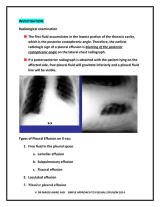
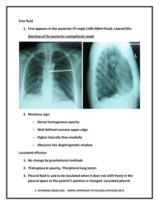
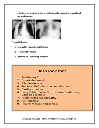
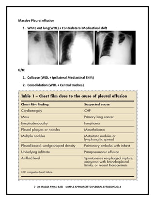
1 DR MAGDI AWAD SASI SIMPLE APPROACH TO PLEURAL EFFUSION 2014
PLEURAL EFFUSION
Normal Physiology :
• Normally pleural space contains a thin layer of fluid.
• The pleural space is not really a space but rather a potential space
between the lung and chest wall.
• There is normally a very thin layer of fluid (from 2 to 10 m thick)
between the two pleural surfaces, the parietal pleura and visceral pleura
• During each respiratory cycle the pleural pressures and the geometry of
the pleural space fluctuate widely. Fluid enters and leaves the pleural
space constantly .
• Fluid enters the pleural space from the capillaries in the parietal pleural
and is removed by the lymphatics in the parietal pleura.
• Fluid can also enter the pleural space from the interstitial spaces of the
lung via the visceral pleura or from the peritoneal cavity through the
diaphragm.
PATHOGENESIS:
• Pleural fluid accumulates when
 Formation increases
 Absorption decreases
• Pleural effusion can be
 Transudative
 Exudative
• Transudative effusion occurs due to systemic factors which either
increase the hydrostatic pressure or decrease the plasma oncotic
pressure( decrease albumin).
2 DR MAGDI AWAD SASI SIMPLE APPROACH TO PLEURAL EFFUSION 2014
• Exudative effusion occurs due to local pathology in the lung or the pleura.
Clinical Manifestations
The symptoms of a patient with a pleural effusion are
to a large extent dictated by the underlying process
causing the effusion.
Many patients have no symptoms referable to the
effusion when effusion is small.
When symptoms are related to the effusion, they arise
either from inflammation of the pleura or from
compromise of pulmonary mechanics.
Pleuritic chest pain is the usual symptom of pleural
inflammation.
Irritation of the pleural surfaces may also result in a dry,
nonproductive cough.
With larger effusions, dyspnea results from lung
compression. 15Dr. Canmao xie
Physical Examinations:
Signs are closely correlated to the volume of pleural effusions. The
volume is larger, the signs is obviously.
 Physical examination of a patient
with pleural effusion reveals :
 Decreased or absent tactile
fremitus, stony dullness to
percussion
 Diminished breath sounds over the
site of the effusion
 Bronchial breath sounds are
frequently present immediately
above the effusion due to lung
collapse.
3 DR MAGDI AWAD SASI SIMPLE APPROACH TO PLEURAL EFFUSION 2014
AETIOLOGY:
• Transudative pleural effusion
 Congestive cardiac failure liver Cirrhosis
 Chronic renal failure Nephrotic syndrome
 Peritoneal dialysis Myxoedema
Exudative Pleural Effusion
• Neoplastic diseases pulmonary embolism
– Metastatic diseases Mesothelioma
• Infectious diseases
– Pneumonia Tuberculosis
• Gastrointestinal diseases
– Pancreatic disease Esophageal perforation
– Intraabdominal abscess Diaphragmatic hernia
• Collagen vascular diseases
– Rheumatoid arthritis SLE
– Drug-induced lupus Immunoblastic lymphadenopathy
– Sjogrens syndrome Wegener’s granulomatosis
– Churg-strauss syndrome Sarcoidosis
• Asbestos exposure
• Sarcoidosis
• Uremia
• Meigs’ syndrome
4 DR MAGDI AWAD SASI SIMPLE APPROACH TO PLEURAL EFFUSION 2014
INVESTIGATION:
Radiological examination
The first fluid accumulates in the lowest portion of the thoracic cavity,
which is the posterior costophrenic angle. Therefore, the earliest
radiologic sign of a pleural effusion is blunting of the posterior
costophrenic angle on the lateral chest radiograph.
If a posteroanterior radiograph is obtained with the patient lying on the
affected side, free pleural fluid will gravitate inferiorly and a pleural fluid
line will be visible.
Types of Pleural Effusion on X-ray:
1. Free fluid in the pleural space
a. Lamellar effusion
b. Subpulmonary effusion
c. Fissural effusion
2. Loculated effusion
3. Massive pleural effusion
5 DR MAGDI AWAD SASI SIMPLE APPROACH TO PLEURAL EFFUSION 2014
Free fluid
1. First appears in the posterior CP angle (100-200ml fluid): Lateral film
blunting of the posterior costophrenic angle
2. Meniscus sign:
– Dense homogenous opacity
– Well defined concave upper edge
– Higher laterally than medially
– Obscures the diaphragmatic shadow
Loculated effusion
1. No change by gravitational methods
2. ?Extrapleural opacity, ?Peripheral lung lesion
3. Pleural fluid is said to be loculated when it does not shift freely in the
pleural space as the patient’s position is changed. Loculated pleural
6 DR MAGDI AWAD SASI SIMPLE APPROACH TO PLEURAL EFFUSION 2014
effusions occur when there are adhesions between the visceral and
parietal pleurae.
Fissural effusion:
1. Lenticular, round or oval shadow
2. “Thickened” fissure
3. ‘Pseudo’ or ‘ Vanishing’ tumors?
Also look for?
1. Positioning?
2. Breast shadows?
3. Rib: Erosions, #
4. Trachea: Shift, Paratracheal shadows
5. Cardiac shadow
6. Lung fields: Cavity, “cotton wool” infiltrates,
Cannon ball mets
7. Hilum: Lymphadenopathy
8. Air-fluid level
9. Pleura: Masses, thickening.
7 DR MAGDI AWAD SASI SIMPLE APPROACH TO PLEURAL EFFUSION 2014
Massive Pleural effusion
1. White out lung(WOL) + Contralateral Mediastinal shift
D/D:
1. Collapse (WOL + Ipsilateral Mediastinal Shift)
2. Consolidation (WOL + Central trachea)
8 DR MAGDI AWAD SASI SIMPLE APPROACH TO PLEURAL EFFUSION 2014
Ultrasonography
• Detects even 5ml of fluid in excess on normal
• Differentiation of pleural thickening from loculated pleural effusion
• Associated abnormalities
Pleural aspiration and Analysis
Transudative or Exudative?
LIGHT’S CRITERIA:
1. Pleural fluid protein/Serum Protein >0.5
2. Pleural fluid LDH/Serum LDH >0.6
3. Pleural fluid LDH > 2/3rd
the upper limit of serum LDH
Tuberculous effusion
1. “Amber” coloured to sero-sanguineous
2. >10%eosinophils; <5%: Mesothelial cells
3. Centrifuged deposits:
– AFB +ve: <10% immunocompetent host
– Culture +ve: 25%
4. ADA elevated (>40U/L)
5. Others: LDH, Soluble IL-2 receptors, IFN-γ
6. Detection of Mycobacteria DNA by PCR
7. Nucleic acid amplification assays
8. Pleural biopsy: Non-caseating granulomas > 80%
9 DR MAGDI AWAD SASI SIMPLE APPROACH TO PLEURAL EFFUSION 2014
MEDICAL MANAGEMNT
 Treatment of underlying cause
 Therapeutic aspiration is necessary in order to relieve dyspnoea
 Precautions:
 Removing more than 1L in one episode in inadvisable
 Can result in re-expansion pulmonary oedema
Should never be aspirated to dryness before the exact etiology is determined
THORACOCENTESIS
 INDICATIONS
 Diagnostic
 Therapeutic
 POSITION
 Sitting position, leaning
forward over a support
 SITE
 Below the scapula,
posteriorly through
the seventh
intercostal space
 PROCEDURE
 Informed consent
 Clean the area with povidine iodine
 Local anesthesia
10 DR MAGDI AWAD SASI SIMPLE APPROACH TO PLEURAL EFFUSION 2014
 Insert the needle and flexible catheter over the needle
 Aspirate pleural fluid
 COMPLICATIONS
 Iatrogenic pneumothorax
 Infection
 Dry tap or bloody tap
 Re-expansion pulmonary oedema
 Pain and respiratory distress
Effusion due to Heart Failure
• Most common cause of pleural effusion
• a diagnostic thoracentesis is done if:
– the effusions are not bilateral and comparable in size
– the patient is febrile
– the patient has pleuritic chest pain to verify that the effusion is
transudative
• Otherwise the patient's heart failure is treated
• If the effusion persists despite therapy, a diagnosticthoracentesis should
be done
• A pleural fluid N-terminal pro-brain natriuretic peptide (NT-proBNP)
>1500 pg/mL is diagnostic of an effusion secondary to congestive heart
failure
11 DR MAGDI AWAD SASI SIMPLE APPROACH TO PLEURAL EFFUSION 2014
Parapneumonic Effusions
• most common cause of exudative pleural effusion (bacterial pneumonias,
lung abscess, bronchiectasis)
• The presence of free pleural fluid can be demonstrated with a lateral
decubitus radiograph, CT of the chest, or ultrasound
• If the free fluid separates the lung from the chest wall by >10 mm, a
therapeutic thoracocentesis should be performed
• A procedure more invasive than thoracocentesis is needed if the following
factors are present:
– Loculated pleural fluid
– Pleural fluid pH <7.20
– Pleural fluid glucose <3.3 mmol/L (<60 mg/dL)
– Positive Gram stain or culture of the pleural fluid
– Presence of gross pus in the pleural space
• If the fluid recurs after the initial therapeutic thoracentesis and if any of
these characteristics are present - a repeat thoracentesis
• If the fluid cannot be completely removed with the therapeutic
thoracentesis,
• insert a chest tube and instill a fibrinolytic agent (e.g., tissue plasminogen
activator, 10 mg)
• perform a thoracoscopy with the breakdown of adhesions
• Decortication (if these measures are ineffective)

Pleural effusion dr magdi sasi

  • 1.
    1 DR MAGDIAWAD SASI SIMPLE APPROACH TO PLEURAL EFFUSION 2014 PLEURAL EFFUSION Normal Physiology : • Normally pleural space contains a thin layer of fluid. • The pleural space is not really a space but rather a potential space between the lung and chest wall. • There is normally a very thin layer of fluid (from 2 to 10 m thick) between the two pleural surfaces, the parietal pleura and visceral pleura • During each respiratory cycle the pleural pressures and the geometry of the pleural space fluctuate widely. Fluid enters and leaves the pleural space constantly . • Fluid enters the pleural space from the capillaries in the parietal pleural and is removed by the lymphatics in the parietal pleura. • Fluid can also enter the pleural space from the interstitial spaces of the lung via the visceral pleura or from the peritoneal cavity through the diaphragm. PATHOGENESIS: • Pleural fluid accumulates when  Formation increases  Absorption decreases • Pleural effusion can be  Transudative  Exudative • Transudative effusion occurs due to systemic factors which either increase the hydrostatic pressure or decrease the plasma oncotic pressure( decrease albumin).
  • 2.
    2 DR MAGDIAWAD SASI SIMPLE APPROACH TO PLEURAL EFFUSION 2014 • Exudative effusion occurs due to local pathology in the lung or the pleura. Clinical Manifestations The symptoms of a patient with a pleural effusion are to a large extent dictated by the underlying process causing the effusion. Many patients have no symptoms referable to the effusion when effusion is small. When symptoms are related to the effusion, they arise either from inflammation of the pleura or from compromise of pulmonary mechanics. Pleuritic chest pain is the usual symptom of pleural inflammation. Irritation of the pleural surfaces may also result in a dry, nonproductive cough. With larger effusions, dyspnea results from lung compression. 15Dr. Canmao xie Physical Examinations: Signs are closely correlated to the volume of pleural effusions. The volume is larger, the signs is obviously.  Physical examination of a patient with pleural effusion reveals :  Decreased or absent tactile fremitus, stony dullness to percussion  Diminished breath sounds over the site of the effusion  Bronchial breath sounds are frequently present immediately above the effusion due to lung collapse.
  • 3.
    3 DR MAGDIAWAD SASI SIMPLE APPROACH TO PLEURAL EFFUSION 2014 AETIOLOGY: • Transudative pleural effusion  Congestive cardiac failure liver Cirrhosis  Chronic renal failure Nephrotic syndrome  Peritoneal dialysis Myxoedema Exudative Pleural Effusion • Neoplastic diseases pulmonary embolism – Metastatic diseases Mesothelioma • Infectious diseases – Pneumonia Tuberculosis • Gastrointestinal diseases – Pancreatic disease Esophageal perforation – Intraabdominal abscess Diaphragmatic hernia • Collagen vascular diseases – Rheumatoid arthritis SLE – Drug-induced lupus Immunoblastic lymphadenopathy – Sjogrens syndrome Wegener’s granulomatosis – Churg-strauss syndrome Sarcoidosis • Asbestos exposure • Sarcoidosis • Uremia • Meigs’ syndrome
  • 4.
    4 DR MAGDIAWAD SASI SIMPLE APPROACH TO PLEURAL EFFUSION 2014 INVESTIGATION: Radiological examination The first fluid accumulates in the lowest portion of the thoracic cavity, which is the posterior costophrenic angle. Therefore, the earliest radiologic sign of a pleural effusion is blunting of the posterior costophrenic angle on the lateral chest radiograph. If a posteroanterior radiograph is obtained with the patient lying on the affected side, free pleural fluid will gravitate inferiorly and a pleural fluid line will be visible. Types of Pleural Effusion on X-ray: 1. Free fluid in the pleural space a. Lamellar effusion b. Subpulmonary effusion c. Fissural effusion 2. Loculated effusion 3. Massive pleural effusion
  • 5.
    5 DR MAGDIAWAD SASI SIMPLE APPROACH TO PLEURAL EFFUSION 2014 Free fluid 1. First appears in the posterior CP angle (100-200ml fluid): Lateral film blunting of the posterior costophrenic angle 2. Meniscus sign: – Dense homogenous opacity – Well defined concave upper edge – Higher laterally than medially – Obscures the diaphragmatic shadow Loculated effusion 1. No change by gravitational methods 2. ?Extrapleural opacity, ?Peripheral lung lesion 3. Pleural fluid is said to be loculated when it does not shift freely in the pleural space as the patient’s position is changed. Loculated pleural
  • 6.
    6 DR MAGDIAWAD SASI SIMPLE APPROACH TO PLEURAL EFFUSION 2014 effusions occur when there are adhesions between the visceral and parietal pleurae. Fissural effusion: 1. Lenticular, round or oval shadow 2. “Thickened” fissure 3. ‘Pseudo’ or ‘ Vanishing’ tumors? Also look for? 1. Positioning? 2. Breast shadows? 3. Rib: Erosions, # 4. Trachea: Shift, Paratracheal shadows 5. Cardiac shadow 6. Lung fields: Cavity, “cotton wool” infiltrates, Cannon ball mets 7. Hilum: Lymphadenopathy 8. Air-fluid level 9. Pleura: Masses, thickening.
  • 7.
    7 DR MAGDIAWAD SASI SIMPLE APPROACH TO PLEURAL EFFUSION 2014 Massive Pleural effusion 1. White out lung(WOL) + Contralateral Mediastinal shift D/D: 1. Collapse (WOL + Ipsilateral Mediastinal Shift) 2. Consolidation (WOL + Central trachea)
  • 8.
    8 DR MAGDIAWAD SASI SIMPLE APPROACH TO PLEURAL EFFUSION 2014 Ultrasonography • Detects even 5ml of fluid in excess on normal • Differentiation of pleural thickening from loculated pleural effusion • Associated abnormalities Pleural aspiration and Analysis Transudative or Exudative? LIGHT’S CRITERIA: 1. Pleural fluid protein/Serum Protein >0.5 2. Pleural fluid LDH/Serum LDH >0.6 3. Pleural fluid LDH > 2/3rd the upper limit of serum LDH Tuberculous effusion 1. “Amber” coloured to sero-sanguineous 2. >10%eosinophils; <5%: Mesothelial cells 3. Centrifuged deposits: – AFB +ve: <10% immunocompetent host – Culture +ve: 25% 4. ADA elevated (>40U/L) 5. Others: LDH, Soluble IL-2 receptors, IFN-γ 6. Detection of Mycobacteria DNA by PCR 7. Nucleic acid amplification assays 8. Pleural biopsy: Non-caseating granulomas > 80%
  • 9.
    9 DR MAGDIAWAD SASI SIMPLE APPROACH TO PLEURAL EFFUSION 2014 MEDICAL MANAGEMNT  Treatment of underlying cause  Therapeutic aspiration is necessary in order to relieve dyspnoea  Precautions:  Removing more than 1L in one episode in inadvisable  Can result in re-expansion pulmonary oedema Should never be aspirated to dryness before the exact etiology is determined THORACOCENTESIS  INDICATIONS  Diagnostic  Therapeutic  POSITION  Sitting position, leaning forward over a support  SITE  Below the scapula, posteriorly through the seventh intercostal space  PROCEDURE  Informed consent  Clean the area with povidine iodine  Local anesthesia
  • 10.
    10 DR MAGDIAWAD SASI SIMPLE APPROACH TO PLEURAL EFFUSION 2014  Insert the needle and flexible catheter over the needle  Aspirate pleural fluid  COMPLICATIONS  Iatrogenic pneumothorax  Infection  Dry tap or bloody tap  Re-expansion pulmonary oedema  Pain and respiratory distress Effusion due to Heart Failure • Most common cause of pleural effusion • a diagnostic thoracentesis is done if: – the effusions are not bilateral and comparable in size – the patient is febrile – the patient has pleuritic chest pain to verify that the effusion is transudative • Otherwise the patient's heart failure is treated • If the effusion persists despite therapy, a diagnosticthoracentesis should be done • A pleural fluid N-terminal pro-brain natriuretic peptide (NT-proBNP) >1500 pg/mL is diagnostic of an effusion secondary to congestive heart failure
  • 11.
    11 DR MAGDIAWAD SASI SIMPLE APPROACH TO PLEURAL EFFUSION 2014 Parapneumonic Effusions • most common cause of exudative pleural effusion (bacterial pneumonias, lung abscess, bronchiectasis) • The presence of free pleural fluid can be demonstrated with a lateral decubitus radiograph, CT of the chest, or ultrasound • If the free fluid separates the lung from the chest wall by >10 mm, a therapeutic thoracocentesis should be performed • A procedure more invasive than thoracocentesis is needed if the following factors are present: – Loculated pleural fluid – Pleural fluid pH <7.20 – Pleural fluid glucose <3.3 mmol/L (<60 mg/dL) – Positive Gram stain or culture of the pleural fluid – Presence of gross pus in the pleural space • If the fluid recurs after the initial therapeutic thoracentesis and if any of these characteristics are present - a repeat thoracentesis • If the fluid cannot be completely removed with the therapeutic thoracentesis, • insert a chest tube and instill a fibrinolytic agent (e.g., tissue plasminogen activator, 10 mg) • perform a thoracoscopy with the breakdown of adhesions • Decortication (if these measures are ineffective)