This document provides an overview of chest anatomy for first year medical students. It includes labeled chest x-rays, diagrams, and images from angiograms and bronchograms to familiarize students with structures like the heart chambers and valves, lungs and lobes, major vessels, and radiologic principles. Key anatomical areas covered are the heart, lungs, pleura, chest wall, diaphragm, and vascular structures in the chest.
Learn Chest X-Ray With Its Normal Positioning & Radio-AnatomyDr.Santosh Atreya
Learn Chest X-Ray With Its Normal Positioning & Radio-Anatomy..For some image description please go through the text book "David Sutton" because i have described these image during my presentation Verbally..There are many animations used inside this presentation so to see all the pictures which are placed layer by layer with the help of animations you simple need to download this presentation first.... Thanx.
The basics of Chest Radiology explained for the undergraduate students. The technical aspects including the various views, exposure, rotation and breath described.
The inside out approach of interpretation explained. The ABCDEFGH description includes Airway, Bones & soft tissue, Cardiac shadow, Diaphragm, Effusion (pleura), Fields (lungs), Gastric bubble and Hila & mediastinum.
The basic cardiac and lung pathologies discussed.
Describes cross sectional anatomy of the mediastinum , and lobar and segmental anatomy of the lung with teaching points and radiological guidelines and multiple examples of lobar and segmental pathologies and how we localize these pathologies .Also the types of chest CT images and indications of chest CT.
Pulmonary Thromboembolism - etilogy, types, medical- Surgical and nursing man...VarunMahajani
Disruption of blood supply to lung alveoli due to blockage of one or more pulmonary blood vessels is called as Pulmonary thromboembolism. In this presentation we will discuss its causes, types and its management in depth.
Lung Cancer: Artificial Intelligence, Synergetics, Complex System Analysis, S...Oleg Kshivets
RESULTS: Overall life span (LS) was 2252.1±1742.5 days and cumulative 5-year survival (5YS) reached 73.2%, 10 years – 64.8%, 20 years – 42.5%. 513 LCP lived more than 5 years (LS=3124.6±1525.6 days), 148 LCP – more than 10 years (LS=5054.4±1504.1 days).199 LCP died because of LC (LS=562.7±374.5 days). 5YS of LCP after bi/lobectomies was significantly superior in comparison with LCP after pneumonectomies (78.1% vs.63.7%, P=0.00001 by log-rank test). AT significantly improved 5YS (66.3% vs. 34.8%) (P=0.00000 by log-rank test) only for LCP with N1-2. Cox modeling displayed that 5YS of LCP significantly depended on: phase transition (PT) early-invasive LC in terms of synergetics, PT N0—N12, cell ratio factors (ratio between cancer cells- CC and blood cells subpopulations), G1-3, histology, glucose, AT, blood cell circuit, prothrombin index, heparin tolerance, recalcification time (P=0.000-0.038). Neural networks, genetic algorithm selection and bootstrap simulation revealed relationships between 5YS and PT early-invasive LC (rank=1), PT N0—N12 (rank=2), thrombocytes/CC (3), erythrocytes/CC (4), eosinophils/CC (5), healthy cells/CC (6), lymphocytes/CC (7), segmented neutrophils/CC (8), stick neutrophils/CC (9), monocytes/CC (10); leucocytes/CC (11). Correct prediction of 5YS was 100% by neural networks computing (area under ROC curve=1.0; error=0.0).
CONCLUSIONS: 5YS of LCP after radical procedures significantly depended on: 1) PT early-invasive cancer; 2) PT N0--N12; 3) cell ratio factors; 4) blood cell circuit; 5) biochemical factors; 6) hemostasis system; 7) AT; 8) LC characteristics; 9) LC cell dynamics; 10) surgery type: lobectomy/pneumonectomy; 11) anthropometric data. Optimal diagnosis and treatment strategies for LC are: 1) screening and early detection of LC; 2) availability of experienced thoracic surgeons because of complexity of radical procedures; 3) aggressive en block surgery and adequate lymph node dissection for completeness; 4) precise prediction; 5) adjuvant chemoimmunoradiotherapy for LCP with unfavorable prognosis.
These simplified slides by Dr. Sidra Arshad present an overview of the non-respiratory functions of the respiratory tract.
Learning objectives:
1. Enlist the non-respiratory functions of the respiratory tract
2. Briefly explain how these functions are carried out
3. Discuss the significance of dead space
4. Differentiate between minute ventilation and alveolar ventilation
5. Describe the cough and sneeze reflexes
Study Resources:
1. Chapter 39, Guyton and Hall Textbook of Medical Physiology, 14th edition
2. Chapter 34, Ganong’s Review of Medical Physiology, 26th edition
3. Chapter 17, Human Physiology by Lauralee Sherwood, 9th edition
4. Non-respiratory functions of the lungs https://academic.oup.com/bjaed/article/13/3/98/278874
Prix Galien International 2024 Forum ProgramLevi Shapiro
June 20, 2024, Prix Galien International and Jerusalem Ethics Forum in ROME. Detailed agenda including panels:
- ADVANCES IN CARDIOLOGY: A NEW PARADIGM IS COMING
- WOMEN’S HEALTH: FERTILITY PRESERVATION
- WHAT’S NEW IN THE TREATMENT OF INFECTIOUS,
ONCOLOGICAL AND INFLAMMATORY SKIN DISEASES?
- ARTIFICIAL INTELLIGENCE AND ETHICS
- GENE THERAPY
- BEYOND BORDERS: GLOBAL INITIATIVES FOR DEMOCRATIZING LIFE SCIENCE TECHNOLOGIES AND PROMOTING ACCESS TO HEALTHCARE
- ETHICAL CHALLENGES IN LIFE SCIENCES
- Prix Galien International Awards Ceremony
Flu Vaccine Alert in Bangalore Karnatakaaddon Scans
As flu season approaches, health officials in Bangalore, Karnataka, are urging residents to get their flu vaccinations. The seasonal flu, while common, can lead to severe health complications, particularly for vulnerable populations such as young children, the elderly, and those with underlying health conditions.
Dr. Vidisha Kumari, a leading epidemiologist in Bangalore, emphasizes the importance of getting vaccinated. "The flu vaccine is our best defense against the influenza virus. It not only protects individuals but also helps prevent the spread of the virus in our communities," he says.
This year, the flu season is expected to coincide with a potential increase in other respiratory illnesses. The Karnataka Health Department has launched an awareness campaign highlighting the significance of flu vaccinations. They have set up multiple vaccination centers across Bangalore, making it convenient for residents to receive their shots.
To encourage widespread vaccination, the government is also collaborating with local schools, workplaces, and community centers to facilitate vaccination drives. Special attention is being given to ensuring that the vaccine is accessible to all, including marginalized communities who may have limited access to healthcare.
Residents are reminded that the flu vaccine is safe and effective. Common side effects are mild and may include soreness at the injection site, mild fever, or muscle aches. These side effects are generally short-lived and far less severe than the flu itself.
Healthcare providers are also stressing the importance of continuing COVID-19 precautions. Wearing masks, practicing good hand hygiene, and maintaining social distancing are still crucial, especially in crowded places.
Protect yourself and your loved ones by getting vaccinated. Together, we can help keep Bangalore healthy and safe this flu season. For more information on vaccination centers and schedules, residents can visit the Karnataka Health Department’s official website or follow their social media pages.
Stay informed, stay safe, and get your flu shot today!
ARTIFICIAL INTELLIGENCE IN HEALTHCARE.pdfAnujkumaranit
Artificial intelligence (AI) refers to the simulation of human intelligence processes by machines, especially computer systems. It encompasses tasks such as learning, reasoning, problem-solving, perception, and language understanding. AI technologies are revolutionizing various fields, from healthcare to finance, by enabling machines to perform tasks that typically require human intelligence.
Couples presenting to the infertility clinic- Do they really have infertility...Sujoy Dasgupta
Dr Sujoy Dasgupta presented the study on "Couples presenting to the infertility clinic- Do they really have infertility? – The unexplored stories of non-consummation" in the 13th Congress of the Asia Pacific Initiative on Reproduction (ASPIRE 2024) at Manila on 24 May, 2024.
Ozempic: Preoperative Management of Patients on GLP-1 Receptor Agonists Saeid Safari
Preoperative Management of Patients on GLP-1 Receptor Agonists like Ozempic and Semiglutide
ASA GUIDELINE
NYSORA Guideline
2 Case Reports of Gastric Ultrasound
Tom Selleck Health: A Comprehensive Look at the Iconic Actor’s Wellness Journeygreendigital
Tom Selleck, an enduring figure in Hollywood. has captivated audiences for decades with his rugged charm, iconic moustache. and memorable roles in television and film. From his breakout role as Thomas Magnum in Magnum P.I. to his current portrayal of Frank Reagan in Blue Bloods. Selleck's career has spanned over 50 years. But beyond his professional achievements. fans have often been curious about Tom Selleck Health. especially as he has aged in the public eye.
Follow us on: Pinterest
Introduction
Many have been interested in Tom Selleck health. not only because of his enduring presence on screen but also because of the challenges. and lifestyle choices he has faced and made over the years. This article delves into the various aspects of Tom Selleck health. exploring his fitness regimen, diet, mental health. and the challenges he has encountered as he ages. We'll look at how he maintains his well-being. the health issues he has faced, and his approach to ageing .
Early Life and Career
Childhood and Athletic Beginnings
Tom Selleck was born on January 29, 1945, in Detroit, Michigan, and grew up in Sherman Oaks, California. From an early age, he was involved in sports, particularly basketball. which played a significant role in his physical development. His athletic pursuits continued into college. where he attended the University of Southern California (USC) on a basketball scholarship. This early involvement in sports laid a strong foundation for his physical health and disciplined lifestyle.
Transition to Acting
Selleck's transition from an athlete to an actor came with its physical demands. His first significant role in "Magnum P.I." required him to perform various stunts and maintain a fit appearance. This role, which he played from 1980 to 1988. necessitated a rigorous fitness routine to meet the show's demands. setting the stage for his long-term commitment to health and wellness.
Fitness Regimen
Workout Routine
Tom Selleck health and fitness regimen has evolved. adapting to his changing roles and age. During his "Magnum, P.I." days. Selleck's workouts were intense and focused on building and maintaining muscle mass. His routine included weightlifting, cardiovascular exercises. and specific training for the stunts he performed on the show.
Selleck adjusted his fitness routine as he aged to suit his body's needs. Today, his workouts focus on maintaining flexibility, strength, and cardiovascular health. He incorporates low-impact exercises such as swimming, walking, and light weightlifting. This balanced approach helps him stay fit without putting undue strain on his joints and muscles.
Importance of Flexibility and Mobility
In recent years, Selleck has emphasized the importance of flexibility and mobility in his fitness regimen. Understanding the natural decline in muscle mass and joint flexibility with age. he includes stretching and yoga in his routine. These practices help prevent injuries, improve posture, and maintain mobilit
micro teaching on communication m.sc nursing.pdfAnurag Sharma
Microteaching is a unique model of practice teaching. It is a viable instrument for the. desired change in the teaching behavior or the behavior potential which, in specified types of real. classroom situations, tends to facilitate the achievement of specified types of objectives.
New Directions in Targeted Therapeutic Approaches for Older Adults With Mantl...i3 Health
i3 Health is pleased to make the speaker slides from this activity available for use as a non-accredited self-study or teaching resource.
This slide deck presented by Dr. Kami Maddocks, Professor-Clinical in the Division of Hematology and
Associate Division Director for Ambulatory Operations
The Ohio State University Comprehensive Cancer Center, will provide insight into new directions in targeted therapeutic approaches for older adults with mantle cell lymphoma.
STATEMENT OF NEED
Mantle cell lymphoma (MCL) is a rare, aggressive B-cell non-Hodgkin lymphoma (NHL) accounting for 5% to 7% of all lymphomas. Its prognosis ranges from indolent disease that does not require treatment for years to very aggressive disease, which is associated with poor survival (Silkenstedt et al, 2021). Typically, MCL is diagnosed at advanced stage and in older patients who cannot tolerate intensive therapy (NCCN, 2022). Although recent advances have slightly increased remission rates, recurrence and relapse remain very common, leading to a median overall survival between 3 and 6 years (LLS, 2021). Though there are several effective options, progress is still needed towards establishing an accepted frontline approach for MCL (Castellino et al, 2022). Treatment selection and management of MCL are complicated by the heterogeneity of prognosis, advanced age and comorbidities of patients, and lack of an established standard approach for treatment, making it vital that clinicians be familiar with the latest research and advances in this area. In this activity chaired by Michael Wang, MD, Professor in the Department of Lymphoma & Myeloma at MD Anderson Cancer Center, expert faculty will discuss prognostic factors informing treatment, the promising results of recent trials in new therapeutic approaches, and the implications of treatment resistance in therapeutic selection for MCL.
Target Audience
Hematology/oncology fellows, attending faculty, and other health care professionals involved in the treatment of patients with mantle cell lymphoma (MCL).
Learning Objectives
1.) Identify clinical and biological prognostic factors that can guide treatment decision making for older adults with MCL
2.) Evaluate emerging data on targeted therapeutic approaches for treatment-naive and relapsed/refractory MCL and their applicability to older adults
3.) Assess mechanisms of resistance to targeted therapies for MCL and their implications for treatment selection
These lecture slides, by Dr Sidra Arshad, offer a quick overview of physiological basis of a normal electrocardiogram.
Learning objectives:
1. Define an electrocardiogram (ECG) and electrocardiography
2. Describe how dipoles generated by the heart produce the waveforms of the ECG
3. Describe the components of a normal electrocardiogram of a typical bipolar leads (limb II)
4. Differentiate between intervals and segments
5. Enlist some common indications for obtaining an ECG
Study Resources:
1. Chapter 11, Guyton and Hall Textbook of Medical Physiology, 14th edition
2. Chapter 9, Human Physiology - From Cells to Systems, Lauralee Sherwood, 9th edition
3. Chapter 29, Ganong’s Review of Medical Physiology, 26th edition
4. Electrocardiogram, StatPearls - https://www.ncbi.nlm.nih.gov/books/NBK549803/
5. ECG in Medical Practice by ABM Abdullah, 4th edition
6. ECG Basics, http://www.nataliescasebook.com/tag/e-c-g-basics
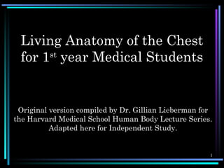
1. Living Anatomy of the Chest
for 1st year Medical Students
Original version compiled by Dr. Gillian Lieberman for
the Harvard Medical School Human Body Lecture Series.
Adapted here for Independent Study.
1
2. Living Anatomy
Radiology is ideally suited to image anatomy in the living patient.
Labeled plain film Chest X-Rays (CXR), Angiograms, Bronchograms,
Computed Tomography (CT) and Magnetic Resonance Images (MRI)
follow, accompanied by anatomic diagrams to help familiarize you with
chest anatomy.
Areas covered include:
The Heart: Chambers, valves, great vessels, coronary arteries
The Lungs: Lobes, pulmonary arteries, bronchial tree
The Pleura
The Azygos-Hemiazygos venous system
Basic Radiologic principles are outlined to facilitate plain film interpretation.
2
3. CXR-PA Anatomy on
1
2
2
4
Normal Chest
X-Ray
13
Key:
• Right 1st rib
3• Right 2nd rib
5
6 • Scapula
7 7
• Trachea
9 8
• Carina
• Bronchus seen end on
• Bilateral hila
• Branch of right main descending
pulmonary artery
• Right minor (horizontal fissure)
• Right hemi diaphragm
10 • Left hemi diaphragm
11
• Gastric air bubble
12 • Left clavicle 3
4. CXR-Left LAT Anatomy on
6
Normal Chest
6
T2
T3
1b
1a 5
X-Ray
4a 4b Key:
T5 1a. Manubrium sternum
8 1b. Body of sternum
T6
4. Right hemi diaphragm
T7 5. Left hemi diaphragm
4a. Right scapula
T8
4b. Left scapula
8. Trachea
T9 9a
9. Soft tissue of the arms
9b
T10 10. Major fissure
11. Minor fissure-little higher in this
3 T11
patient than the usual
9a. 9th left rib
2
9b. 9th right rib
T2-11 Thoracic vertebrae 4
5. Radiologic Principles: I
Right Upper Lobe Pneumonia with
Partial Volume Loss Pneumoperitoneum This film is helpful to demonstrate some
basic radiologic principles which are
essential to understanding x-ray
1
interpretation.
2
Key:
2. Denser and smaller right upper lobe due to
pneumonia
3. Elevated minor fissure
5 4. Top surface of liver
4
7
3 5. Undersurface of diaphragm
5
4 6. Top surface of diaphragm
6 7
7. Top surface of spleen
8. Free air in the abdominal cavity =
pneumoperitoneum 5
6. Radiologic Principles: II
Fat
The plain films are made up of four densities-
Bone Black Air e.g. in lungs,stomach
Fat
Soft tissue which include muscle,
organs e.g. liver, fluid
e.g. blood
Air
White Bone heavy metal e.g. calcium,
iron
Soft Tissue
A line or border is seen only when there is an
interface between two of these densities. E.g.
The right heart outline is usually seen because
soft tissue density of the heart is next to air
density of the right middle lobe of the lung.
6
7. Radiologic Principles: III
Pneumoperitoneum
Free air in the abdomen (always abnormal)
Pneumonia
(pneumoperitoneum) rises to a position under the
diaphragm when the patient is upright. It
therefore outlines the top of the liver on the right,
the top of the spleen on the left, and
undersurfaces on both hemi diaphragms. These
are usually hot seen because liver, spleen and
diaphragm are all soft tissue and therefore no
interface is present. The upper border of the
diaphragms are usually seen because air in the
lower lung lobes abut the soft tissues of the
diaphragm.
Pneumonia
In pneumonia, the air in the lung gets
Pneumoperitoneum replaced with fluid which shows up soft
tissue density on x-ray. The lung lobe
often also gets smaller or consolidated
so the fissures move. Bacterial
infection commonly respects the lobar
boundaries as in this case. 7
8. Lobes of the Lung
Lobes and Fissures of the Lung (from the front) IMPORTANT FACTS:
The right lung has 3 lobes (separated
by the major Oblique fissure &
minor Horizontal fissure)
-Right upper lobe
-Right middle lobe
-Right lower lobe
The left lung has 2 lobes separated
by major (oblique) fissure
-Left upper lobe
(medial portion is called the lingula)
-Left lower lobe
“Man’s Anatomy” by Tobias & Arnold 8
9. The Pleura
The pleura is the lining of the lungs.
There are 2 layers -1. The visceral pleura hugs the lung lobes
-2. The parietal pleura hugs the chest wall
The pleural space is a potential space between the two.
A pneumothorax is the presence of air (always abnormal) in the pleural space.
A pleural effusion is the presence of detectable fluid (always abnormal) in the pleural space.
A hydropneumothorax is air and fluid in the pleural space.
Coronal Section of Pleural Sacs
(schematic)
“Man’s Anatomy”
by Tobias &
Arnold
9
10. Right Tension Pneumothorax
Lobes of the
Lung
This film is included because it helps demonstrate
the 3 lobes of the right lung, the pleura and the
pleural space.
4
Key:
2. Normal pointy left costophrenic angle
3
3. Blunted denser right costophrenic angle
5 due to fluid in pleural space = pleural
2 effusion
7
4. Air in pleural space = pneumothorax
5. … partially collapsed right upper lobe
6 6. ---partially collapsed right middle lobe
7. -.-partially collapsed right lower lobe
2 8. Visceral pleura of right middle lobe
9. (Position of parietal pleura – not seen)
9 1
10. Left breast shadow. (Notice the right
Common causes for hydropneumothorax include rib fractures breast has been removed = right
penetrating chest wounds e.g. stab or bullet wounds and mastectomy)
iatrogenic causes e.g. lung biopsies or effusion drainages. 10
Pneumothorax and pleural effusion =
hydropneumothorax
11. Anatomy on Normal Chest X-Ray
Heart borders and chambers of the heart on PA and lateral views.
11
12. Heart Chambers and Valves
The heart is made up of 4 chambers. The right side which handles deoxygenated blood is
separated from the left side which handles oxygenated blood by septa, the top is separated
from the bottom by valves.
Simplistic view: 1
12
13. Venous Return to the Heart
The atria receives blood from the body and lungs.
The SVC and IVC bring deoxygenated (blue) blood to the right atrium from the
body. The pulmonary veins bring oxygenated (red) blood to the left atrium from
the lungs.
Simplistic view: 2
13
14. Arterial Output from the Heart
The ventricles receive blood from their respective atria.
The right ventricle pumps deoxygenated blood via the pulmonary
artery to the lungs.
The left ventricle pumps oxygenated blood via the aorta to the body.
The entrance to the aorta and the pulmonary artery have aortic and
pulmonary valves respectively.
Simplistic view: 3
Pulmonary valve Aortic valve
14
15. Heart Valves
This patient had a malfunctioning mitral valve (between left atrium and left ventricle) and aortic valve
(between left ventricle and aorta) and prosthetic valves were inserted (better seen on lateral)
Frontal CXR LAT CXR
Key:
2. Suture material
used for repair
1 of vertical
incision thru
sternum (median
sternotomy)
1 2 3. Aortic valve
2 prosthesis
3 4. Mitral valve
3 prosthesis
5. Left hemi
diaphragm
6. Right hemi
diaphragm
5
4 4
5
15
16. Schema of great vessels
connected to the heart
The pulmonary artery and aorta cross one another in the mediastinum.
16
“Man’s Anatomy by Tobias & Arnold
19. Angiograms-Aortic arch angiogram
10
9
6
An angiogram is an x-ray examination of blood
vessels following contrast administration.
5
Arteriogram = Arterial Study
4
7 Venogram = Venous Study
8
2
1
3
19
20. Angiograms-Pulmonary arteriogram
(PA gram)
Pulmonary Art #1
Key:
2. Right main pulmonary artery branch
3. Right upper lobe pulmonary artery branch
4. Right middle lobe pulmonary artery branch
2 5
2 7 5. Right lower lobe pulmonary artery branch
1
6. Left main pulmonary artery
3
7. Left upper lobe pulmonary artery branch
8. Left lower lobe pulmonary artery branch
9. Pulmonary veins
10. Left atrium
11. Left ventricle
12. Ascending aorta
13. Descending aorta 20
21. Angiograms-Pulmonary
arteriogram (PA gram)
Pulmonary Art #2
Key:
2. Right main pulmonary artery branch
3. Right upper lobe pulmonary artery branch
4. Right middle lobe pulmonary artery branch
11 5. Right lower lobe pulmonary artery branch
12
6. Left main pulmonary artery
7. Left upper lobe pulmonary artery branch
9
8. Left lower lobe pulmonary artery branch
9. Pulmonary veins
10. Left atrium
10
11. Left ventricle
12. Ascending aorta
13. Descending aorta 21
22. Angiograms-Pulmonary
arteriogram (PA gram)
Pulmonary Art #3
Key:
2. Right main pulmonary artery branch
3. Right upper lobe pulmonary artery branch
4. Right middle lobe pulmonary artery branch
11 12
5. Right lower lobe pulmonary artery branch
6. Left main pulmonary artery
7. Left upper lobe pulmonary artery branch
8. Left lower lobe pulmonary artery branch
9. Pulmonary veins
10
10. Left atrium
11. Left ventricle
12. Ascending aorta
13. Descending aorta 22
23. Heart and Vessels
Cardiomegaly plus early Congestive Heart Failure (CHF) Key:
2. Inferior vena cava (IVC)
3. Superior vena cava (SVC)
*3. Azygos vein
7 5 7 7 5. Carina
7 2
6. Trachea
4
3 7. Right main stem bronchus
8. Prominent pulmonary vessels
Any and or all heart chambers may enlarge when the
heart becomes diseased. Cardiomegaly = a big heart.
A patient’s heart enlarges due to a number of
diseases e.g. valve disease, high blood pressure,
congestive heart failure.
1 If the heart fails, the lung often become congested.
Early on the pulmonary vessels appear more
prominent as in this case. More advanced failure can
result in a condition of pulmonary edema which is
fluid flooding into the alveoli of the lungs causing
the patient marked shortness of breath.
23
24. Azygos-Hemiazygos venous system
The Azygos vein receives
tributaries from intercostal
veins as outlined. It is seen
as an oval density to the
right of the trachea just
above the right main stem
bronchus on all chest x-rays
(*3 on the earlier film)
This is the portion that
travels forward to join the
SVC.
In CHF, the Azygos vein
dilates and this density
becomes prominent as seen
on the previous patient’s
CXR.
24
“Man’s Anatomy by Tobias & Arnold
27. Coronary Angiograms
LT Coronary Art LAO
Left main
coronary
LAO
artery
Diagonal The coronary arteries can be
artery outlined in the living patient by
Left circumflex
artery injecting contrast into them. A
catheter (tube) is threaded
Obtuse through the Patients vessels to the
Marginal heart, to gain access- called
Artery “cardiac catheterization”
27
28. Coronary Angiograms
LT Coronary Art LAO
Left main coronary
artery
Sinus Node Artery
LAD
AV Diagonal
Groove artery
Left IV Groove
circumflex Obtuse
artery marginal Septal
artery perforator
28
29. Coronary Angiograms
RT Coronary Art LAO
Conus
Bronch
RCA
AV Node A
AV Crux
Groove
Acute Posterior LV
marginal Bronch
artery
29
32. Normal Bronchogram
Frontal CXR Contrast agent can be instilled or inhaled into the bronchial tree Lateral CXR
outlining the walls of the trachea, main stem bronchi, segmental and
even subsegmental bronchi
32
33. Abnormal Bronchogram: Bronchiectasis
Bronchiectasis = localized irreversible dilatation of the bronchial tree
Contrast agent can be instilled or
inhaled into the bronchial tree
outlining the walls of the
trachea, main stem bronchi,
segmental and even
subsegmental bronchi
33
34. Computed Tomography
Computer tomography (CT) scanning obtains multiple cross sectional images through a patient
using x-rays and computer enhancement. (Imagine slicing a sausage crosswise into many round
equal thickness slices and then looking at these to see what’s in the sausage)
CT, ultrasound and magnetic resonance imaging (MRI) all allow imaging of the body in different
planes.
TERMINOLOGY:
The following description considers the body in the anatomical position
Axial plane (=cross section) a plane of the body parallel to the horizon
Median/Midline Sagittal plane – the vertical plane which passes through the sagittal suture of the
skull and through the midline of the body dividing the body into right and left halves.
ParaSagittal plane –any vertical plane parallel to the median sagittal plane.
Coronal plane –any vertical plane perpendicular to the median sagittal plane and parallel to the
vertical plane through the coronal suture of the skull.
With CT scanning, factors can be altered for better resolution of different body parts.
e.g. Referring to the images enclosed, the scanner was set to optimally visualize mediastinal
structures (1-4A), and lung parenchyma in (1-4B) 34
35. Normal Chest anatomy on
Key:
Axial Computed Tomography
2. Pectoralis major muscle 16. Superior vena cava (SVC 29. Diaphragm
3. Pectoralis minor muscle • Aortic arch • Liver
4. Sternum • Ascending aorta 3. Spleen
5. Clavicle • Descending aorta 4. Stomach
6. Rib • Azygos vein 5. Kidney
7. Humeral head * Carina (tracheal bifurcation) 6. Lung –upper lobe
8. Scapula 7. Pulmonary artery 7. Lung –right middle lobe
9. Vertebral body 8. Main stem bronchus 8. Lung –lower lobe
10. Thyroid gland 9. Right ventricular outflow 9. Major (oblique) fissure
tract
11. Trachea 10. Minor (horizontal) fissure
10. Left atrium
12. Esophagus 11. Segmental bronchus
11. Right atrium
13. Subclavian artery
12. Left ventricle
14. Carotid artery
13. Right ventricle
15. Innominate (brachialcephalic)
artery 28A. Pulmonary veins
35
16. Innominate vein 28B. Inferior vena cava (IVC)
50. Normal MRI Chest
Magnetic Resonance Imaging (MRI) utilizes changing magnetic and
electrical fields to obtain images of a patient. Factors can be altered to
enhance resolution of different structures thus blood for example can look
bright white or dark black.
Among the advantages of MRI are:
1. X-rays and the attendant hazards of ionizing radiation are not present.
2. Scans in multiple different projections e.g. oblique, sagittal, coronal, axial
can be obtained with ease.
Refer to films:
Film 1 -Sagittal oblique MRI angiogram chosen to best demonstrate the aortic arch.
Film 2&3 -Axial sections
Film 4&5 -Sagittal oblique MRI angiogram chosen to best demonstrate the coronary arteries
50
62. Conclusion of
Living anatomy of the chest
Congratulations! You have completed this module.
You worked through many anatomic diagrams and labeled
chest x-rays, bronchograms, angiograms, CT scans & MRI
images. You saw the normal and also some Abnormal images to
peak your interest. Radiology is ideally suited to image not
only normal anatomy, but more importantly from a clinical
diagnostic standpoint, abnormal anatomy & pathology.
Wishing you a joy-filled career and
life long love of learning. Gill
With grateful thanks to Pamela Lepkowski, Education Coordinator, Harvard
Medical School & Assistant extraordinaire for her outstanding work on this 62
Independent study module.