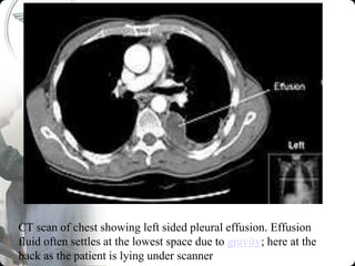
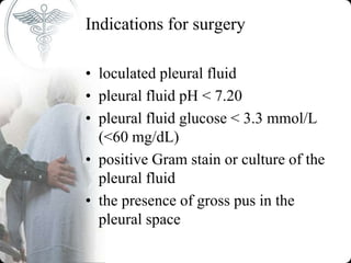
Shree sahAjanand institute of nursing,
PLEURAL EFFUSION
Presented by
Mr. AKRAM KHAN
M.S.N. (HOD)
ASST. PROFESSOR
SSIN, BHAVNAGAR
Introduction
• Normally, 10-20 mL of fluid is
spread thinly over the visceral and
parietal pleurae.
• The fluid is similar in composition to
plasma except that it is lower in
protein (< 1.5 g/dL).
• Incidence – 1 million cases per year
• Prevalence – 320/100,000 in
industrialised countries.
DEFINITION
• A pleural effusion is present when
there is an excess quantity of fluid in
the pleural space (space lies between
the lung and chest wall and normally
contains a very thin layer of fluid,
which serves as a coupling system).
AETIOLOGY
• In the normal pleural space, there is a steady state
in which there is a roughly equal rate of the
formation (entry) and absorption (exit) of liquid
• This balance must be disturbed in order to produce
a pleural effusion.
• It is likely that both mechanisms contribute to
effusion formation for the following reasons
• An isolated increase in entry rate, unless massive,
is unlikely to cause effusion because the absorbing
pleural lymphatics have a large reserve capacity to
deal with excess pleural liquid. 20 times
• An isolated decrease in exit rate is also unlikely to
cause effusions because the normal entry rate is
low.
CLASSIFICATION
• Classified into unilateral or bilateral
• Into transudative and exudative.
• Causes of bilateral effusion
– Hypoproteinemia
– Connective tissue diseases
– CCF
TRANSUDATE
• Due to systemic factors
• Transudates are due to an imbalance
between hydrostatic and oncotic
pressures .
• -Incr. Hydrostatic factors(pleural
fluid)
• -decr. Osmotic pressure(plasma)
• -decr. Oncotic pressure(plasma)
AETIOLOGY OF TRANSUDATIVE
• Nephrotic syndrome
• CCF
• Liver cirrhosis
• Myxedema
• SVC Obstruction
• Hypoproteinemia – malnutrition,
malabsorption
• Biliothorax
• Peritoneal dialysis
EXUDATE
• Due to local factors
• Change in pleural surface permeability
• -Incr. microvascular permeability- protein
leakage
• -Injury to adjacent lung tissue
• Exudates are secondary to a disturbance of
the systems regulating pleural fluid
formation and absorption/drainage (as in
bacterial, viral, or fungal infection,
rheumatologic disease, or malignancy).
AETIOLOGY OF EXUDATIVE
– TB, pneumonia, fungal
– Pulmonary embolism
– Malignancy-lung cancer,breast ca, lymphoma, metastatic
– Hemothorax
– Lymphatic obstruction
– Ureamia
– CT dse- SLE, rheumatoid arthritis
– Meig’s syndrome (triad of ovarian tumour w ascites and p effusion)
– Asbestos exposure
– Radiation
– Drugs- nitrofurantoin, phenytoin, methotrexate, amiodarone.
– Trauma
– Sarcoidosis
– Subdiaphragmatic abscess
– Acute pancreatitis
• Exudative pleural effusions meet at least one of
the following criteria, whereas transudative
pleural effusions meet none of these ratios
• Pleural fluid protein/serum protein >0.5
• Pleural fluid LDH/serum LDH >0.6
• Pleural fluid LDH more than two-thirds normal
upper limit for serum
• If one or more of the exudative criteria are met
and the patient is clinically thought to have a
condition producing a transudative effusion, the
difference between the protein levels in the serum
and the pleural fluid should be measured. If this
gradient is greater than 31 g/L (3.1 g/dL), the
exudative categorization by the above criteria can
be ignored because almost all such patients have a
transudative pleural effusion.
PATHOGENESIS
• INCREASED FLUID ENTRY
• -Increase in permeability
• -Increase in microvascular
pressure
• -Decrease in pleural pressure
• -Decrease in plasma osmotic
pressure
DECREASED FLUID EXIT
• -Intrinsic factors
• -Cytokines & prod. of inflame
• - endotoxins
• - Endocrine abnormalities
• -hypothyroidism
• -Injury due to radiation /chemo
• -Infiltration of lymphatics by cancer
• -Anatomic abnormalities
• -yellow nail syndrome (reduced lymphatic drainage)
DECREASED FLUID EXIT
• -Extrinsic factors
• Limitation of respiratory motion- diaphragm
paralysis
• Extrinsic compression of lymphatics- pleural
fibrosis,
• Blockage of lymphatic stomata - pleural
malignancy
• Decreased intrapleural pressure -fibrous rind on the
visceral pleura
• Increased systemic venous pressure – decr.
lymphatic flow
• Decreased liquid availability - pneumothorax
CLINICAL FEATURES
• Symptoms
• -Dyspnea(most common) - because of distortion of
diaphragm and chest wall during resp more than from
hypoxemia
• -mild, nonproductive cough.
• -pleuritic chest pain
• - chest wall splinting(stiffening of body part to avoid
pain)
• -tachypnea esp with lung compression or more severe
infxn.
• -Fever
• -hemoptysis
• -Constitutional features ( wt loss, myalgia, general
malaise, headache)
Cont….
• Other symptoms may suggest the etiology
of the pleural effusion.
– More severe cough or production of purulent
or bloody sputum suggests an underlying
pneumonia or endobronchial lesion.
– Constant chest wall pain may reflect chest
wall invasion by bronchogenic carcinoma or
malignant mesothelioma.
– Pleuritic chest pain suggests either pulmonary
embolism or an inflammatory pleural process.
– Systemic toxicity evidenced by fever, weight
loss, and inanition suggests empyema.
Cont……• SIGNS
• -Resp distress( FAN, ICR, SCR, use of acces muscles)
• -Decr. Chest movement. Decreased motion of the
hemithorax
• -Trachea shifted to opp. Side (mostly if massive more
than 1000ml)
• -Tenderness
• -Tactile fremitus decr
• -Stony dullness
• -Diminished or absent breath sounds
• -Vocal fremitus decr.
• - Basal crepitations
• -Friction rub
• -Egophony (E-to-A change)
INVESTIGATIONS
• Chest x-ray –PA-Lateral
– Homogenous opacities with meniscus
– Blunting of the costo-phrenic angle
– Incr. upper lung field vasculature
– Mediastinal shift if large unilateral (>1000ml)
• CT scan of chest – mass,
• Pleural biopsy
• Thoracocentesis - pleural tap
• Thoracoscopy (provides direct view of both
parietal and visceral aspects of pleura)
Massive right-sided pleural effusion (white area) in a
patient presenting with lung cancer
CT scan of chest showing left sided pleural effusion. Effusion
fluid often settles at the lowest space due to gravity; here at the
back as the patient is lying under scanner
Contraindication to Pleural tap (relative)
• - Low volume of effusion <1cm thickness on
lateral decubitus Xray
– Bleeding diathesis or systemic anticoagulation
– Mechanical ventilation
– Cutaneous disease over the puncture site
COMPLICATIONS
– pain at site
– Spleen/liver puncture
– Pneumothorax -Use of needles larger
than 20 gauge increases the risk
– Cutaneous or internal bleeding
Pleural tap
Colour
• -pale yellow/straw-transudate
• -hrrghic-malignancy, trauma, TB,
• -milky/whitish-chylothorax
• -Brown-long standing hrrg(amoebic liver abscess)
• -Black- aspergillosis
• -Yellow green –arthritis
• -Dark green- biliothorax
• Fluid may be clear yellow (serous), milky
(chylous), blood-tinged (serosanguineous), grossly
bloody (sanguineous), or translucent or opaque and
thick (purulent).
– Character
presence of pus (empyema)
• -viscous(mesothelioma)
• -Debris (rheumatoid arthritis)
• -Turbid (inflamm. conditions)
Biochemistry (lights criteria)
• - All exudates evaluated for amylase
level, differential cell count,
glucose level.
• -Culture & sensitivity
• -Microbiology -Gram stain
• -ZN stain (TB)
• Cytology (Tumour markers: CEA,VIM)
– Pleural fluid lymphocytosis, with lymphocytes greater
than 85% of the total nucleated cells, suggests
tuberculosis (TB), lymphoma, sarcoidosis, chronic
rheumatoid pleurisy, yellow nail syndrome, or
chylothorax. Pleural lymphocytes of 50-70% of the
nucleated cells suggests malignancy.
– Pleural fluid eosinophilia, with eosinophils greater than
10% of nucleated cells, is seen in hydropneumothorax,
hemothorax, pulmonary infarction, benign asbestos
pleural effusion, parasitic disease, fungal infection,
drugs, and malignancy.
– Mesothelial cells are found in variable numbers in most
effusions, but their presence at more than 5% of total
nucleated cells makes a diagnosis of TB unlikely.
– Markedly increased numbers of mesothelial cells,
especially in bloody or eosinophilic effusions, suggests
pulmonary embolism as the cause.
Difference between transudate and exudate
(Lights criteria)
Two-test rule
• - Pleural fluid cholesterol greater than 45 mg/dL
• - Pleural fluid LDH greater than 0.45 times the
upper limit of the laboratory's normal serum LDH
• Three-test rule
• - Pleural fluid protein greater than 2.9 g/dL
• - Pleural fluid cholesterol greater than 45 mg/Dl
• - Pleural fluid LDH greater than 0.45 times the
upper limit of the laboratory's normal serum LDH
Test required
• CBC
• ESR
• UEC-nephrotic syndrome
• LFT-liver cirrhosis
• Urinalysis
• ANA, RF
• Pleural biopsy
• Thoracoscopy
• Bronchoscopy
• Suspect TB pleuritis in patients with a history of exposure or a
positive purified protein derivative (PPD) finding and in
patients with lymphocytic exudative effusions, especially if
less than 5% mesothelial cells are detected on differential cell
counts.
– Because most tuberculous pleural effusions probably result from a
hypersensitivity reaction to the mycobacterium rather than from
microbial invasion of the pleura, acid-fast bacillus stains of pleural
fluid are rarely diagnostic (<10% of cases), and pleural fluid
cultures grow Mycobacterium tuberculosis in less than 65% of
cases.
– In contrast, the combination of histology and culture of pleural
tissue obtained by pleural biopsy increases the diagnostic yield to
90%.
– Adenosine deaminase (ADA) activity of more than 43 U/mL in
pleural fluid supports the diagnosis of TB pleuritis. However, the
test has a sensitivity of only 78%; therefore, pleural ADA values
less than 43 U/mL do not exclude the diagnosis of TB pleuritis.
– Interferon-gamma concentrations in pleural fluid greater than 140
pg/mL also support the diagnosis of TB pleuritis, but this test is not
routinely available.
• Measure pleural fluid amylase if a pancreatic
origin or ruptured esophagus is suspected or if a
unilateral left pleural effusion remains
undiagnosed after initial testing. An additional
assay of amylase isoenzymes can help distinguish
a pancreatic source from other etiologies.
• Measure triglycerides and cholesterol on milky
pleural fluids when chylothorax or
pseudochylothorax is suspected.
• Consider immunologic studies, including pleural
fluid antinuclear antibody and rheumatoid factor,
when collagen-vascular diseases are suspected.
Management
• Supportive
• -Supplemental oxygen
• -IV fluid hydration
• -Chest physiotherapy
• -Therapeutic/diagnostic thoracentesis
• -Antibiotics
• -Empirically by age/social
circumstances and modified by blood
and pleural effusion fluid culture
results
Definitive
• Treat underlying cause
• chest tube for continuous drainage
• pleuroperitoneal shunt and chemical pleurodesis
• Chemical pleurodesis -Doxycycline 500 mg,
• Bleomycin 60 IU
• TALC in a slurry(rarely used)
• Empyema: -Antibiotics alone with close monitoring in children
• Antibiotics with chest tube drainage in adults
• Pleurectomy - trapped lung (Excision of pleura, usually parietal)
• Pleural fluid deloculation (If unsuccessful, then either thoracoscopic
adhesiolysis or decortications via thoracotomy are indicated)
• Diuresis as appropriate for effusions secondary to congestive heart failure
and ascites
• May inject 250,000 units of streptokinase or 100,000 units of urokinase
intrapleurally to dissolve fibrin meshes creating loculation.
• Indication for chest tube
Recurrent pleural effusion
Empyema
Pneumothorax & hydrothorax
• Malignant pleural effusion
– Repeated effusions- pleurodesis
Indications for surgery
• loculated pleural fluid
• pleural fluid pH < 7.20
• pleural fluid glucose < 3.3 mmol/L
(<60 mg/dL)
• positive Gram stain or culture of the
pleural fluid
• the presence of gross pus in the
pleural space
• Thanks to all

PLEURAL EFFUSION BY Mr. AKRAM KHAN

  • 1.
    Shree sahAjanand instituteof nursing, PLEURAL EFFUSION Presented by Mr. AKRAM KHAN M.S.N. (HOD) ASST. PROFESSOR SSIN, BHAVNAGAR
  • 2.
    Introduction • Normally, 10-20mL of fluid is spread thinly over the visceral and parietal pleurae. • The fluid is similar in composition to plasma except that it is lower in protein (< 1.5 g/dL). • Incidence – 1 million cases per year • Prevalence – 320/100,000 in industrialised countries.
  • 3.
    DEFINITION • A pleuraleffusion is present when there is an excess quantity of fluid in the pleural space (space lies between the lung and chest wall and normally contains a very thin layer of fluid, which serves as a coupling system).
  • 4.
    AETIOLOGY • In thenormal pleural space, there is a steady state in which there is a roughly equal rate of the formation (entry) and absorption (exit) of liquid • This balance must be disturbed in order to produce a pleural effusion. • It is likely that both mechanisms contribute to effusion formation for the following reasons • An isolated increase in entry rate, unless massive, is unlikely to cause effusion because the absorbing pleural lymphatics have a large reserve capacity to deal with excess pleural liquid. 20 times • An isolated decrease in exit rate is also unlikely to cause effusions because the normal entry rate is low.
  • 5.
    CLASSIFICATION • Classified intounilateral or bilateral • Into transudative and exudative. • Causes of bilateral effusion – Hypoproteinemia – Connective tissue diseases – CCF
  • 6.
    TRANSUDATE • Due tosystemic factors • Transudates are due to an imbalance between hydrostatic and oncotic pressures . • -Incr. Hydrostatic factors(pleural fluid) • -decr. Osmotic pressure(plasma) • -decr. Oncotic pressure(plasma)
  • 7.
    AETIOLOGY OF TRANSUDATIVE •Nephrotic syndrome • CCF • Liver cirrhosis • Myxedema • SVC Obstruction • Hypoproteinemia – malnutrition, malabsorption • Biliothorax • Peritoneal dialysis
  • 8.
    EXUDATE • Due tolocal factors • Change in pleural surface permeability • -Incr. microvascular permeability- protein leakage • -Injury to adjacent lung tissue • Exudates are secondary to a disturbance of the systems regulating pleural fluid formation and absorption/drainage (as in bacterial, viral, or fungal infection, rheumatologic disease, or malignancy).
  • 9.
    AETIOLOGY OF EXUDATIVE –TB, pneumonia, fungal – Pulmonary embolism – Malignancy-lung cancer,breast ca, lymphoma, metastatic – Hemothorax – Lymphatic obstruction – Ureamia – CT dse- SLE, rheumatoid arthritis – Meig’s syndrome (triad of ovarian tumour w ascites and p effusion) – Asbestos exposure – Radiation – Drugs- nitrofurantoin, phenytoin, methotrexate, amiodarone. – Trauma – Sarcoidosis – Subdiaphragmatic abscess – Acute pancreatitis
  • 10.
    • Exudative pleuraleffusions meet at least one of the following criteria, whereas transudative pleural effusions meet none of these ratios • Pleural fluid protein/serum protein >0.5 • Pleural fluid LDH/serum LDH >0.6 • Pleural fluid LDH more than two-thirds normal upper limit for serum • If one or more of the exudative criteria are met and the patient is clinically thought to have a condition producing a transudative effusion, the difference between the protein levels in the serum and the pleural fluid should be measured. If this gradient is greater than 31 g/L (3.1 g/dL), the exudative categorization by the above criteria can be ignored because almost all such patients have a transudative pleural effusion.
  • 11.
    PATHOGENESIS • INCREASED FLUIDENTRY • -Increase in permeability • -Increase in microvascular pressure • -Decrease in pleural pressure • -Decrease in plasma osmotic pressure
  • 12.
    DECREASED FLUID EXIT •-Intrinsic factors • -Cytokines & prod. of inflame • - endotoxins • - Endocrine abnormalities • -hypothyroidism • -Injury due to radiation /chemo • -Infiltration of lymphatics by cancer • -Anatomic abnormalities • -yellow nail syndrome (reduced lymphatic drainage)
  • 13.
    DECREASED FLUID EXIT •-Extrinsic factors • Limitation of respiratory motion- diaphragm paralysis • Extrinsic compression of lymphatics- pleural fibrosis, • Blockage of lymphatic stomata - pleural malignancy • Decreased intrapleural pressure -fibrous rind on the visceral pleura • Increased systemic venous pressure – decr. lymphatic flow • Decreased liquid availability - pneumothorax
  • 14.
    CLINICAL FEATURES • Symptoms •-Dyspnea(most common) - because of distortion of diaphragm and chest wall during resp more than from hypoxemia • -mild, nonproductive cough. • -pleuritic chest pain • - chest wall splinting(stiffening of body part to avoid pain) • -tachypnea esp with lung compression or more severe infxn. • -Fever • -hemoptysis • -Constitutional features ( wt loss, myalgia, general malaise, headache)
  • 15.
    Cont…. • Other symptomsmay suggest the etiology of the pleural effusion. – More severe cough or production of purulent or bloody sputum suggests an underlying pneumonia or endobronchial lesion. – Constant chest wall pain may reflect chest wall invasion by bronchogenic carcinoma or malignant mesothelioma. – Pleuritic chest pain suggests either pulmonary embolism or an inflammatory pleural process. – Systemic toxicity evidenced by fever, weight loss, and inanition suggests empyema.
  • 16.
    Cont……• SIGNS • -Respdistress( FAN, ICR, SCR, use of acces muscles) • -Decr. Chest movement. Decreased motion of the hemithorax • -Trachea shifted to opp. Side (mostly if massive more than 1000ml) • -Tenderness • -Tactile fremitus decr • -Stony dullness • -Diminished or absent breath sounds • -Vocal fremitus decr. • - Basal crepitations • -Friction rub • -Egophony (E-to-A change)
  • 17.
    INVESTIGATIONS • Chest x-ray–PA-Lateral – Homogenous opacities with meniscus – Blunting of the costo-phrenic angle – Incr. upper lung field vasculature – Mediastinal shift if large unilateral (>1000ml) • CT scan of chest – mass, • Pleural biopsy • Thoracocentesis - pleural tap • Thoracoscopy (provides direct view of both parietal and visceral aspects of pleura)
  • 18.
    Massive right-sided pleuraleffusion (white area) in a patient presenting with lung cancer
  • 19.
    CT scan ofchest showing left sided pleural effusion. Effusion fluid often settles at the lowest space due to gravity; here at the back as the patient is lying under scanner
  • 20.
    Contraindication to Pleuraltap (relative) • - Low volume of effusion <1cm thickness on lateral decubitus Xray – Bleeding diathesis or systemic anticoagulation – Mechanical ventilation – Cutaneous disease over the puncture site
  • 21.
    COMPLICATIONS – pain atsite – Spleen/liver puncture – Pneumothorax -Use of needles larger than 20 gauge increases the risk – Cutaneous or internal bleeding
  • 22.
    Pleural tap Colour • -paleyellow/straw-transudate • -hrrghic-malignancy, trauma, TB, • -milky/whitish-chylothorax • -Brown-long standing hrrg(amoebic liver abscess) • -Black- aspergillosis • -Yellow green –arthritis • -Dark green- biliothorax • Fluid may be clear yellow (serous), milky (chylous), blood-tinged (serosanguineous), grossly bloody (sanguineous), or translucent or opaque and thick (purulent).
  • 23.
    – Character presence ofpus (empyema) • -viscous(mesothelioma) • -Debris (rheumatoid arthritis) • -Turbid (inflamm. conditions)
  • 24.
    Biochemistry (lights criteria) •- All exudates evaluated for amylase level, differential cell count, glucose level. • -Culture & sensitivity • -Microbiology -Gram stain • -ZN stain (TB)
  • 25.
    • Cytology (Tumourmarkers: CEA,VIM) – Pleural fluid lymphocytosis, with lymphocytes greater than 85% of the total nucleated cells, suggests tuberculosis (TB), lymphoma, sarcoidosis, chronic rheumatoid pleurisy, yellow nail syndrome, or chylothorax. Pleural lymphocytes of 50-70% of the nucleated cells suggests malignancy. – Pleural fluid eosinophilia, with eosinophils greater than 10% of nucleated cells, is seen in hydropneumothorax, hemothorax, pulmonary infarction, benign asbestos pleural effusion, parasitic disease, fungal infection, drugs, and malignancy. – Mesothelial cells are found in variable numbers in most effusions, but their presence at more than 5% of total nucleated cells makes a diagnosis of TB unlikely. – Markedly increased numbers of mesothelial cells, especially in bloody or eosinophilic effusions, suggests pulmonary embolism as the cause.
  • 26.
    Difference between transudateand exudate (Lights criteria)
  • 27.
    Two-test rule • -Pleural fluid cholesterol greater than 45 mg/dL • - Pleural fluid LDH greater than 0.45 times the upper limit of the laboratory's normal serum LDH • Three-test rule • - Pleural fluid protein greater than 2.9 g/dL • - Pleural fluid cholesterol greater than 45 mg/Dl • - Pleural fluid LDH greater than 0.45 times the upper limit of the laboratory's normal serum LDH
  • 28.
    Test required • CBC •ESR • UEC-nephrotic syndrome • LFT-liver cirrhosis • Urinalysis • ANA, RF • Pleural biopsy • Thoracoscopy • Bronchoscopy
  • 29.
    • Suspect TBpleuritis in patients with a history of exposure or a positive purified protein derivative (PPD) finding and in patients with lymphocytic exudative effusions, especially if less than 5% mesothelial cells are detected on differential cell counts. – Because most tuberculous pleural effusions probably result from a hypersensitivity reaction to the mycobacterium rather than from microbial invasion of the pleura, acid-fast bacillus stains of pleural fluid are rarely diagnostic (<10% of cases), and pleural fluid cultures grow Mycobacterium tuberculosis in less than 65% of cases. – In contrast, the combination of histology and culture of pleural tissue obtained by pleural biopsy increases the diagnostic yield to 90%. – Adenosine deaminase (ADA) activity of more than 43 U/mL in pleural fluid supports the diagnosis of TB pleuritis. However, the test has a sensitivity of only 78%; therefore, pleural ADA values less than 43 U/mL do not exclude the diagnosis of TB pleuritis. – Interferon-gamma concentrations in pleural fluid greater than 140 pg/mL also support the diagnosis of TB pleuritis, but this test is not routinely available.
  • 30.
    • Measure pleuralfluid amylase if a pancreatic origin or ruptured esophagus is suspected or if a unilateral left pleural effusion remains undiagnosed after initial testing. An additional assay of amylase isoenzymes can help distinguish a pancreatic source from other etiologies. • Measure triglycerides and cholesterol on milky pleural fluids when chylothorax or pseudochylothorax is suspected. • Consider immunologic studies, including pleural fluid antinuclear antibody and rheumatoid factor, when collagen-vascular diseases are suspected.
  • 31.
    Management • Supportive • -Supplementaloxygen • -IV fluid hydration • -Chest physiotherapy • -Therapeutic/diagnostic thoracentesis • -Antibiotics • -Empirically by age/social circumstances and modified by blood and pleural effusion fluid culture results
  • 32.
    Definitive • Treat underlyingcause • chest tube for continuous drainage • pleuroperitoneal shunt and chemical pleurodesis • Chemical pleurodesis -Doxycycline 500 mg, • Bleomycin 60 IU • TALC in a slurry(rarely used) • Empyema: -Antibiotics alone with close monitoring in children • Antibiotics with chest tube drainage in adults • Pleurectomy - trapped lung (Excision of pleura, usually parietal) • Pleural fluid deloculation (If unsuccessful, then either thoracoscopic adhesiolysis or decortications via thoracotomy are indicated) • Diuresis as appropriate for effusions secondary to congestive heart failure and ascites • May inject 250,000 units of streptokinase or 100,000 units of urokinase intrapleurally to dissolve fibrin meshes creating loculation.
  • 33.
    • Indication forchest tube Recurrent pleural effusion Empyema Pneumothorax & hydrothorax • Malignant pleural effusion – Repeated effusions- pleurodesis
  • 34.
    Indications for surgery •loculated pleural fluid • pleural fluid pH < 7.20 • pleural fluid glucose < 3.3 mmol/L (<60 mg/dL) • positive Gram stain or culture of the pleural fluid • the presence of gross pus in the pleural space
  • 35.