SlideShare a Scribd company logo
1 of 4
Phase contrast microscopy
From Wikipedia, the freeencyclopedia
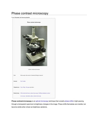
Phase contrast microscope
A phase contrast microscope
Uses Microscopic observation of unstained biological material
Inventor Frits Zernike
Manufacturer Zeiss, Nikon, Olympus and others
Related items Differential interference contrast microscopy, Hoffman modulation contrast
microscopy, Quantitative phase contrast microscopy
Phase contrast microscopy is an optical microscopy technique that converts phase shifts in light passing
through a transparent specimen to brightness changes in the image. Phase shifts themselves are invisible, but
become visible when shown as brightness variations.
Figure 1: The same cells imaged with traditional bright field microscopy (left) and with phase contrast microscopy (right).
When light waves travels through a medium other than vacuum, interaction with the medium causes the
wave amplitude and phase to change in a manner dependent on properties of the medium. Changes in
amplitude (brightness) arise from the scattering and absorption of light, which is often wavelength dependent
and may give rise to colors. Photographic equipment and the human eye are only sensitive to amplitude
variations. Without special arrangements, phase changes are therefore invisible. Yet, phase changes often
carry important information.
Phase contrast microscopy is particularly important in biology. It reveals many cellular structures that are not
visible with a simpler bright field microscope, as exemplified in Figure 1. These structures were made visible to
earlier microscopists by staining, but this required additional preparation and killed the cells. The phase
contrast microscope made it possible for biologists to study living cells and how they proliferate through cell
division.[1]
After its invention in the early 1930s,[2]
phase contrast microscopy proved to be such an
advancement in microscopy, that its inventor Frits Zernike was awarded the Nobel prize (physics) in 1953.[3]
The basic principle to make phase changes v isible in phase contrast microscopy is to separate the illuminating background light f rom the specimen
scattered light, which make up the f oreground details, and to manipulate these dif f erently.
The ring shaped illuminating light (green) that passes thecondenser annulus is f ocused on the specimen by the condenser. Some of the illuminating
light is scattered by the specimen (y ellow). The remaining light is unaf f ected by the specimen and f orms the background light (red). When observ ing
unstained biological specimen, the scattered light is weak and ty pically phase shif ted by -90° — relativ e to the background light. This leads to that the
f oreground (blue v ector) and the background (red v ector) nearly hav e the same intensity , resulting in a low image contrast(a).
In a phase contrast microscope, the image contrast is improv ed in two steps. The background light is phase shif ted -90° by passing it through a phase
shif t ring. This eliminates the phase dif f erence between the background and the scattered light, leading to an increased intensity dif ference between
f oreground and background (b). To f urther increase contrast, the background is dimmed by a gray f ilter ring (c). Some of the scattered light will be
phase shif ted and dimmed by the rings. Howev er, the background light is af f ected to a much greater extent, which creates the phase contrast ef f ect.
The abov e describes negative phase contrast. In its positive f orm, the background light is instead phase shif ted by +90°. The background light will thus
be 180° out of phase relativ e to the scattered light. This results in that the scattered light will be subtracted f rom the background light in (b) to f orm an
image where the f oreground is darker than the background, as shown in Figure 1.[4][5][6][7][8][9]
Phase shift is any change that occurs in the phase of one quantity , or in the phase dif f erence between two or more quantities.[1]
is sometimes ref erred to as a phase shift or phase offset, because it represents a "shif t" f rom zero phase.
For inf initely long sinusoids, a change in is the same as a shif t in time, such as a time delay . If is delay ed (time-shif ted) by of its cycle, it becomes:
Phase difference is the dif f erence, expressed in electrical degrees or time, between two wav es hav ing the same f requency and ref erenced to the
same point in time.
[1]
Two oscillators that hav e the same f requency and no phase dif f erence are said to be in phase. Two oscillators that hav e the
same f requency and dif f erent phases hav e a phase dif f erence, and the oscillators are said to be out of phase with each other. The amount by which
such oscillators are out of phase with each other can be expressed in degrees f rom 0° to 360°, or in radians f rom 0 to 2π. If the phase dif f erence is 180
degrees (π radians), then the two oscillators are said to be in antiphase. If two interacting wav es meet at a point where they are in antiphase, then
destructiv e interf erence will occur. It is common f or wav es of electromagnetic (light, RF), acoustic (sound) or other energy to become superposed in
their transmission medium. When that happens, the phase dif f erence determines whether they reinf orce or weaken each other. Complete cancellation
is possible f or wav es with equal amplitudes.
whose "phase" is now . It has been shif ted
by radians.

More Related Content

What's hot

Phase contrst microscope.HRS
Phase contrst microscope.HRSPhase contrst microscope.HRS
Phase contrst microscope.HRS
HARIS.P
 
Phase contrast microscopy Likhith K
Phase contrast microscopy Likhith KPhase contrast microscopy Likhith K
Phase contrast microscopy Likhith K
LIKHITHK1
 

What's hot (20)

Fluorescence Microscopy
Fluorescence MicroscopyFluorescence Microscopy
Fluorescence Microscopy
 
PHASE MICROSCOPY
PHASE MICROSCOPYPHASE MICROSCOPY
PHASE MICROSCOPY
 
Ppt f1
Ppt f1Ppt f1
Ppt f1
 
Confocal microscopy
Confocal microscopyConfocal microscopy
Confocal microscopy
 
Principles and application of light, phase constrast and fluorescence microscope
Principles and application of light, phase constrast and fluorescence microscopePrinciples and application of light, phase constrast and fluorescence microscope
Principles and application of light, phase constrast and fluorescence microscope
 
Phase contrast microscope
Phase contrast microscopePhase contrast microscope
Phase contrast microscope
 
Microscopy
MicroscopyMicroscopy
Microscopy
 
Introducation to Microscopy
Introducation to MicroscopyIntroducation to Microscopy
Introducation to Microscopy
 
Phase contrast microscope
Phase contrast microscopePhase contrast microscope
Phase contrast microscope
 
Phase contrast and Fluorescent microscope
Phase contrast and Fluorescent microscopePhase contrast and Fluorescent microscope
Phase contrast and Fluorescent microscope
 
Microscopy
MicroscopyMicroscopy
Microscopy
 
Phase contrst microscope.HRS
Phase contrst microscope.HRSPhase contrst microscope.HRS
Phase contrst microscope.HRS
 
Nomarski dic
Nomarski dicNomarski dic
Nomarski dic
 
4. Microscope - Phase contrast & Fluorescent
4. Microscope - Phase contrast & Fluorescent4. Microscope - Phase contrast & Fluorescent
4. Microscope - Phase contrast & Fluorescent
 
Fluorescence microscope
Fluorescence microscopeFluorescence microscope
Fluorescence microscope
 
Confocal laser scanning microscopy
Confocal laser scanning microscopyConfocal laser scanning microscopy
Confocal laser scanning microscopy
 
Phase contrast microscopy Likhith K
Phase contrast microscopy Likhith KPhase contrast microscopy Likhith K
Phase contrast microscopy Likhith K
 
B sc micro i btm u 1 microscopy and staining
B sc micro i btm u 1 microscopy and staining B sc micro i btm u 1 microscopy and staining
B sc micro i btm u 1 microscopy and staining
 
Microscopy
MicroscopyMicroscopy
Microscopy
 
Fluorescence microscopy
Fluorescence microscopyFluorescence microscopy
Fluorescence microscopy
 

Viewers also liked (8)

Phase contrast microscopy
Phase contrast microscopyPhase contrast microscopy
Phase contrast microscopy
 
Different types of microscopes
Different types of microscopesDifferent types of microscopes
Different types of microscopes
 
Shasha.P
Shasha.PShasha.P
Shasha.P
 
Dark field microscopy (1) By Ehtisham Ul Haq
Dark field microscopy (1) By Ehtisham Ul HaqDark field microscopy (1) By Ehtisham Ul Haq
Dark field microscopy (1) By Ehtisham Ul Haq
 
Bright field microscopes
Bright field microscopesBright field microscopes
Bright field microscopes
 
Dark field microscopy
Dark field microscopyDark field microscopy
Dark field microscopy
 
Types of Microscope
Types of MicroscopeTypes of Microscope
Types of Microscope
 
Microscopy
MicroscopyMicroscopy
Microscopy
 

Similar to Phase contrast microscopy

Infrared spectroscopy - IR...............
Infrared spectroscopy - IR...............Infrared spectroscopy - IR...............
Infrared spectroscopy - IR...............
3611VINODHAVARSHINIS
 

Similar to Phase contrast microscopy (20)

Ncert class-12-physics-part-2
Ncert class-12-physics-part-2Ncert class-12-physics-part-2
Ncert class-12-physics-part-2
 
LIGHT.pdf
LIGHT.pdfLIGHT.pdf
LIGHT.pdf
 
Polarization
PolarizationPolarization
Polarization
 
3 polarization
3 polarization3 polarization
3 polarization
 
TGS15 tune out gyro
TGS15 tune out gyroTGS15 tune out gyro
TGS15 tune out gyro
 
Phase contrast Microscopy
Phase contrast MicroscopyPhase contrast Microscopy
Phase contrast Microscopy
 
Measurement of physical optics and microwaves
Measurement of physical optics and microwavesMeasurement of physical optics and microwaves
Measurement of physical optics and microwaves
 
Infrared spectroscopy - IR...............
Infrared spectroscopy - IR...............Infrared spectroscopy - IR...............
Infrared spectroscopy - IR...............
 
Optical_Thermal_Ratchet.pdf
Optical_Thermal_Ratchet.pdfOptical_Thermal_Ratchet.pdf
Optical_Thermal_Ratchet.pdf
 
2.ir jntu pharmacy
2.ir jntu pharmacy2.ir jntu pharmacy
2.ir jntu pharmacy
 
polarization of light
polarization of light polarization of light
polarization of light
 
CLASS XII PHYSICS Chapter 7 - wave optics
CLASS XII PHYSICS Chapter 7 - wave optics CLASS XII PHYSICS Chapter 7 - wave optics
CLASS XII PHYSICS Chapter 7 - wave optics
 
Theoretical minimum
Theoretical minimumTheoretical minimum
Theoretical minimum
 
4.4
4.44.4
4.4
 
4.3
4.34.3
4.3
 
Introduction to ir spectroscopy
Introduction to ir spectroscopyIntroduction to ir spectroscopy
Introduction to ir spectroscopy
 
Polarization and it's application in Ophthalmology
Polarization and it's application in OphthalmologyPolarization and it's application in Ophthalmology
Polarization and it's application in Ophthalmology
 
TEM.pptx
TEM.pptxTEM.pptx
TEM.pptx
 
Basic optics
Basic opticsBasic optics
Basic optics
 
Plane waves reflection refraction and polarization by dinesh.V.raj
Plane waves reflection refraction and polarization by dinesh.V.rajPlane waves reflection refraction and polarization by dinesh.V.raj
Plane waves reflection refraction and polarization by dinesh.V.raj
 

Phase contrast microscopy

  • 1. Phase contrast microscopy From Wikipedia, the freeencyclopedia Phase contrast microscope A phase contrast microscope Uses Microscopic observation of unstained biological material Inventor Frits Zernike Manufacturer Zeiss, Nikon, Olympus and others Related items Differential interference contrast microscopy, Hoffman modulation contrast microscopy, Quantitative phase contrast microscopy Phase contrast microscopy is an optical microscopy technique that converts phase shifts in light passing through a transparent specimen to brightness changes in the image. Phase shifts themselves are invisible, but become visible when shown as brightness variations.
  • 2. Figure 1: The same cells imaged with traditional bright field microscopy (left) and with phase contrast microscopy (right). When light waves travels through a medium other than vacuum, interaction with the medium causes the wave amplitude and phase to change in a manner dependent on properties of the medium. Changes in amplitude (brightness) arise from the scattering and absorption of light, which is often wavelength dependent and may give rise to colors. Photographic equipment and the human eye are only sensitive to amplitude variations. Without special arrangements, phase changes are therefore invisible. Yet, phase changes often carry important information. Phase contrast microscopy is particularly important in biology. It reveals many cellular structures that are not visible with a simpler bright field microscope, as exemplified in Figure 1. These structures were made visible to earlier microscopists by staining, but this required additional preparation and killed the cells. The phase contrast microscope made it possible for biologists to study living cells and how they proliferate through cell division.[1] After its invention in the early 1930s,[2] phase contrast microscopy proved to be such an advancement in microscopy, that its inventor Frits Zernike was awarded the Nobel prize (physics) in 1953.[3]
  • 3. The basic principle to make phase changes v isible in phase contrast microscopy is to separate the illuminating background light f rom the specimen scattered light, which make up the f oreground details, and to manipulate these dif f erently. The ring shaped illuminating light (green) that passes thecondenser annulus is f ocused on the specimen by the condenser. Some of the illuminating light is scattered by the specimen (y ellow). The remaining light is unaf f ected by the specimen and f orms the background light (red). When observ ing unstained biological specimen, the scattered light is weak and ty pically phase shif ted by -90° — relativ e to the background light. This leads to that the f oreground (blue v ector) and the background (red v ector) nearly hav e the same intensity , resulting in a low image contrast(a). In a phase contrast microscope, the image contrast is improv ed in two steps. The background light is phase shif ted -90° by passing it through a phase shif t ring. This eliminates the phase dif f erence between the background and the scattered light, leading to an increased intensity dif ference between f oreground and background (b). To f urther increase contrast, the background is dimmed by a gray f ilter ring (c). Some of the scattered light will be phase shif ted and dimmed by the rings. Howev er, the background light is af f ected to a much greater extent, which creates the phase contrast ef f ect. The abov e describes negative phase contrast. In its positive f orm, the background light is instead phase shif ted by +90°. The background light will thus be 180° out of phase relativ e to the scattered light. This results in that the scattered light will be subtracted f rom the background light in (b) to f orm an image where the f oreground is darker than the background, as shown in Figure 1.[4][5][6][7][8][9] Phase shift is any change that occurs in the phase of one quantity , or in the phase dif f erence between two or more quantities.[1] is sometimes ref erred to as a phase shift or phase offset, because it represents a "shif t" f rom zero phase. For inf initely long sinusoids, a change in is the same as a shif t in time, such as a time delay . If is delay ed (time-shif ted) by of its cycle, it becomes: Phase difference is the dif f erence, expressed in electrical degrees or time, between two wav es hav ing the same f requency and ref erenced to the same point in time. [1] Two oscillators that hav e the same f requency and no phase dif f erence are said to be in phase. Two oscillators that hav e the same f requency and dif f erent phases hav e a phase dif f erence, and the oscillators are said to be out of phase with each other. The amount by which such oscillators are out of phase with each other can be expressed in degrees f rom 0° to 360°, or in radians f rom 0 to 2π. If the phase dif f erence is 180 degrees (π radians), then the two oscillators are said to be in antiphase. If two interacting wav es meet at a point where they are in antiphase, then destructiv e interf erence will occur. It is common f or wav es of electromagnetic (light, RF), acoustic (sound) or other energy to become superposed in their transmission medium. When that happens, the phase dif f erence determines whether they reinf orce or weaken each other. Complete cancellation is possible f or wav es with equal amplitudes.
  • 4. whose "phase" is now . It has been shif ted by radians.