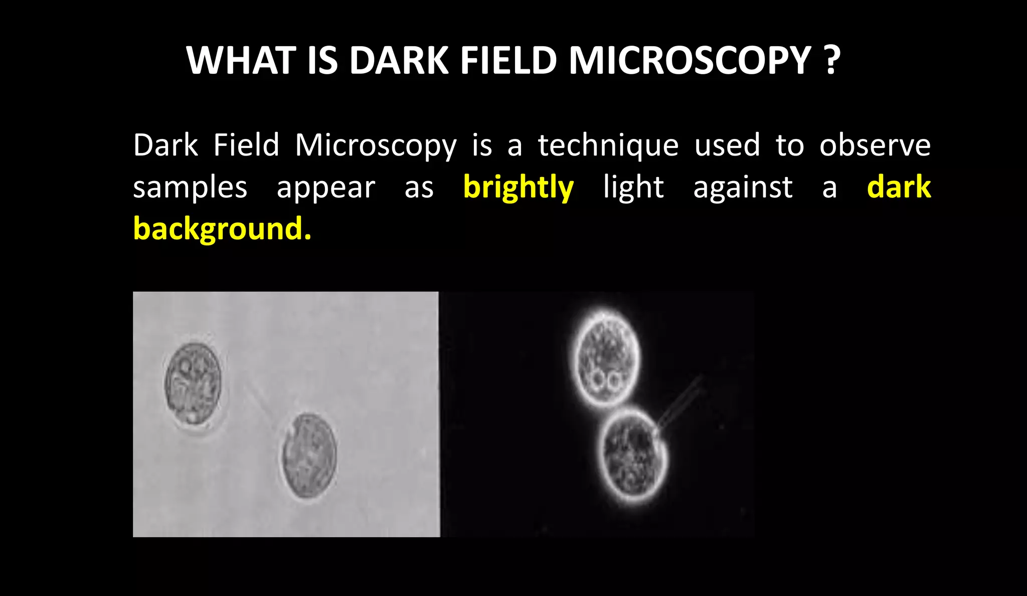
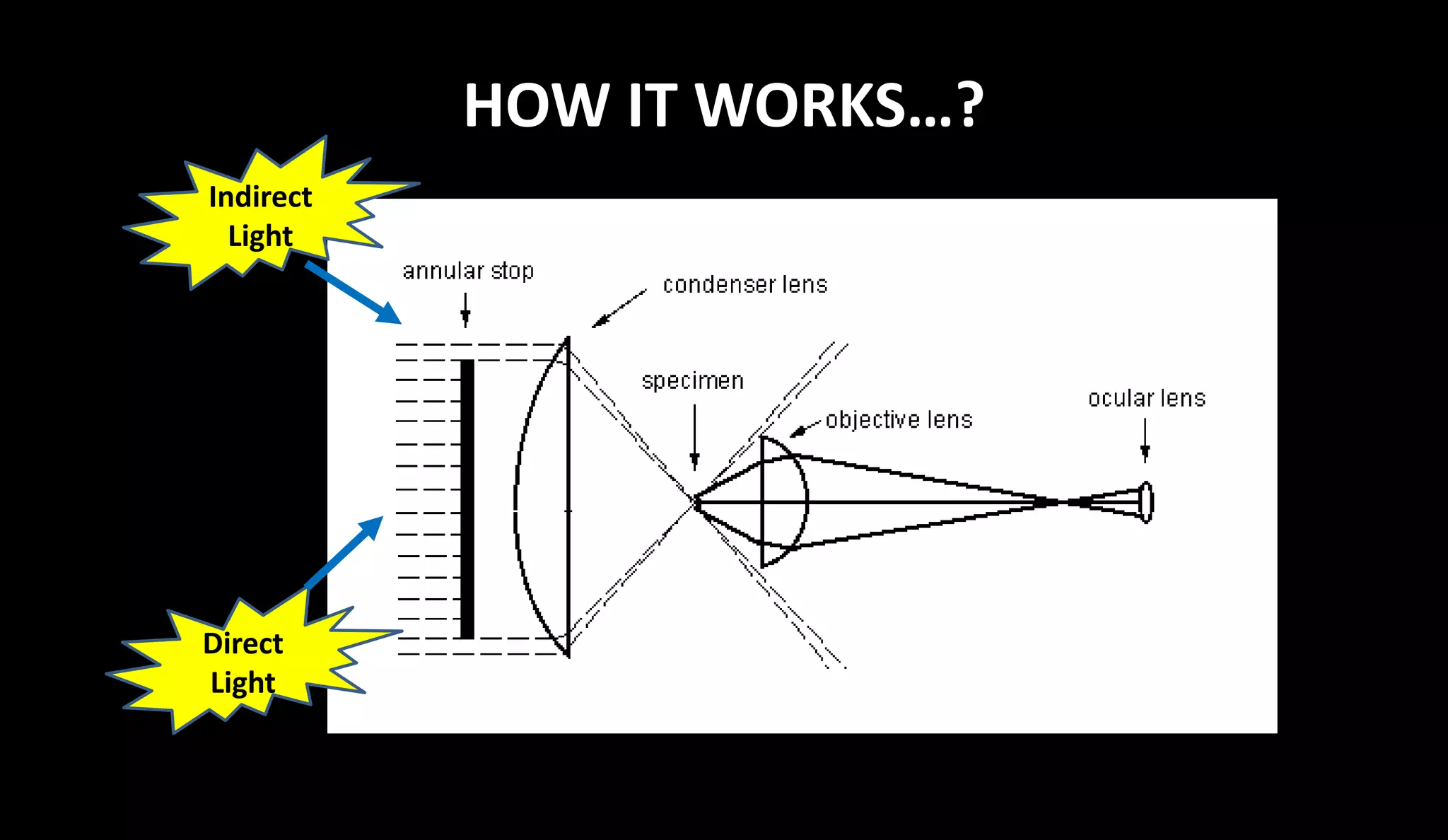
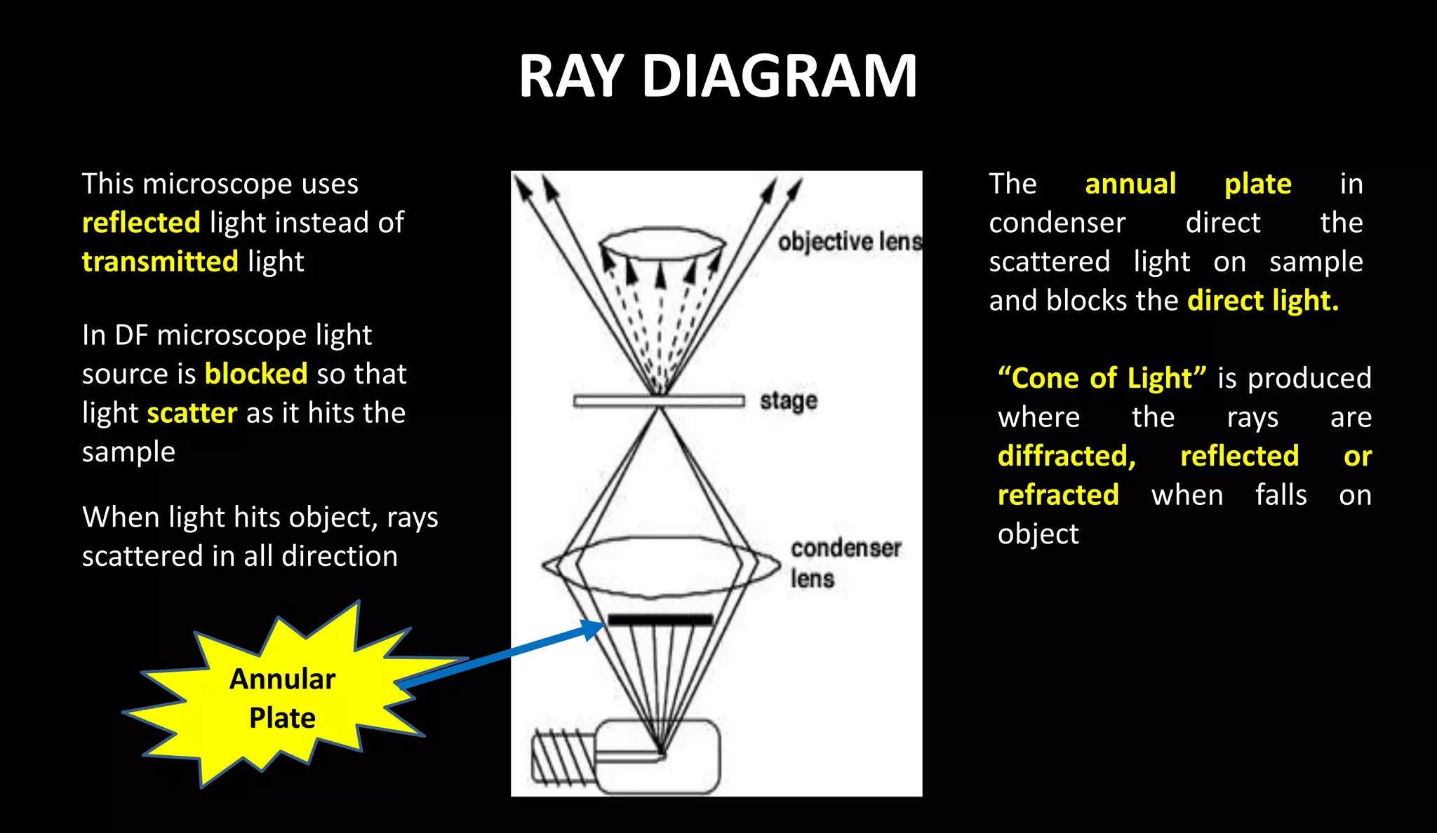
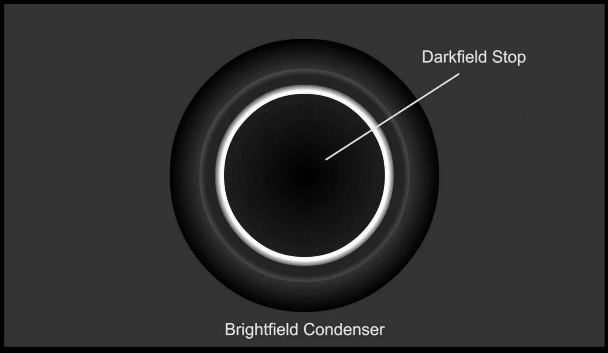
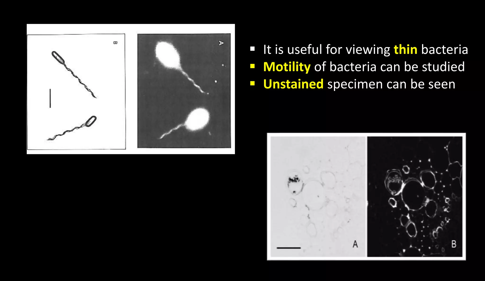
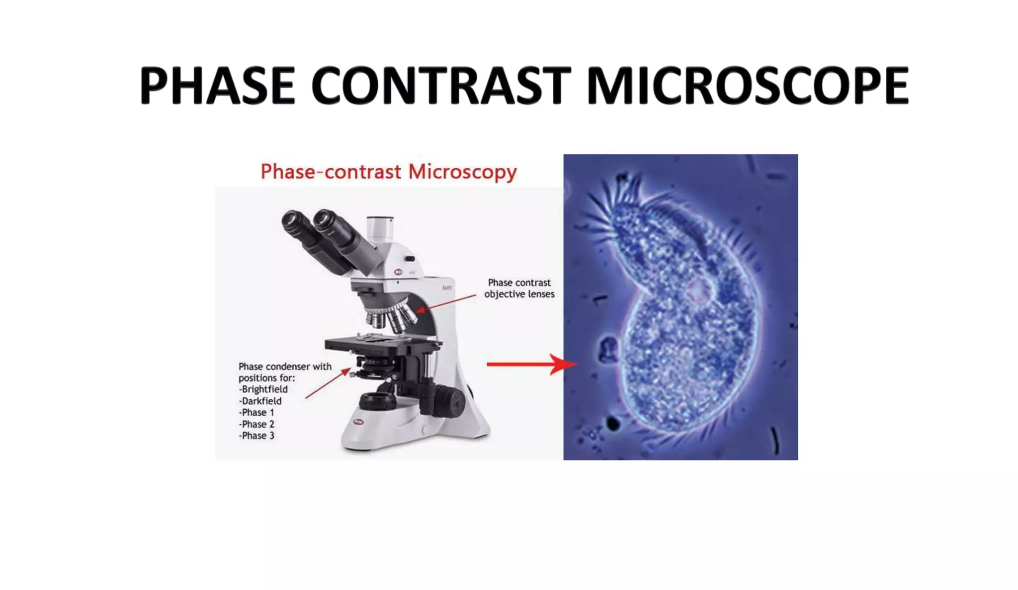
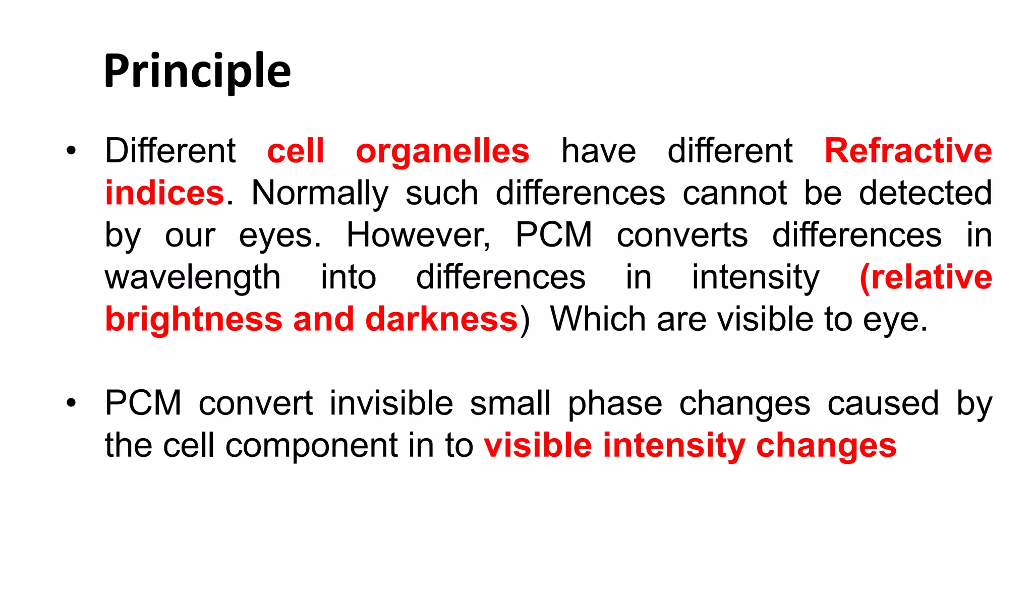
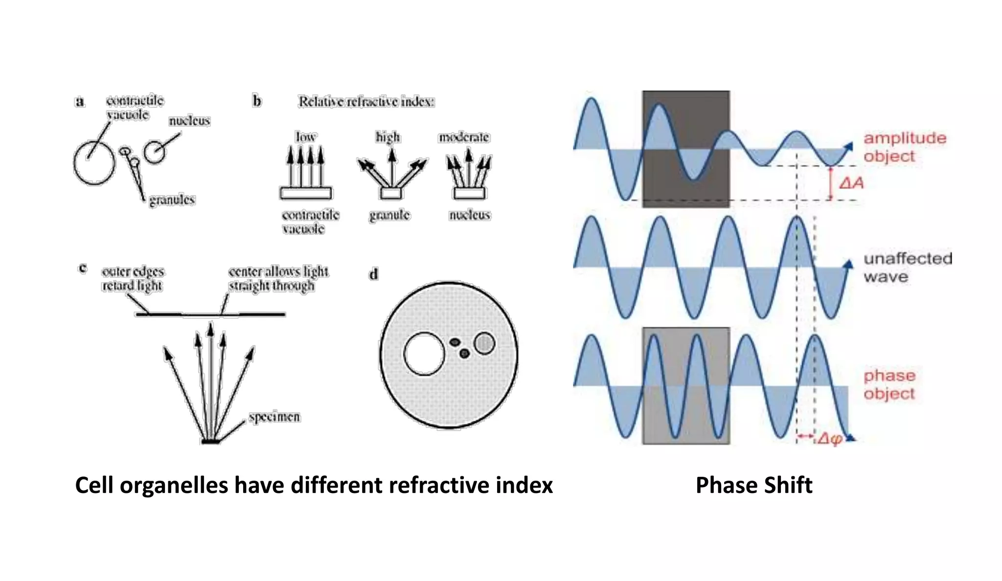
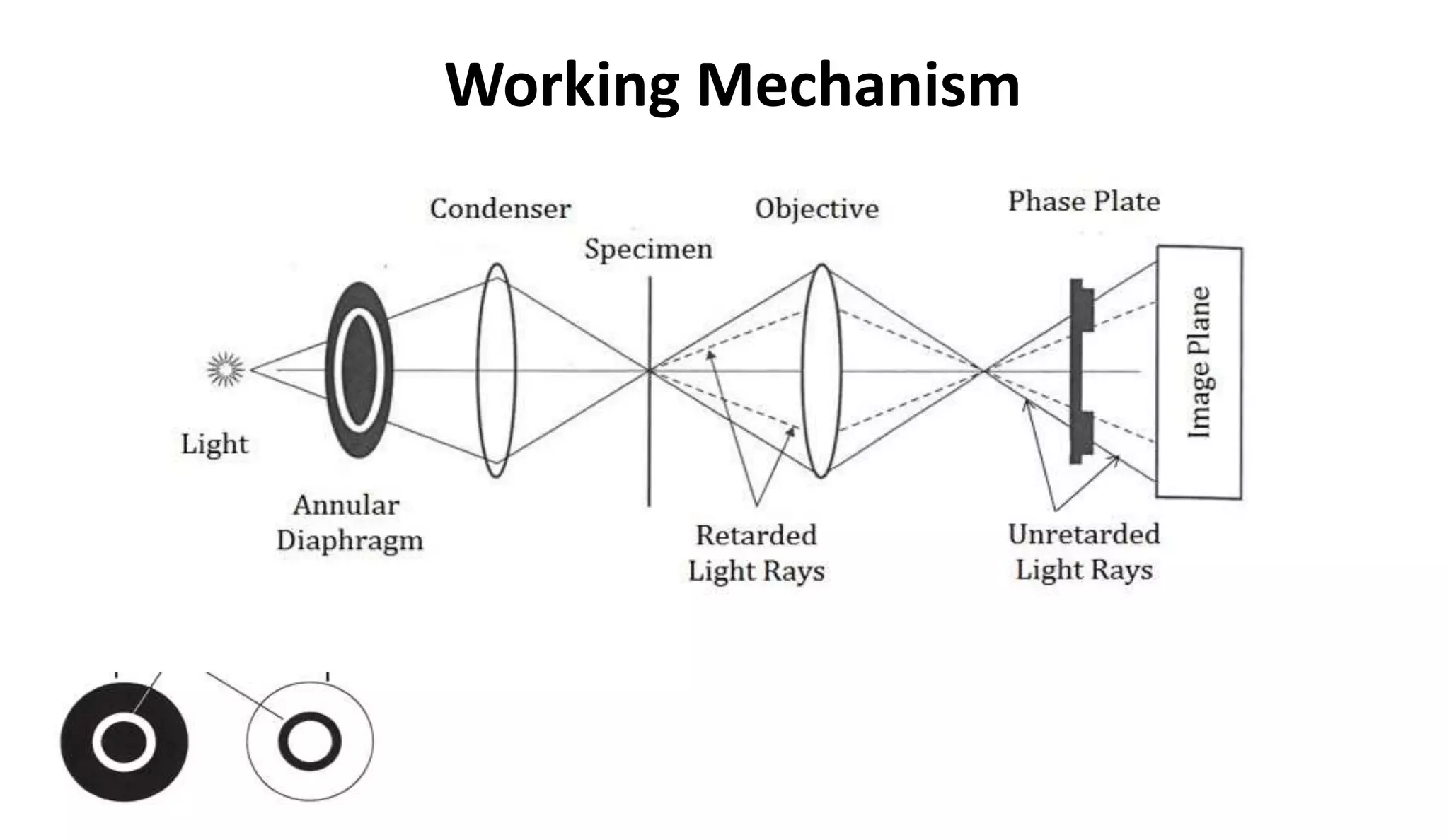
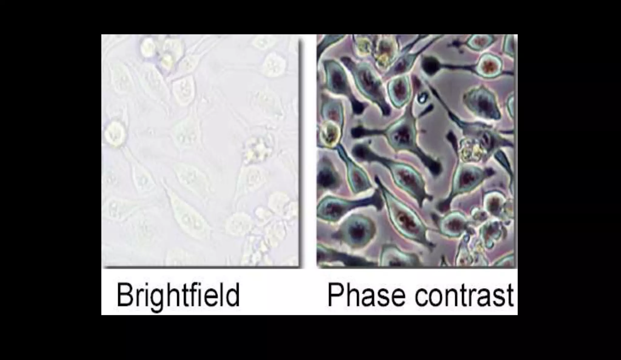
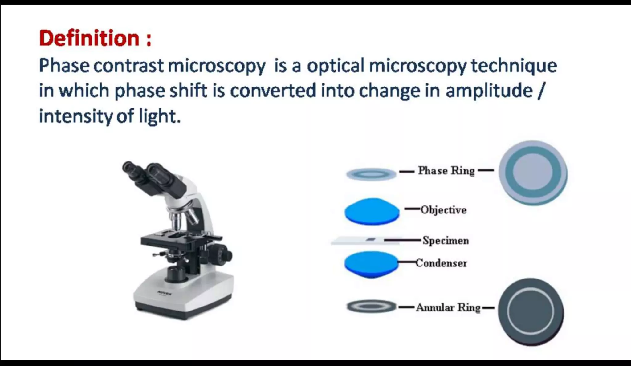
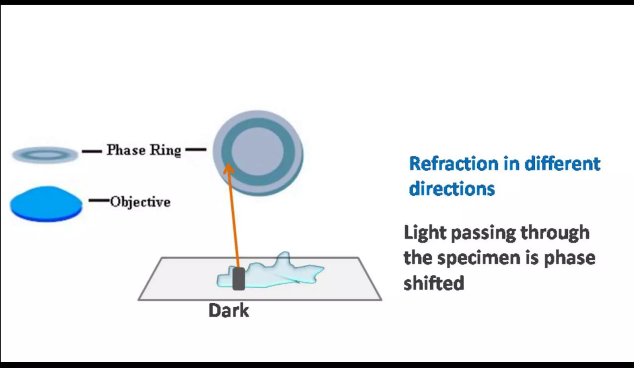
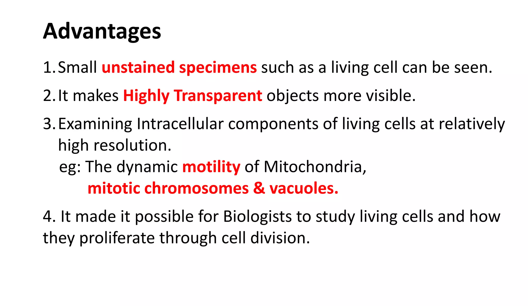
Dark Field Microscope
WHAT IS DARK FIELD MICROSCOPY ?
Dark Field Microscopy is a technique used to observe
samples appear as brightly light against a dark
background.
HOW IT WORKS…?
Indirect
Light
Direct
Light
RAY DIAGRAM
Annular
Plate
In DF microscope light
source is blocked so that
light scatter as it hits the
sample
When light hits object, rays
scattered in all direction
The annual plate in
condenser direct the
scattered light on sample
and blocks the direct light.
“Cone of Light” is produced
where the rays are
diffracted, reflected or
refracted when falls on
object
This microscope uses
reflected light instead of
transmitted light
5
CHARLIE 6
7
 It is useful for viewing thin bacteria
 Motility of bacteria can be studied
 Unstained specimen can be seen
 Unstained living cells absorb no light. Poor light
absorption results in extremely small difference In
intensity distribution in image. Therefore image
cant be seen clearly.
 Phase contrast microscope convert phase shift in
light passing through a transparent specimen to
brightness change in the image
• Different cell organelles have different Refractive
indices. Normally such differences cannot be detected
by our eyes. However, PCM converts differences in
wavelength into differences in intensity (relative
brightness and darkness) Which are visible to eye.
• PCM convert invisible small phase changes caused by
the cell component in to visible intensity changes
Principle
12
Cell organelles have different refractive index Phase Shift
Working Mechanism
CHARLIE 16
CHARLIE 17
1.Small unstained specimens such as a living cell can be seen.
2.It makes Highly Transparent objects more visible.
3.Examining Intracellular components of living cells at relatively
high resolution.
eg: The dynamic motility of Mitochondria,
mitotic chromosomes & vacuoles.
4. It made it possible for Biologists to study living cells and how
they proliferate through cell division.
Advantages
CHARLIE 19

Dark field and Phase Contrast Microscope

  • 1.
  • 2.
    WHAT IS DARKFIELD MICROSCOPY ? Dark Field Microscopy is a technique used to observe samples appear as brightly light against a dark background.
  • 3.
  • 4.
    RAY DIAGRAM Annular Plate In DFmicroscope light source is blocked so that light scatter as it hits the sample When light hits object, rays scattered in all direction The annual plate in condenser direct the scattered light on sample and blocks the direct light. “Cone of Light” is produced where the rays are diffracted, reflected or refracted when falls on object This microscope uses reflected light instead of transmitted light
  • 5.
  • 6.
  • 7.
    7  It isuseful for viewing thin bacteria  Motility of bacteria can be studied  Unstained specimen can be seen
  • 10.
     Unstained livingcells absorb no light. Poor light absorption results in extremely small difference In intensity distribution in image. Therefore image cant be seen clearly.  Phase contrast microscope convert phase shift in light passing through a transparent specimen to brightness change in the image
  • 11.
    • Different cellorganelles have different Refractive indices. Normally such differences cannot be detected by our eyes. However, PCM converts differences in wavelength into differences in intensity (relative brightness and darkness) Which are visible to eye. • PCM convert invisible small phase changes caused by the cell component in to visible intensity changes Principle
  • 12.
    12 Cell organelles havedifferent refractive index Phase Shift
  • 13.
  • 16.
  • 17.
  • 18.
    1.Small unstained specimenssuch as a living cell can be seen. 2.It makes Highly Transparent objects more visible. 3.Examining Intracellular components of living cells at relatively high resolution. eg: The dynamic motility of Mitochondria, mitotic chromosomes & vacuoles. 4. It made it possible for Biologists to study living cells and how they proliferate through cell division. Advantages
  • 19.

Editor's Notes

  • #3 When light hits an object, rays are scattered in all azimuths or directions. The design of the dark field microscope is such that it removes the dispersed light, or zeroth order, so that only the scattered beams hit the sample.The introduction of a condenser and/or stop below the stage ensures that these light rays will hit the specimen at different angles, rather than as a direct light source above/below the object. The result is a “cone of light” where rays are diffracted, reflected and/or refracted off the object, ultimately, allowing you to view a specimen in dark field.
  • #4 Darkfield microscopy relies on a different illumination system. Rather than illuminating the sample with a filled cone of light, the condenser is designed to form a hollow cone of light. The light at the apex of the cone is focused at the plane of the specimen; as this light moves past the specimen plane it spreads again into a hollow cone. The objective lens sits in the dark hollow of this cone; although the light travels around and past the objective lens, no rays enter it The entire field appears dark when there is no sample on the microscope stage; thus the name darkfield microscopy. When a sample is on the stage, the light at the apex of the cone strikes it. The image is made only by those rays scattered by the sample and captured in the objective lens (note the rays scattered by the specimen in Figure 1). The image appears bright against the dark background. This situation can be compared to the glittery appearance of dust particles in a dark room illuminated by strong shafts of light coming in through a side window. The dust particles are very small, but are easily seen when they scatter the light rays. This is the working principle of darkfield microscopy and explains how the image of low contrast material is created: an object will be seen against a dark background if it scatters light which is captured with the proper device such as an objective lens.
  • #5 1.Light enters the microscope for illumination of the sample. 2.A specially sized disc, the patch stop (see figure) blocks some light from the light source, leaving an outer ring of illumination. A wide phase annulus can also be reasonably substituted at low magnification. 3.The condenser lens focuses the light towards the sample. 4.The light enters the sample. Most is directly transmitted, while some is scattered from the sample. 5.The scattered light enters the objective lens, while the directly transmitted light simply misses the lens and is not collected due to a direct illumination block (see figure). 6.Only the scattered light goes on to produce the image, while the directly transmitted light is omitted.
  • #8 LEFT:(A) S. typhimurium during translational movement. Theperitrichous flagella form apolar bundle (see cartoon, Fig. 1B) which could be mistaken forpolarflagellation. Classifications should therefore be based on the appearance offlagella on stationary cells. Dark-fieldphotomicrograph taken with high-intensity pulsed xenon arc. Bar equals 5 gm. RIGHT: Polar flagellation in pseudomonads. (A) Stationary cell ofP. stutzeri. (B) Same negative [overex- posed duringprinting] to show cell outline. (C) Cartoon combining information from (A) and (B). (D-F) As for (A-C) but with P. aeruginosa (note presence oftwo flagella at pole, although species is predominantly monotrichous). Bar equals 5 ,um.