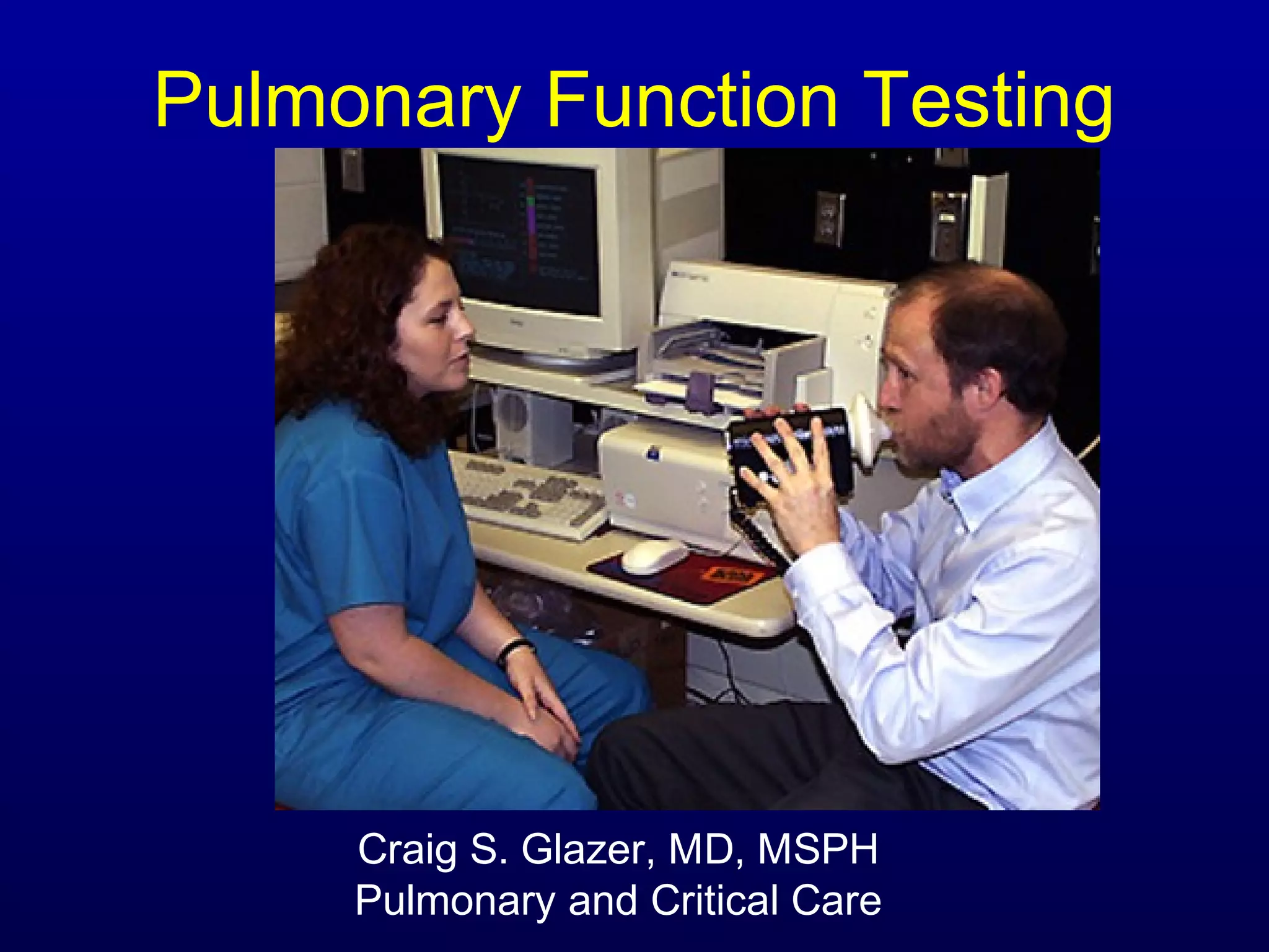
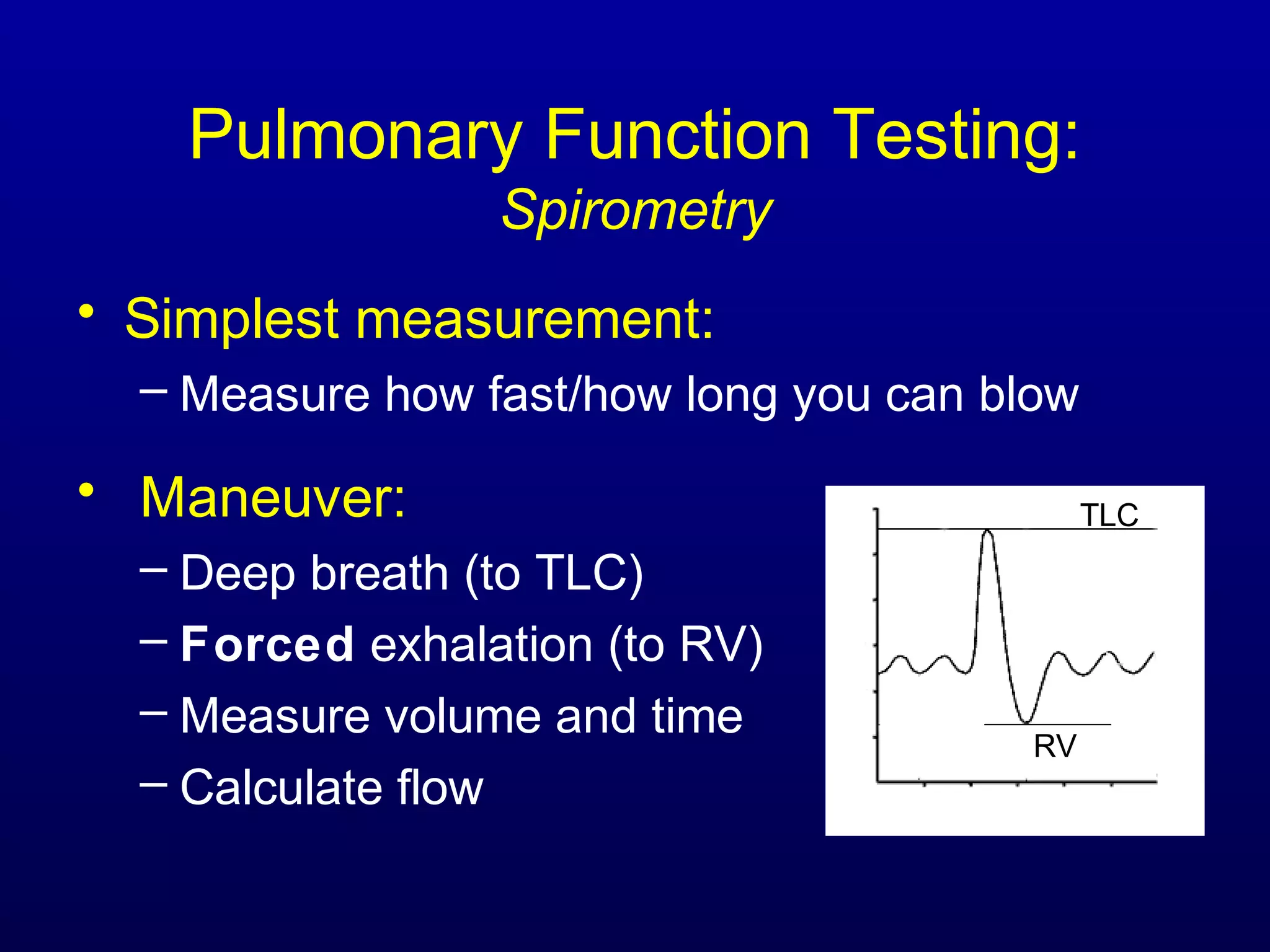
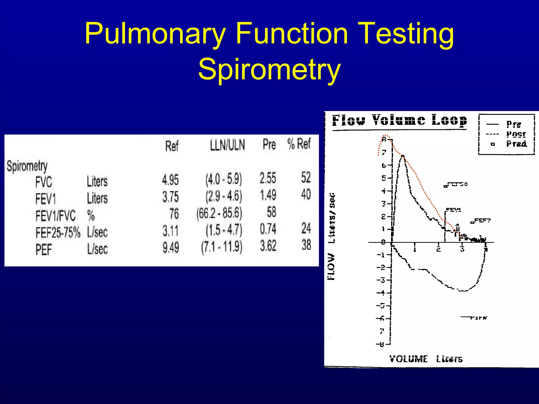
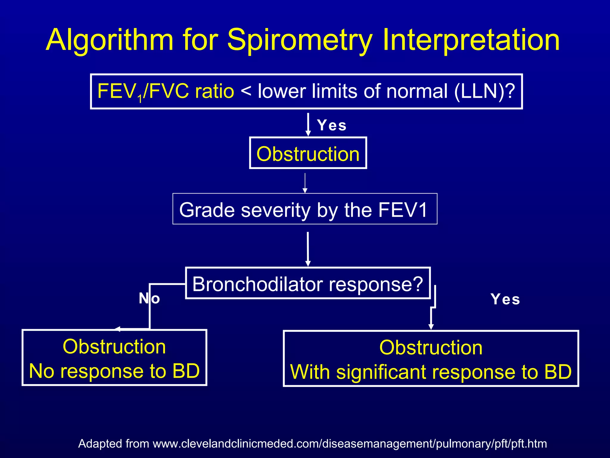
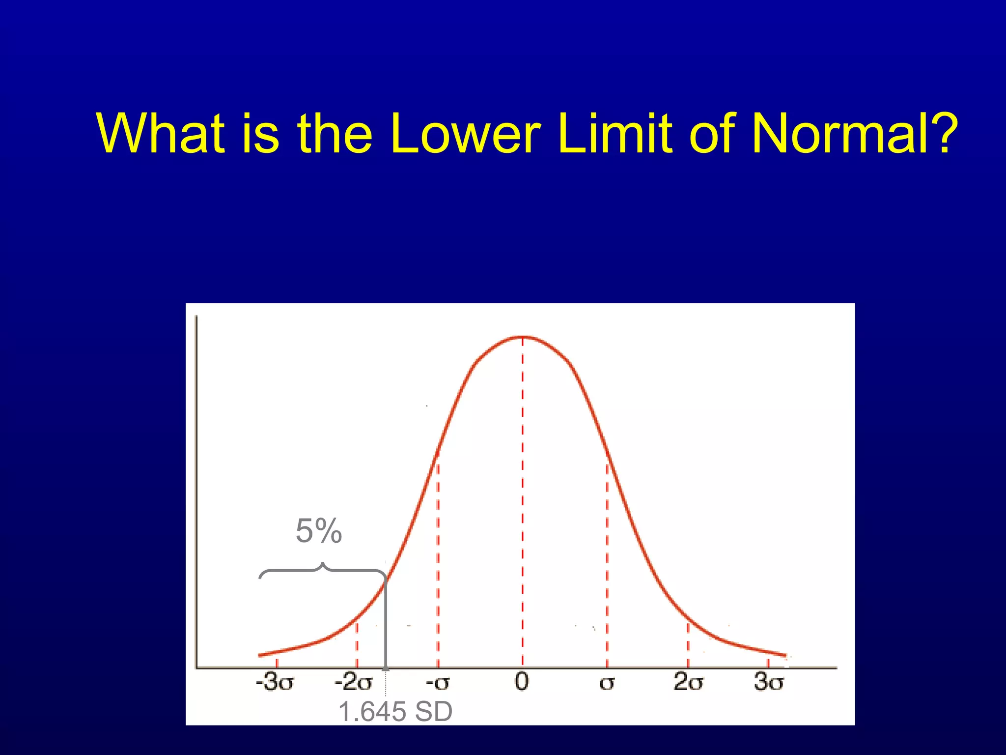
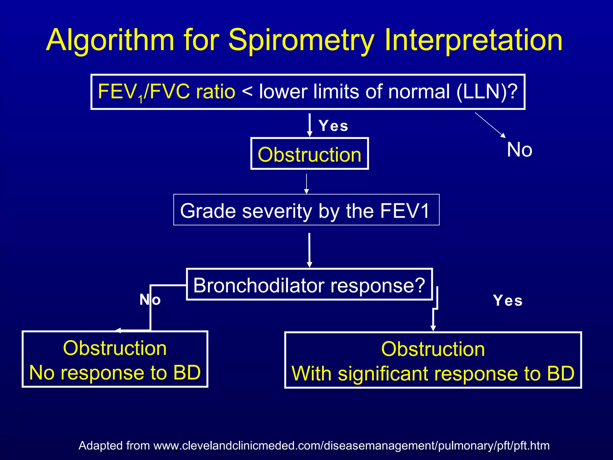
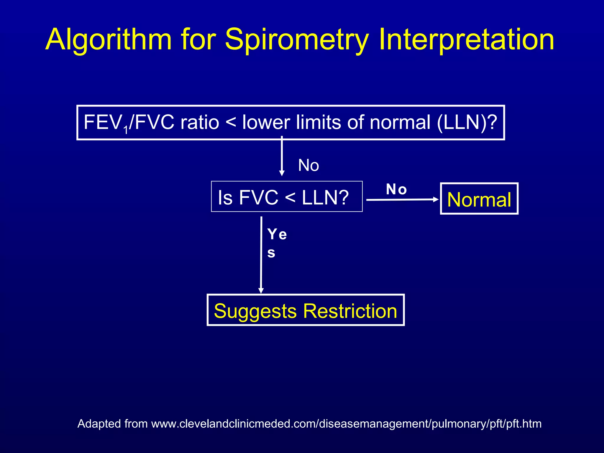
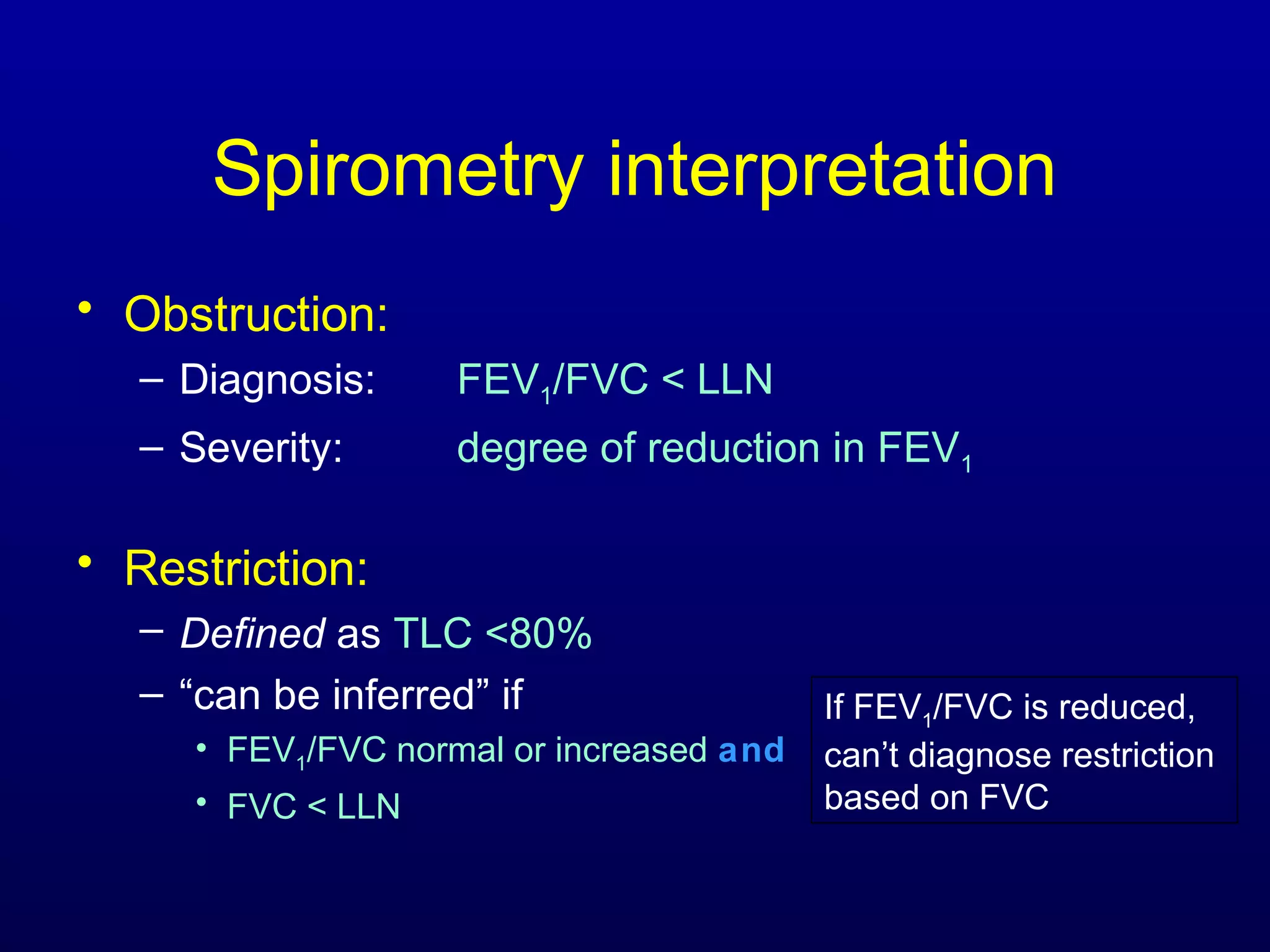
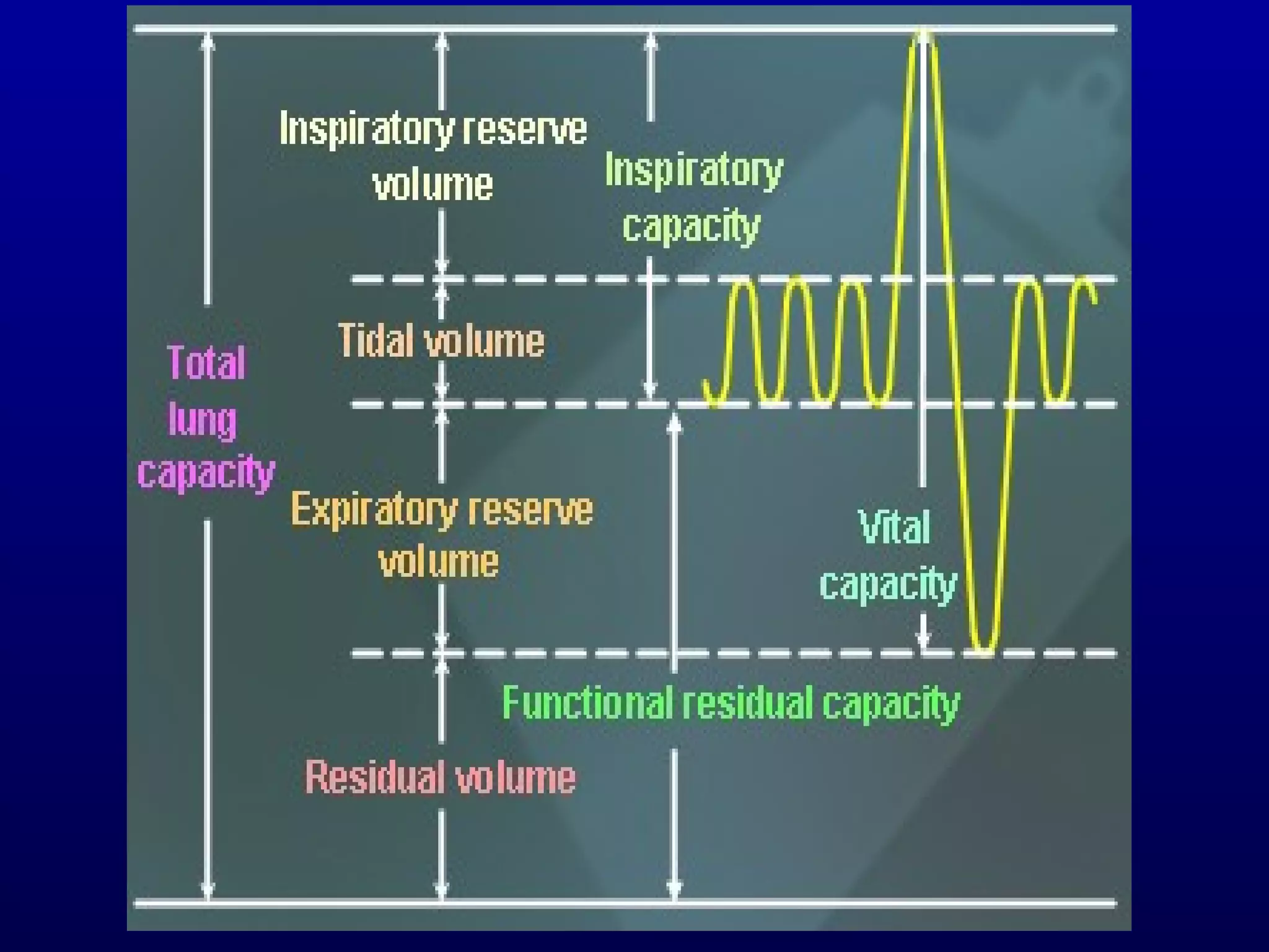
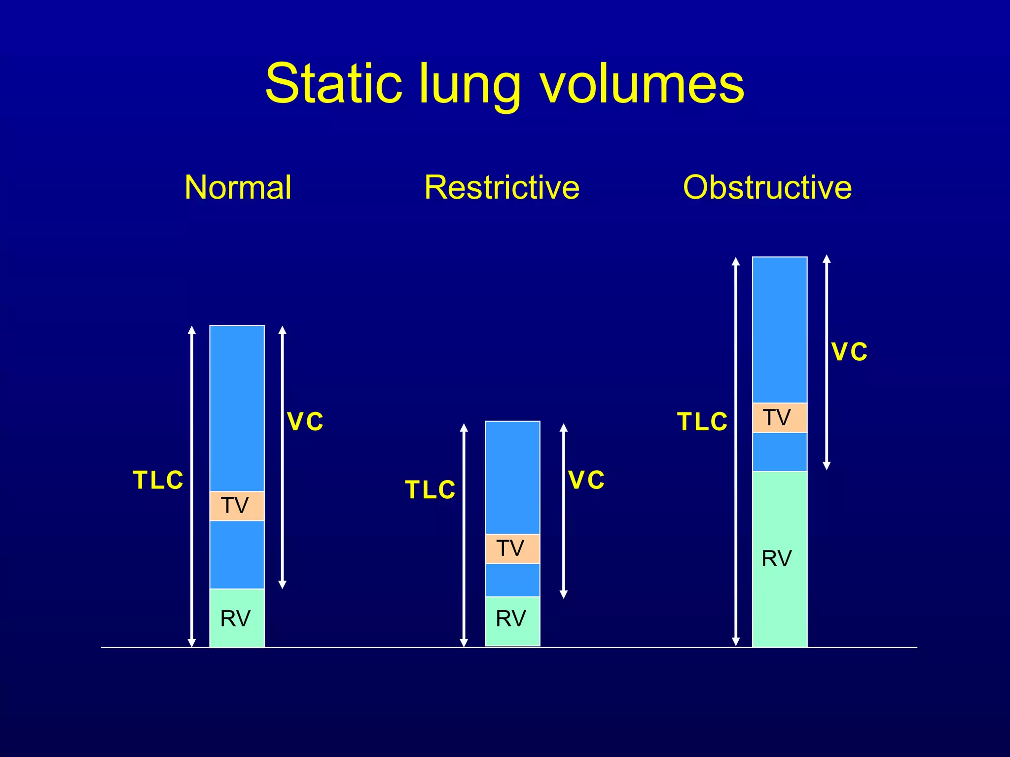
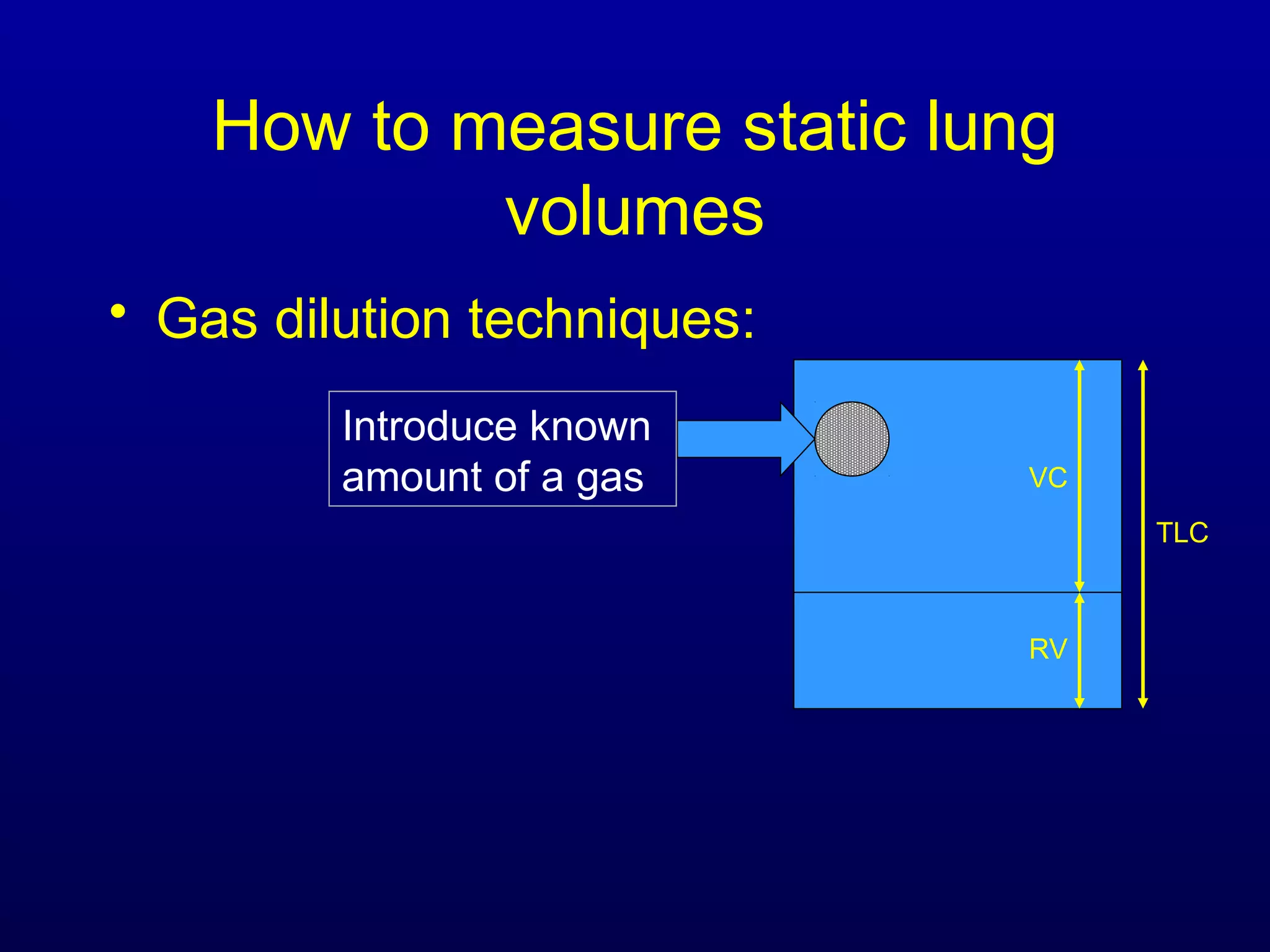
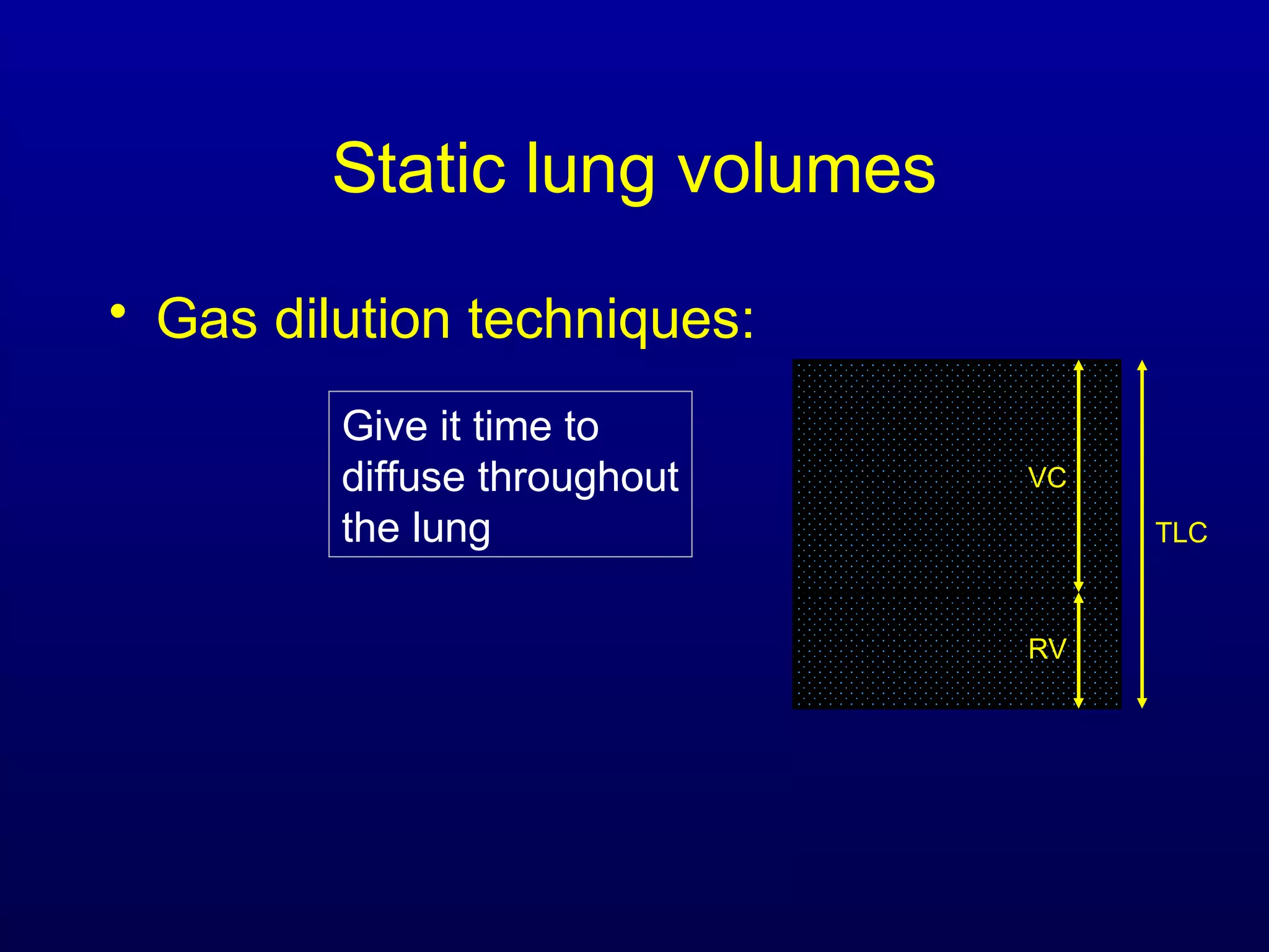
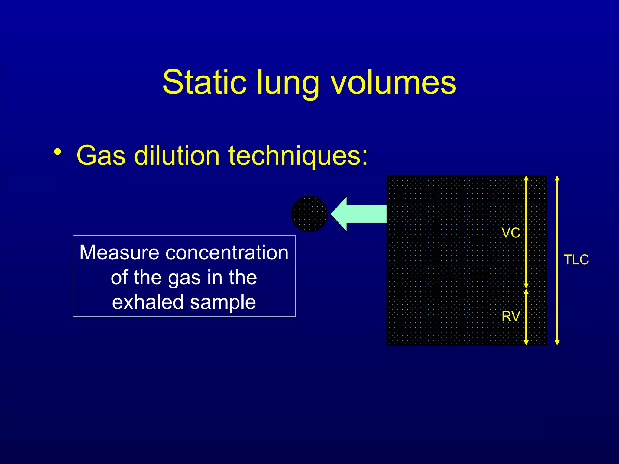
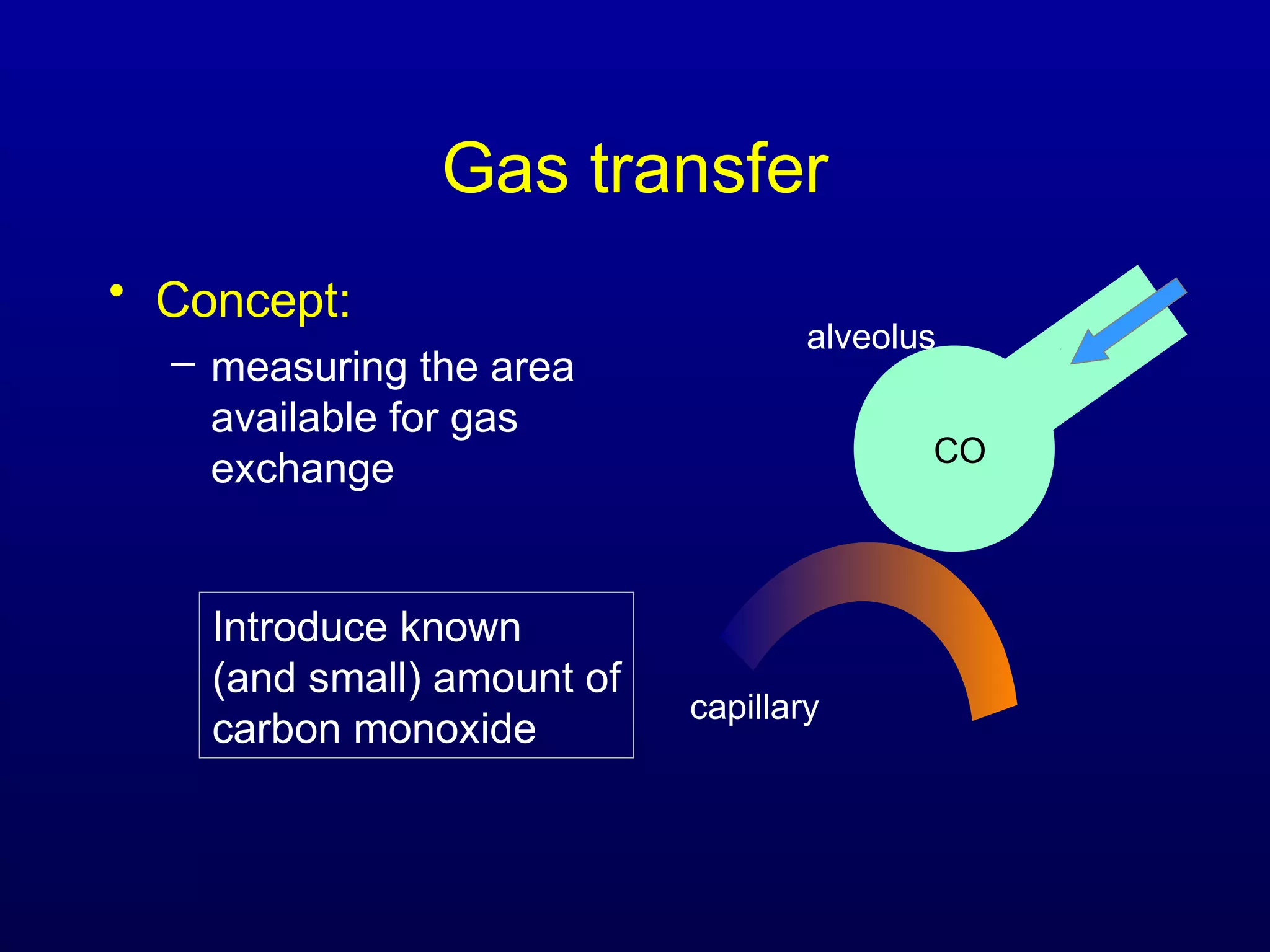
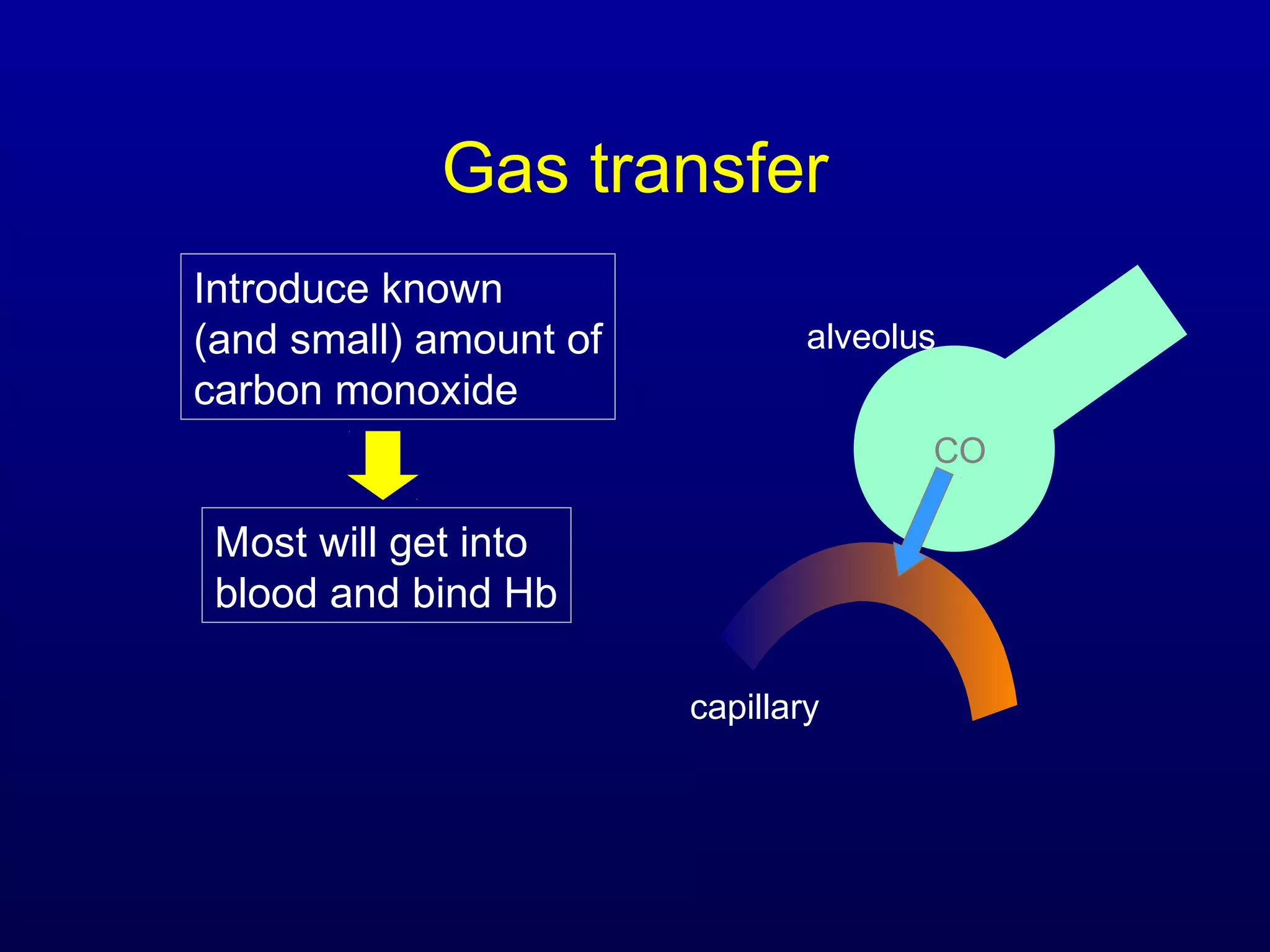
Pulmonary Function Testing
Craig S. Glazer, MD, MSPH
Pulmonary and Critical Care
What Are PFTs?
• Series of maneuvers designed to
measure lung size and function
• Elements of the test:
– Spirometry – dynamic flows and volumes
– Static lung volumes
– Gas transfer
Uses of PFTs
• Diagnosis
– Patterns of abnormalities
• Obstruction – COPD, asthma, bronchiectasis,
bronchiolitis
• Restriction – ILD, pleural disease, neuromuscular
disease, thoracic cage abnormalities
– Identify subtle physiologic abnormalities
• Isolated low DLCO – pulmonary hypertension,
emphysema, early ILD
• Prognosis
– Measure of disease severity – COPD
– Assess risk – neuromuscular disease
• Assess treatment response
What does the test measure?
• Flow and volume during maximal inspiration
and forced expiration - spirometry
• Total lung volumes
• Surface area of gas exchange
• Respiratory muscle function
• Cardiopulmonary function during exercise
Definition of volumes
time
volume
Definition of volumes
time
volume
Tidal volume
Vital capacity (VC)
Definition of volumes
time
volume Total lung capacity
(TLC)
Reserve volume
(RV)
Pulmonary Function Testing:
Spirometry
• Simplest measurement:
– Measure how fast/how long you can blow
• Maneuver:
– Deep breath (to TLC)
– Forced exhalation (to RV)
– Measure volume and time
– Calculate flow
TLC
RV
Pulmonary Function Testing:
Spirometry
TLC
RV
A given patient
can never get out
of the envelope of
the flow-volume
loop
•WHY?
Dynamic airway
collapse
Pulmonary Function Testing:
Spirometry
TLC RV
Dynamic ventilatory mechanics:
dynamic airway compression
Alveolus
Palv= 0
outside
PB= 0
End-expiration:
(No air flow)
Negative pleural pressure
(- 5 cmH2O)
Dynamic ventilatory mechanics:
dynamic airway compression
Alveolus
Palv= - 3 cmH2O
Pleural pressure more negative
(- 8 cmH2O)
Inspiration:
outside
PB= 0
Dynamic ventilatory mechanics:
dynamic airway compression
Alveolus
Palv= - 3 cmH2O
Inspiration:
Air flow down pressure gradient
outside
PB= 0
Pleural pressure more negative
(- 8 cmH2O)
Dynamic ventilatory mechanics:
dynamic airway compression
Alveolus
Palv= +30 cmH2O
Forced expiration:
outside
PB= 0
Positive pleural pressure
(+ 25 cmH2O)
Dynamic ventilatory mechanics:
dynamic airway compression
Alveolus
Palv= +30 cmH2O
Forced expiration:
Air flow down pressure gradient
outside
PB= 0
Positive pleural pressure
(+ 25 cmH2O)
Dynamic ventilatory mechanics:
dynamic airway compression
Alveolus
Palv= +30 cmH2O
Forced expiration:
30 29 28 27 26 ...
outside
PB= 0
Positive pleural pressure
(+ 25 cmH2O)
Dynamic ventilatory mechanics:
dynamic airway compression
Forced expiration:
30 29 28 27 26 … 25
outside
PB= 0
Positive pleural pressure
(+ 25 cmH2O)
Alveolus
Palv= +30 cmH2O
Dynamic ventilatory mechanics:
dynamic airway compression
Forced expiration:
Equal pressure point
Closing volume
Dynamic ventilatory mechanics:
dynamic airway compression
• Distal movement of equal pressure
point exaggerated with:
– Increased airway resistance
• Asthma, etc.
– Reduced lung elastic recoil
• Emphysema
• Aging
Pulmonary Function Testing
Spirometry
Spirometry Interpretation: Step 1
Is the test of adequate quality?
1. Reproducibilty Criteria
2. Acceptability Criteria
Spirometry Interpretation:
Acceptability Criteria
• Flow Volume Loop
– Good Start
– No artifacts
• Volume Time Curve
– 6 seconds exhalation
• Or plateau
TLC RV
Spirometry Interpretation:
Acceptability Criteria
Algorithm for Spirometry
Interpretation
1. Assess test quality
2. Is there obstruction?
1. Is there a bronchodilator response?
3. Is there evidence for restriction?
Algorithm for Spirometry Interpretation
FEV1/FVC ratio < lower limits of normal (LLN)?
Obstruction
Yes
Bronchodilator response?
Obstruction
No response to BD
No
Obstruction
With significant response to BD
Yes
Adapted from www.clevelandclinicmeded.com/diseasemanagement/pulmonary/pft/pft.htm
Grade severity by the FEV1
What is the Lower Limit of Normal?
5%
1.645 SD
Algorithm for Spirometry Interpretation
FEV1/FVC ratio < lower limits of normal (LLN)?
Obstruction
Yes
Bronchodilator response?
Obstruction
No response to BD
No
Obstruction
With significant response to BD
Yes
Adapted from www.clevelandclinicmeded.com/diseasemanagement/pulmonary/pft/pft.htm
Grade severity by the FEV1
No
Algorithm for Spirometry Interpretation
FEV1/FVC ratio < lower limits of normal (LLN)?
Normal
No
Suggests Restriction
Ye
s
Adapted from www.clevelandclinicmeded.com/diseasemanagement/pulmonary/pft/pft.htm
Is FVC < LLN?
No
Algorithm for Spirometry Interpretation
FEV1/FVC ratio < lower limits of normal (LLN)?
Possible Mixed
Process
Yes
Obstruction
Yes
Bronchodilator response?
Obstruction
No response to BD
No
Obstruction
With significant response to BD
Yes
Is FVC < LLN?
Isolated Obstruction
No
Severity of impairment as determined
by spirometry:
Normal > LLN
Mild 70-80% predicted
Moderate 60-69%
Moderate-Severe 50-59%
Severe 34-49%
Very Severe < 34%
Case #1
• Obstructive or Restrictive
Process?
Obstructive
• What is the severity?
– moderate
• 45 y/o man complaining of
cough and dyspnea for
months. Smoked 10 pack-
yrs
Case #1 Spirometry
• Bronchodilator response:
– Spirometry repeated after inhaled beta-agonists
– “Significant” with a 12% and a 200ml
improvement in FEV-1 or FVC
BD response = obstructive defect but doesn’t equal
asthma
No response does not mean adequate Rx
Case #1 Spirometry with BD
challenge
Pre-BD
%
Pred
Post-
BD
%
Pred
%
Chang
e
FEV1 1.33 60% 1.89 80% 42%
FVC 2.61 95% 2.97 108% 14%
FEV1
/FVC
50% 64%
Case #2
• 64 y/o man referred for
shortness of breath on
exertion
Suggests restrictive process
- good start
- smooth contour
- effort/reproducibility
Spirometry interpretation
• Obstruction:
– Diagnosis: FEV1/FVC < LLN
– Severity: degree of reduction in FEV1
• Restriction:
– Defined as TLC <80%
– “can be inferred” if
• FEV1/FVC normal or increased and
• FVC < LLN
If FEV1/FVC is reduced,
can’t diagnose restriction
based on FVC
Static lung volumes
TLC
VC
RV
TV
TLC
VC
RV
TV
TLC VC
RV
TV
Normal ObstructiveRestrictive
How to measure static lung
volumes
• Gas dilution techniques:
Introduce known
amount of a gas
TLC
VC
RV
Static lung volumes
• Gas dilution techniques:
Give it time to
diffuse throughout
the lung TLC
VC
RV
Static lung volumes
• Gas dilution techniques:
Measure concentration
of the gas in the
exhaled sample
TLC
VC
RV
Static lung volumes
• Gas dilution techniques:
– Easy to do
– Extra equipment cheap
– Only measures volume of
areas in free communication
with the mouth
TLC
VC
RVbulla
Static lung volumes
• Body plethysmography
Patient makes panting
movements against a
closed mouth shutter
Static lung volumes
• Body plethysmography
Measure pressure at
the mouth and in the box
Use Boyle’s law to
calculate the
intra-thoracic volume
Case #2
• 64 y/o man referred for
shortness of breath on
exertion
Restrictive process
CO
RBC
Capillary
CO CO + Hb
Hb.CO
Alveolus
Measuring gas transfer
CO
RBC
Capillary
CO CO + Hb
Hb.CO
Alveolus
Amount of CO taken
up is proportional to
surface area available
for gas exchange
Gas transfer
alveolus
capillary
CO
Introduce known
(and small) amount of
carbon monoxide
• Concept:
– measuring the area
available for gas
exchange
Gas transfer
alveolus
capillary
CO
Introduce known
(and small) amount of
carbon monoxide
Most will get into
blood and bind Hb
Gas transfer
Introduce known
(and small) amount of
carbon monoxide
Most will get into
Blood and bind Hb
Measure amount of
exhaled CO
alveolus
CO
capillary
Gas transfer
• Reduced DLCO • Increased DLCO
Gas transfer
• Reduced DLCO
– Fewer alveoli
•
•
– Fewer working alveoli
•
•
– Not enough blood
•
•
• Increased DLCO
Gas transfer
• Reduced DLCO
– Fewer alveoli
• Lobectomy
• Pleural effusion
– Fewer working alveoli
• Emphysema
• IPF
– Not enough blood
• Anemia
• Vasculitis
• Pulmonary Hypertension
• Increased DLCO
Gas transfer
• Reduced DLCO
– Fewer alveoli
• Lobectomy
• Pleural effusion
– Fewer working alveoli
• Emphysema
• IPF
– Not enough blood
• Anemia
• Vasculitis
• Pulmonary
Hypertension
• Increased DLCO
– Too much blood
– Faster transit of blood
Gas transfer
• Reduced DLCO
– Fewer alveoli
• Lobectomy
• Pleural effusion
– Fewer working alveoli
• Emphysema
• IPF
– Not enough blood
• Anemia
• Vasculitis
• Pulmonary
Hypertension
• Increased DLCO
– Too much blood
• Polycythemia
• Alveolar hemorrhage
– Faster transit of blood
• High cardiac output
• L -> R shunt
Case #3
• 62 y/o woman referred for
shortness of breath
Obstructive Defect
- very severe
????
- good start
- smooth contour
- effort/reproducibility
Case #3
• 62 y/o woman referred for
shortness of breath
Severe obstruction
Hyperexpansion and air-trapping
Moderately reduced DLCO
- good start
- smooth contour
- effort/reproducibility
Case #4
• 53 y/o woman with chest
tightness
• FVC 5.08 103%pred
FEV-1 2.66 74%pred
FEV-1/FVC 52% 72%
Uninterpretable study
0 1 2 3 4 5 6 7
time
Case #5:
• 32 y/o LVN with poorly-controlled
asthma on chronic steroids.
• Admitted to Zale-Lipshy University
Hospital with a diagnosis of status
asthmaticus.
• Physical exam: Retractions, audible
wheezing, and respiratory distress.
“Asthma attack” No attack
Vocal Cord Dysfunction:
• Mimicker of asthma
• Predominantly seen in women
• Conversion disorder
– History of physical or sexual abuse
– Pre-existing psychiatric illness
• Diagnosis:
– Flow-volume loops
– Direct laryngoscopy
• Treatment:
– Heli-Ox
– Speech therapy and psychotherapy
– ?Self-hypnosis and bio-feedback self-regulation
training
Vocal Cord Paralysis
• Most common cause of
extra-thoracic airflow
obstruction
• Due to trauma or a
laryngeal or intrathoracic
tumor
• Speech might be
preserved
• Acute treatment with Heli-
Ox or emergency
tracheostomy
Diseases of the larynx and trachea:
The Flow-Volume loop
St. John RC. Journal of General Internal Medicine 1993; 8:564-572
and Cherniack RM. Pulmonary Function Testing 1992, 209-230.
Case #6
• 38 y/o man with a
“wheeze”
FVC 3.66 103%pred
FEV-1 2.30 83%pred
FEV-1/FVC 63% 78%
Fixed large airway obstruction
Tracheal lesions:
Post-intubation stenosis

Pft lecture for residents mine

  • 1.
    Pulmonary Function Testing CraigS. Glazer, MD, MSPH Pulmonary and Critical Care
  • 2.
    What Are PFTs? •Series of maneuvers designed to measure lung size and function • Elements of the test: – Spirometry – dynamic flows and volumes – Static lung volumes – Gas transfer
  • 3.
    Uses of PFTs •Diagnosis – Patterns of abnormalities • Obstruction – COPD, asthma, bronchiectasis, bronchiolitis • Restriction – ILD, pleural disease, neuromuscular disease, thoracic cage abnormalities – Identify subtle physiologic abnormalities • Isolated low DLCO – pulmonary hypertension, emphysema, early ILD • Prognosis – Measure of disease severity – COPD – Assess risk – neuromuscular disease • Assess treatment response
  • 4.
    What does thetest measure? • Flow and volume during maximal inspiration and forced expiration - spirometry • Total lung volumes • Surface area of gas exchange • Respiratory muscle function • Cardiopulmonary function during exercise
  • 5.
  • 6.
    Definition of volumes time volume Tidalvolume Vital capacity (VC)
  • 7.
    Definition of volumes time volumeTotal lung capacity (TLC) Reserve volume (RV)
  • 8.
    Pulmonary Function Testing: Spirometry •Simplest measurement: – Measure how fast/how long you can blow • Maneuver: – Deep breath (to TLC) – Forced exhalation (to RV) – Measure volume and time – Calculate flow TLC RV
  • 9.
  • 10.
    A given patient cannever get out of the envelope of the flow-volume loop •WHY? Dynamic airway collapse Pulmonary Function Testing: Spirometry TLC RV
  • 11.
    Dynamic ventilatory mechanics: dynamicairway compression Alveolus Palv= 0 outside PB= 0 End-expiration: (No air flow) Negative pleural pressure (- 5 cmH2O)
  • 12.
    Dynamic ventilatory mechanics: dynamicairway compression Alveolus Palv= - 3 cmH2O Pleural pressure more negative (- 8 cmH2O) Inspiration: outside PB= 0
  • 13.
    Dynamic ventilatory mechanics: dynamicairway compression Alveolus Palv= - 3 cmH2O Inspiration: Air flow down pressure gradient outside PB= 0 Pleural pressure more negative (- 8 cmH2O)
  • 14.
    Dynamic ventilatory mechanics: dynamicairway compression Alveolus Palv= +30 cmH2O Forced expiration: outside PB= 0 Positive pleural pressure (+ 25 cmH2O)
  • 15.
    Dynamic ventilatory mechanics: dynamicairway compression Alveolus Palv= +30 cmH2O Forced expiration: Air flow down pressure gradient outside PB= 0 Positive pleural pressure (+ 25 cmH2O)
  • 16.
    Dynamic ventilatory mechanics: dynamicairway compression Alveolus Palv= +30 cmH2O Forced expiration: 30 29 28 27 26 ... outside PB= 0 Positive pleural pressure (+ 25 cmH2O)
  • 17.
    Dynamic ventilatory mechanics: dynamicairway compression Forced expiration: 30 29 28 27 26 … 25 outside PB= 0 Positive pleural pressure (+ 25 cmH2O) Alveolus Palv= +30 cmH2O
  • 18.
    Dynamic ventilatory mechanics: dynamicairway compression Forced expiration: Equal pressure point Closing volume
  • 19.
    Dynamic ventilatory mechanics: dynamicairway compression • Distal movement of equal pressure point exaggerated with: – Increased airway resistance • Asthma, etc. – Reduced lung elastic recoil • Emphysema • Aging
  • 20.
  • 21.
    Spirometry Interpretation: Step1 Is the test of adequate quality? 1. Reproducibilty Criteria 2. Acceptability Criteria
  • 22.
    Spirometry Interpretation: Acceptability Criteria •Flow Volume Loop – Good Start – No artifacts • Volume Time Curve – 6 seconds exhalation • Or plateau TLC RV
  • 23.
  • 24.
    Algorithm for Spirometry Interpretation 1.Assess test quality 2. Is there obstruction? 1. Is there a bronchodilator response? 3. Is there evidence for restriction?
  • 25.
    Algorithm for SpirometryInterpretation FEV1/FVC ratio < lower limits of normal (LLN)? Obstruction Yes Bronchodilator response? Obstruction No response to BD No Obstruction With significant response to BD Yes Adapted from www.clevelandclinicmeded.com/diseasemanagement/pulmonary/pft/pft.htm Grade severity by the FEV1
  • 26.
    What is theLower Limit of Normal? 5% 1.645 SD
  • 27.
    Algorithm for SpirometryInterpretation FEV1/FVC ratio < lower limits of normal (LLN)? Obstruction Yes Bronchodilator response? Obstruction No response to BD No Obstruction With significant response to BD Yes Adapted from www.clevelandclinicmeded.com/diseasemanagement/pulmonary/pft/pft.htm Grade severity by the FEV1 No
  • 28.
    Algorithm for SpirometryInterpretation FEV1/FVC ratio < lower limits of normal (LLN)? Normal No Suggests Restriction Ye s Adapted from www.clevelandclinicmeded.com/diseasemanagement/pulmonary/pft/pft.htm Is FVC < LLN? No
  • 29.
    Algorithm for SpirometryInterpretation FEV1/FVC ratio < lower limits of normal (LLN)? Possible Mixed Process Yes Obstruction Yes Bronchodilator response? Obstruction No response to BD No Obstruction With significant response to BD Yes Is FVC < LLN? Isolated Obstruction No
  • 30.
    Severity of impairmentas determined by spirometry: Normal > LLN Mild 70-80% predicted Moderate 60-69% Moderate-Severe 50-59% Severe 34-49% Very Severe < 34%
  • 31.
    Case #1 • Obstructiveor Restrictive Process? Obstructive • What is the severity? – moderate • 45 y/o man complaining of cough and dyspnea for months. Smoked 10 pack- yrs
  • 32.
    Case #1 Spirometry •Bronchodilator response: – Spirometry repeated after inhaled beta-agonists – “Significant” with a 12% and a 200ml improvement in FEV-1 or FVC BD response = obstructive defect but doesn’t equal asthma No response does not mean adequate Rx
  • 33.
    Case #1 Spirometrywith BD challenge Pre-BD % Pred Post- BD % Pred % Chang e FEV1 1.33 60% 1.89 80% 42% FVC 2.61 95% 2.97 108% 14% FEV1 /FVC 50% 64%
  • 34.
    Case #2 • 64y/o man referred for shortness of breath on exertion Suggests restrictive process - good start - smooth contour - effort/reproducibility
  • 35.
    Spirometry interpretation • Obstruction: –Diagnosis: FEV1/FVC < LLN – Severity: degree of reduction in FEV1 • Restriction: – Defined as TLC <80% – “can be inferred” if • FEV1/FVC normal or increased and • FVC < LLN If FEV1/FVC is reduced, can’t diagnose restriction based on FVC
  • 37.
    Static lung volumes TLC VC RV TV TLC VC RV TV TLCVC RV TV Normal ObstructiveRestrictive
  • 38.
    How to measurestatic lung volumes • Gas dilution techniques: Introduce known amount of a gas TLC VC RV
  • 39.
    Static lung volumes •Gas dilution techniques: Give it time to diffuse throughout the lung TLC VC RV
  • 40.
    Static lung volumes •Gas dilution techniques: Measure concentration of the gas in the exhaled sample TLC VC RV
  • 41.
    Static lung volumes •Gas dilution techniques: – Easy to do – Extra equipment cheap – Only measures volume of areas in free communication with the mouth TLC VC RVbulla
  • 42.
    Static lung volumes •Body plethysmography Patient makes panting movements against a closed mouth shutter
  • 43.
    Static lung volumes •Body plethysmography Measure pressure at the mouth and in the box Use Boyle’s law to calculate the intra-thoracic volume
  • 44.
    Case #2 • 64y/o man referred for shortness of breath on exertion Restrictive process
  • 45.
    CO RBC Capillary CO CO +Hb Hb.CO Alveolus Measuring gas transfer
  • 46.
    CO RBC Capillary CO CO +Hb Hb.CO Alveolus Amount of CO taken up is proportional to surface area available for gas exchange
  • 47.
    Gas transfer alveolus capillary CO Introduce known (andsmall) amount of carbon monoxide • Concept: – measuring the area available for gas exchange
  • 48.
    Gas transfer alveolus capillary CO Introduce known (andsmall) amount of carbon monoxide Most will get into blood and bind Hb
  • 49.
    Gas transfer Introduce known (andsmall) amount of carbon monoxide Most will get into Blood and bind Hb Measure amount of exhaled CO alveolus CO capillary
  • 50.
    Gas transfer • ReducedDLCO • Increased DLCO
  • 51.
    Gas transfer • ReducedDLCO – Fewer alveoli • • – Fewer working alveoli • • – Not enough blood • • • Increased DLCO
  • 52.
    Gas transfer • ReducedDLCO – Fewer alveoli • Lobectomy • Pleural effusion – Fewer working alveoli • Emphysema • IPF – Not enough blood • Anemia • Vasculitis • Pulmonary Hypertension • Increased DLCO
  • 53.
    Gas transfer • ReducedDLCO – Fewer alveoli • Lobectomy • Pleural effusion – Fewer working alveoli • Emphysema • IPF – Not enough blood • Anemia • Vasculitis • Pulmonary Hypertension • Increased DLCO – Too much blood – Faster transit of blood
  • 54.
    Gas transfer • ReducedDLCO – Fewer alveoli • Lobectomy • Pleural effusion – Fewer working alveoli • Emphysema • IPF – Not enough blood • Anemia • Vasculitis • Pulmonary Hypertension • Increased DLCO – Too much blood • Polycythemia • Alveolar hemorrhage – Faster transit of blood • High cardiac output • L -> R shunt
  • 55.
    Case #3 • 62y/o woman referred for shortness of breath Obstructive Defect - very severe ???? - good start - smooth contour - effort/reproducibility
  • 56.
    Case #3 • 62y/o woman referred for shortness of breath Severe obstruction Hyperexpansion and air-trapping Moderately reduced DLCO - good start - smooth contour - effort/reproducibility
  • 57.
    Case #4 • 53y/o woman with chest tightness • FVC 5.08 103%pred FEV-1 2.66 74%pred FEV-1/FVC 52% 72% Uninterpretable study 0 1 2 3 4 5 6 7 time
  • 58.
    Case #5: • 32y/o LVN with poorly-controlled asthma on chronic steroids. • Admitted to Zale-Lipshy University Hospital with a diagnosis of status asthmaticus. • Physical exam: Retractions, audible wheezing, and respiratory distress.
  • 61.
  • 62.
    Vocal Cord Dysfunction: •Mimicker of asthma • Predominantly seen in women • Conversion disorder – History of physical or sexual abuse – Pre-existing psychiatric illness • Diagnosis: – Flow-volume loops – Direct laryngoscopy • Treatment: – Heli-Ox – Speech therapy and psychotherapy – ?Self-hypnosis and bio-feedback self-regulation training
  • 63.
    Vocal Cord Paralysis •Most common cause of extra-thoracic airflow obstruction • Due to trauma or a laryngeal or intrathoracic tumor • Speech might be preserved • Acute treatment with Heli- Ox or emergency tracheostomy
  • 64.
    Diseases of thelarynx and trachea: The Flow-Volume loop St. John RC. Journal of General Internal Medicine 1993; 8:564-572 and Cherniack RM. Pulmonary Function Testing 1992, 209-230.
  • 65.
    Case #6 • 38y/o man with a “wheeze” FVC 3.66 103%pred FEV-1 2.30 83%pred FEV-1/FVC 63% 78% Fixed large airway obstruction
  • 66.