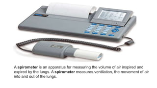
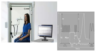
PULMONARY FUNCTION TEST – A
DETAILED STUDY
PREPARED BY
MARTIN SHAJI
PHARM D
The primary function of the respiratory system is to maintain
normality of arterial blood gases, that is, arterial pressure of oxygen
(PaO2) and arterial pressure of carbon dioxide (PaCO2). To achieve
this goal, several processes must be accomplished, including alveolar
ventilation, pulmonary perfusion, ventilation–perfusion matching,
and gas transfer across the alveolar–capillary membrane.
Different pulmonary function tests (PFTs) are used to evaluate the
physiologic processes of the respiratory system. Physiologic
abnormalities that can be measured by pulmonary function testing
include obstruction to airflow, restriction of lung size, and decrease in
transfer of gas across the alveolar–capillary membrane.
Potential uses of pulmonary function testing include evaluation of
patients with known or suspected lung disease; evaluation of symptoms
such as chronic cough, dyspnea, or chest tightness; monitoring of the
effects of exposure to dust, chemicals, or pulmonary toxic drugs; risk
stratification prior to surgery; monitoring of the effectiveness of
therapeutic interventions.
PFTs can help diagnose:
Asthma
Allergies
chronic bronchitis
respiratory infections
lung fibrosis
bronchiectasis, a condition in which the airways in the lungs stretch
and widen
COPD, which used to be called emphysema
asbestosis, a condition caused by exposure to asbestos
sarcoidosis, an inflammation of your lungs, liver, lymph nodes, eyes,
skin, or other tissues
scleroderma, a disease that affects your connective tissue
pulmonary tumor
lung cancer
weaknesses of the chest wall muscles
Definitions of lung volumes and capacities :
The air within the lung at the end of a forced inspiration can be
divided into four compartments or lung volumes.
The volume of air exhaled during normal quiet breathing is the tidal
volume (VT).
The maximal volume of air inhaled above tidal volume is the
inspiratory reserve volume (IRV).
The maximal air exhaled below tidal volume is the expiratory
reserve volume (ERV).
The residual volume (RV) is the amount of air remaining in the
lungs after a maximal exhalation.
The combinations or sums of two or more lung volumes are termed
capacities
Vital capacity (VC) is the maximal amount of air that can be exhaled
after a maximal inspiration. It is equal to the sum of IRV, VT, and
ERV.
When measured on a forced expiration, it is called the forced vital
capacity (FVC).
When measured over an exhalation of at least 30 seconds, it is
called the slow vital capacity (SVC).
 The VC is approximately 75% of the total lung capacity (TLC), and when the
SVC is within the normal range, a significant restrictive disorder is unlikely.
Normally, the values for SVC and FVC are very similar unless
airway obstruction is present.
TLC is the volume of air in the lung after the maximal
inspiration and is the sum of the four primary lung volumes (IRV,
VT, ERV, and RV). Its measurement is difficult because the
amount of air remaining in the chest after maximal exhalation
(RV) must be measured by indirect methods.
TOTAL LUNG CAPACITY
The functional residual capacity (FRC) is the volume of air remaining in
the lungs at the end of a quiet expiration. FRC is the normal resting
position of the lung; it occurs when there is no contraction of either
inspiratory or expiratory muscles and normally is 40% of TLC.
Inspiratory capacity (IC) is the maximal volume of air that can be
inhaled from the end of a quiet expiration and is the sum of VT and IRV.
FVC which represents the total amount of air that can be exhaled, can
be expressed as a series of timed volumes.
The forced expiratory volume in the first second of expiration (FEV1)
is the volume of air exhaled during the first second of the FVC
maneuver. Although FEV1 is a volume, it conveys information on
obstruction because it is measured over a known time interval. FEV1
depends on the volume of air within the lung and the effort during
exhalation;
The forced expiratory flow (FEF) during 25% to 75% of FVC
(FEF25%–75%) represents the mean flow during the middle half of the
FVC. FEF25%–75%, formerly called the maximal midexpiratory flow,
is reported frequently in the assessment of small airways.
What happens during pulmonary function tests?
You may have your procedure as an outpatient. This means you
go home the same day. Or it may be done as part of a longer
stay in the hospital. The way the procedure is done may vary. It
depends on your condition and your healthcare provider's
methods. In most cases, the procedure will follow this process:
1.You’ll be asked to loosen tight clothing, jewelry, or other things
that may cause a problem with the procedure.
2.If you wear dentures, you will need to wear them during the
procedure.
3.You’ll need to empty your bladder before the procedure.
4.You’ll sit in a chair. A soft clip will be put on your nose. This
is so all of your breathing is done through your mouth, not
your nose.
5.You’ll be given a sterile mouthpiece that is attached to a
spirometer.
6.You’ll form a tight seal over the mouthpiece with your
mouth. You’ll be instructed to inhale and exhale in different
ways.
7.You will be watched carefully during the procedure for
dizziness, trouble breathing, or other problems.
8.You may be given a bronchodilator after certain tests. The
tests will then be repeated several minutes later, after the
bronchodilator has taken effect.
SPIROMETRY/FLOW–VOLUME LOOP:
Spirometry is used to diagnose asthma, chronic obstructive
pulmonary disease (COPD) and other conditions that affect
breathing. Spirometry may also be used periodically to monitor your
lung condition and check whether a treatment for a chronic lung
condition is helping you breathe better
Spirometry is the most widely available and useful PFT. It takes only 15
to 20 minutes, carries no risks, and provides information about
obstructive and restrictive disease. Spirometry allows for measurement
of all lung volumes and capacities except RV, FRC, and TLC; it also
allows assessment of FEV1 and FEF25%–75%. Spirometry
measurements can be reported in two different formats—standard
spirometry and the flow–volume loop.
A spirometer is an apparatus for measuring the volume of air inspired and
expired by the lungs. A spirometer measures ventilation, the movement of air
into and out of the lungs.
In standard spirometry, the volumes are recorded on the vertical (y)
axis and the time on the horizontal (x) axis. In flow–volume loops,
volume is plotted on the horizontal (x) axis, and flow (derived from
volume/ time) is plotted on the vertical (y) axis. The shape of the
flow–volume loop can be helpful in differentiating obstructive and
restrictive defects and in diagnosing upper airway obstruction. This
curve gives a visual representation of obstruction because the
expiratory descent becomes more concave with worsening
obstruction.
Spirometry measures three of the four basic lung volumes but cannot
measure RV. RV must be measured to determine TLC. TLC should be
measured anytime VC is reduced.
The four methods for measuring TLC are
helium dilution,
nitrogen washout,
body plethysmography,
and chest x-ray measurement (planimetry).
The first two methods are called dilution techniques and only
measure lung volumes in communication with the upper airway.
In patients with airway obstruction who have trapped air,
dilution techniques will underestimate the actual volume of the
lungs.
Body plethysmography:
Body plethysmography, or body box, is the most accurate
technique for lung volume determinations. It measures all the air in
the lungs, including trapped air. The principle of the measurement of
the body box is Boyle’s gas law (P1V1 = P2V2): A volume of gas in
a closed system varies inversely with the pressure applied to it.
Diffusion capacity test
This test evaluates how well the small air sacks inside the lungs,
called alveoli, work. For this part of a pulmonary function test, you
will be asked to breathe in certain gases such as oxygen, helium, or
carbon dioxide.
You may also breathe in a “tracer gas” for one breath. The machine
can detect when you breathe out this gas. This tests how well your
lungs are able to transfer oxygen and carbon dioxide to and from
your bloodstream
CARBON MONOXIDE DIFFUSING CAPACITY:
The diffusing capacity of the lungs (DL) is a measurement of the
ability of a gas to diffuse across the alveolar–capillary membrane.
Carbon monoxide is the usual test gas because normally it is not
present in the lungs and is much more soluble in blood than in lung
tissue. When the diffusing capacity is determined with carbon
monoxide, the test is called the diffusing capacity of lung for carbon
monoxide (DLCO). Because DLCO is directly related to alveolar
volume (VA), it frequently is normalized to the value DL/VA, which
allows for its interpretation in the presence of abnormal lung volumes
(e.g., after surgical lung resection).
The diffusing capacity will be reduced in all clinical situations where gas
transfer from the alveoli to capillary blood is impaired.3 Common
conditions that reduce DLCO include lung resection, emphysema (loss
of functioning alveolar–capillary units), and interstitial lung disease
(thickening of the alveolar–capillary membrane). Normal PFTs with
reduced DLCO should suggest the possibility of pulmonary vascular
disease (e.g., pulmonary embolus) but also can be seen with anaemia,
early interstitial lung disease, and mild Pneumocystis carinii pneumonia
(PCP) infection in patients with acquired immune deficiency syndrome.
What are the risks of pulmonary function tests?
A PFT can cause problems if:
you’ve recently had a heart attack
you’ve recently had eye surgery
you’ve recently had chest surgery
you’ve recently had abdominal surgery
you have a severe respiratory infection
you have unstable heart disease
PFTs are usually safe for most people. However, because the test may
require you to breathe in and out quickly, you may feel dizzy and
there’s a risk that you may faint. If you feel lightheaded, tell your
doctor. If you have asthma, the test may cause you to have an asthma
attack. In very rare cases, PFTs may cause a collapsed lung.
a detailed study on pulmonary function test

a detailed study on pulmonary function test

  • 1.
    PULMONARY FUNCTION TEST– A DETAILED STUDY PREPARED BY MARTIN SHAJI PHARM D
  • 2.
    The primary functionof the respiratory system is to maintain normality of arterial blood gases, that is, arterial pressure of oxygen (PaO2) and arterial pressure of carbon dioxide (PaCO2). To achieve this goal, several processes must be accomplished, including alveolar ventilation, pulmonary perfusion, ventilation–perfusion matching, and gas transfer across the alveolar–capillary membrane. Different pulmonary function tests (PFTs) are used to evaluate the physiologic processes of the respiratory system. Physiologic abnormalities that can be measured by pulmonary function testing include obstruction to airflow, restriction of lung size, and decrease in transfer of gas across the alveolar–capillary membrane.
  • 3.
    Potential uses ofpulmonary function testing include evaluation of patients with known or suspected lung disease; evaluation of symptoms such as chronic cough, dyspnea, or chest tightness; monitoring of the effects of exposure to dust, chemicals, or pulmonary toxic drugs; risk stratification prior to surgery; monitoring of the effectiveness of therapeutic interventions.
  • 4.
    PFTs can helpdiagnose: Asthma Allergies chronic bronchitis respiratory infections lung fibrosis bronchiectasis, a condition in which the airways in the lungs stretch and widen
  • 5.
    COPD, which usedto be called emphysema asbestosis, a condition caused by exposure to asbestos sarcoidosis, an inflammation of your lungs, liver, lymph nodes, eyes, skin, or other tissues scleroderma, a disease that affects your connective tissue pulmonary tumor lung cancer weaknesses of the chest wall muscles
  • 6.
    Definitions of lungvolumes and capacities : The air within the lung at the end of a forced inspiration can be divided into four compartments or lung volumes. The volume of air exhaled during normal quiet breathing is the tidal volume (VT). The maximal volume of air inhaled above tidal volume is the inspiratory reserve volume (IRV). The maximal air exhaled below tidal volume is the expiratory reserve volume (ERV).
  • 7.
    The residual volume(RV) is the amount of air remaining in the lungs after a maximal exhalation.
  • 8.
    The combinations orsums of two or more lung volumes are termed capacities Vital capacity (VC) is the maximal amount of air that can be exhaled after a maximal inspiration. It is equal to the sum of IRV, VT, and ERV. When measured on a forced expiration, it is called the forced vital capacity (FVC). When measured over an exhalation of at least 30 seconds, it is called the slow vital capacity (SVC).  The VC is approximately 75% of the total lung capacity (TLC), and when the SVC is within the normal range, a significant restrictive disorder is unlikely.
  • 9.
    Normally, the valuesfor SVC and FVC are very similar unless airway obstruction is present. TLC is the volume of air in the lung after the maximal inspiration and is the sum of the four primary lung volumes (IRV, VT, ERV, and RV). Its measurement is difficult because the amount of air remaining in the chest after maximal exhalation (RV) must be measured by indirect methods. TOTAL LUNG CAPACITY
  • 10.
    The functional residualcapacity (FRC) is the volume of air remaining in the lungs at the end of a quiet expiration. FRC is the normal resting position of the lung; it occurs when there is no contraction of either inspiratory or expiratory muscles and normally is 40% of TLC. Inspiratory capacity (IC) is the maximal volume of air that can be inhaled from the end of a quiet expiration and is the sum of VT and IRV. FVC which represents the total amount of air that can be exhaled, can be expressed as a series of timed volumes.
  • 11.
    The forced expiratoryvolume in the first second of expiration (FEV1) is the volume of air exhaled during the first second of the FVC maneuver. Although FEV1 is a volume, it conveys information on obstruction because it is measured over a known time interval. FEV1 depends on the volume of air within the lung and the effort during exhalation; The forced expiratory flow (FEF) during 25% to 75% of FVC (FEF25%–75%) represents the mean flow during the middle half of the FVC. FEF25%–75%, formerly called the maximal midexpiratory flow, is reported frequently in the assessment of small airways.
  • 12.
    What happens duringpulmonary function tests? You may have your procedure as an outpatient. This means you go home the same day. Or it may be done as part of a longer stay in the hospital. The way the procedure is done may vary. It depends on your condition and your healthcare provider's methods. In most cases, the procedure will follow this process: 1.You’ll be asked to loosen tight clothing, jewelry, or other things that may cause a problem with the procedure. 2.If you wear dentures, you will need to wear them during the procedure.
  • 13.
    3.You’ll need toempty your bladder before the procedure. 4.You’ll sit in a chair. A soft clip will be put on your nose. This is so all of your breathing is done through your mouth, not your nose. 5.You’ll be given a sterile mouthpiece that is attached to a spirometer. 6.You’ll form a tight seal over the mouthpiece with your mouth. You’ll be instructed to inhale and exhale in different ways.
  • 14.
    7.You will bewatched carefully during the procedure for dizziness, trouble breathing, or other problems. 8.You may be given a bronchodilator after certain tests. The tests will then be repeated several minutes later, after the bronchodilator has taken effect.
  • 15.
  • 16.
    Spirometry is usedto diagnose asthma, chronic obstructive pulmonary disease (COPD) and other conditions that affect breathing. Spirometry may also be used periodically to monitor your lung condition and check whether a treatment for a chronic lung condition is helping you breathe better
  • 17.
    Spirometry is themost widely available and useful PFT. It takes only 15 to 20 minutes, carries no risks, and provides information about obstructive and restrictive disease. Spirometry allows for measurement of all lung volumes and capacities except RV, FRC, and TLC; it also allows assessment of FEV1 and FEF25%–75%. Spirometry measurements can be reported in two different formats—standard spirometry and the flow–volume loop.
  • 18.
    A spirometer isan apparatus for measuring the volume of air inspired and expired by the lungs. A spirometer measures ventilation, the movement of air into and out of the lungs.
  • 19.
    In standard spirometry,the volumes are recorded on the vertical (y) axis and the time on the horizontal (x) axis. In flow–volume loops, volume is plotted on the horizontal (x) axis, and flow (derived from volume/ time) is plotted on the vertical (y) axis. The shape of the flow–volume loop can be helpful in differentiating obstructive and restrictive defects and in diagnosing upper airway obstruction. This curve gives a visual representation of obstruction because the expiratory descent becomes more concave with worsening obstruction.
  • 20.
    Spirometry measures threeof the four basic lung volumes but cannot measure RV. RV must be measured to determine TLC. TLC should be measured anytime VC is reduced. The four methods for measuring TLC are helium dilution, nitrogen washout, body plethysmography, and chest x-ray measurement (planimetry).
  • 21.
    The first twomethods are called dilution techniques and only measure lung volumes in communication with the upper airway. In patients with airway obstruction who have trapped air, dilution techniques will underestimate the actual volume of the lungs.
  • 22.
    Body plethysmography: Body plethysmography,or body box, is the most accurate technique for lung volume determinations. It measures all the air in the lungs, including trapped air. The principle of the measurement of the body box is Boyle’s gas law (P1V1 = P2V2): A volume of gas in a closed system varies inversely with the pressure applied to it.
  • 24.
    Diffusion capacity test Thistest evaluates how well the small air sacks inside the lungs, called alveoli, work. For this part of a pulmonary function test, you will be asked to breathe in certain gases such as oxygen, helium, or carbon dioxide. You may also breathe in a “tracer gas” for one breath. The machine can detect when you breathe out this gas. This tests how well your lungs are able to transfer oxygen and carbon dioxide to and from your bloodstream
  • 25.
    CARBON MONOXIDE DIFFUSINGCAPACITY: The diffusing capacity of the lungs (DL) is a measurement of the ability of a gas to diffuse across the alveolar–capillary membrane. Carbon monoxide is the usual test gas because normally it is not present in the lungs and is much more soluble in blood than in lung tissue. When the diffusing capacity is determined with carbon monoxide, the test is called the diffusing capacity of lung for carbon monoxide (DLCO). Because DLCO is directly related to alveolar volume (VA), it frequently is normalized to the value DL/VA, which allows for its interpretation in the presence of abnormal lung volumes (e.g., after surgical lung resection).
  • 26.
    The diffusing capacitywill be reduced in all clinical situations where gas transfer from the alveoli to capillary blood is impaired.3 Common conditions that reduce DLCO include lung resection, emphysema (loss of functioning alveolar–capillary units), and interstitial lung disease (thickening of the alveolar–capillary membrane). Normal PFTs with reduced DLCO should suggest the possibility of pulmonary vascular disease (e.g., pulmonary embolus) but also can be seen with anaemia, early interstitial lung disease, and mild Pneumocystis carinii pneumonia (PCP) infection in patients with acquired immune deficiency syndrome.
  • 27.
    What are therisks of pulmonary function tests? A PFT can cause problems if: you’ve recently had a heart attack you’ve recently had eye surgery you’ve recently had chest surgery you’ve recently had abdominal surgery you have a severe respiratory infection you have unstable heart disease PFTs are usually safe for most people. However, because the test may require you to breathe in and out quickly, you may feel dizzy and there’s a risk that you may faint. If you feel lightheaded, tell your doctor. If you have asthma, the test may cause you to have an asthma attack. In very rare cases, PFTs may cause a collapsed lung.