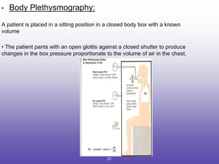
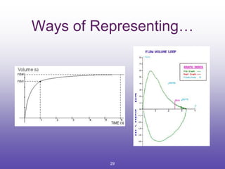
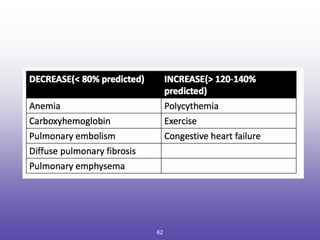
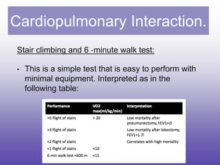
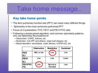
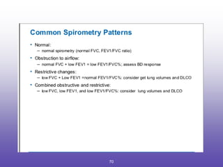
SlideShare a Scribd company logo
PULMONARY FUNCTION
TESTS
Presented by : Dr. Prasant N
Moderated by : Dr. ( Prof) Ranjita Acharya.
1
Table of Contents:
• Introduction
• Classification of PFT’s.
• Bedside PFT’s.
• Lung Volumes and Capacities.( Static and Dynamic).
• Spirometry
• Interpretation of a Spirometry.
• Flow volume loops.
• Gas Exchange Function Tests.
• Cardiopulmonary Interaction
• Summary.
2
Introduction.
• Pulmonary function tests is a generic term used to indicate a series
of studies or maneuvers that may be performed to measure lung
function.
• Evaluates one or more aspects of the respiratory system:
1. Respiratory mechanics.
2. Lung parenchymal function/ Gas exchange
3. Cardiopulmonary interaction.
3
Indications for a PFT : ( American College of Physicians)
Patients under going:
1. Lung resection.
2. Cardiac Surgery.
3. Upper Abdominal Surgeries.
4. Lower Abdominal Surgeries.
• Patients with a history of exposure to lung irritants. ( asbestos).
• H/o prolonged cough or excessive sputum production.
• C/o : cough , dyspnea , orthopnea, wheezing.
• Known smokers.
• Uncharacterized pulmonary disease(defined as H/o Pulmonary Disease or symptoms and no PFT in last
60 days)
4
Assessing disease severity and progression:
• Pulmonary disease- COPD, Cystic fibrosis, Interstitial lung disease,
Sarcoidosis.
• Cardiac Disease- CHF, Congenital heart disease.
• Neuromuscular diseases- Guillain-Barre Syndrome, Multiple Sclerosis,
Myasthenia gravis
5
Contraindications:
• Recent eye surgery
• Thoracic , abdominal and cerebral aneurysms
• Active hemoptysis
• Pneumothorax
• Unstable angina/ recent MI within 1 month
6
Classification of PFT’
A. Mechanical Ventilatory Functions of Lung/ Chest wall.
B. Gas Exchange Tests.
C. Cardiopulmonary Interaction.
7
Mechanical and ventilatory functions of lung/chest wall
Bedside Pulmonary Function Tests:
1.History:
• Daily activity.
• Talking time- whether patient is able to finish his sentence in one
breath or has to catch his breath in between.
2. Examination:
• Orthopnea, dyspnea, cyanosis, chest wall retraction, accessory
muscles of respiration, tracheal tug etc
8
Mechanical Ventilatory functions of lung/chest
wall.
Tests:
1. Sabrasez Breath Holding Test:
Ask the patient to take a full but not too deep breath & hold it as
long as possible.
• Normal > 40 seconds.
• 20-25 sec: Mild reduction in cardiopulmonary reserve.
• 15-20 sec : Moderate reduction in cardiopulmonary reserve.
• < 15 sec : Severe reduction and is a contraindication for surgery.
9
2.Snider’s match blowing test / Modified Snider’s test:
• It is an indirect measure of FEV1.
Patient is asked to extinguish a lit match held at 15 cm distance with
mouth open and nose pinched.
It reflects MBC > 60L/min, FEV1 > 1.6 L.
• If the patient is not able to extinguish at 8 cm, reflects FEV1 <1.6 L,
MBC <40 L/minute.
10
3.Forced expiratory time:
• It is done for obstructive diseases.
• Patient has to take a deep breath and then exhale as forcibly and
completely as possible through mouth.
• Normally it is completed in 3 seconds.
• Obstructive Lung Disease - > 6 SEC
• Restructive Lung Disease- < 3 SEC
11
4. Debono’s whistle:
• It measures the peak expiratory flow rate.
• Patient blows down a wide bore tube at the end of which is a whistle.
• On the other side is a hole with adjustable knob.
• As subject blows → whistle blows.
• Leak hole is gradually increased till the intensity of whistle disappears.
• At the last position at which the whistle can be blown , the PEFR can be
read off the scale.
12
Static and Dynamic Lung
Volumes
• Static lung volumes reflect the elastic properties of the
lungs and chest wall.
• These include all the capacity measurements.
• Not affected by the rate of air movement in and out of
the lungs
• Dynamic volumes and capacities are based on time and
these tests reflect the caliber and integrity of the airways.
13
Lung Volumes and Capacities
VOLUMES CAPACITIES
Tidal Volume (TV)
Inspiratory Capacity
(IC)
Inspiratory Reserve Volume ( IRV)
Expiratory Capacity
(EC)
Expiratory Reserve Volume.( ERV)
Vital Capacity
(VC)
Residual Volume (RV) Functional Residual Volume (FRC)
Total Lung Capacity (TLC)
14
LUNG VOLUMES:
1. Tidal Volume (TV):
• Volume of air inhaled or exhaled with each breath during quiet
breathing (6‐8 ml/kg).
• Normal : 500 mL
2. Inspiratory Reserve Volume (IRV)
• Maximum volume of air inhaled from the end‐ inspiratory tidal
position.
• Normal : 3000 mL
15
3. Expiratory Reserve Volume ( ERV):
• Maximum volume of air that can be exhaled from resting end‐expiratory tidal position.
• Normal :1500 ml
4. Residual Volume (RV):
– Volume of air remaining in lungs after maximum exhalation .(20‐25 ml/kg)
Normal :200 ml
– Indirectly measured by (FRC‐ ERV).
– It can not be measured by spirometry .
16
Lung Capacities:
1. Total Lung Capacity (TLC):
• Sum of all volume compartments or volume of air in lungs after
maximal inspiration.
• Normal :4‐6 L
2. Vital Capacity (VC) :
• Maximum volume of air exhaled from maximal inspiratory level.
• VC = IRV + TV+ ERV
• Normal : (60‐70 ml/kg)
17
3. Inspiratory Capacity (IC):
• The maximum volume of air that can be inhaled from the end‐expiratory tidal position.
• IC = IRV + TV
• Normal :(2400‐3800ml)
4. Expiratory Capacity (EC):
• EC =TV+ ERV
5.Functional Residual Capacity:
• Is the volume of gas remaining in lungs after passive expiration.
• FRC= RV + ERV.
• It ranges between 1.8-3.4 liters.
• Body plethysmography is the gold standard for measuring FRC.
• Other methods include helium dilution technique and nitrogen washout method.
18
• Normal : 30‐35 ml/kg . ( around 50% of TLC)
• Decreases in :
1. Induction of Anesthesia by 16-20%.
2. Supine position.
3. Obese Patients.
Importance of FRC:
• Oxygen store
• Buffer for maintaining a steady arterial po2.
• Partial inflation helps prevent atelectasis.
• Minimizes the work of breathing.
19
20
• Body Plethysmography:
A patient is placed in a sitting position in a closed body box with a known
volume
• The patient pants with an open glottis against a closed shutter to produce
changes in the box pressure proportionate to the volume of air in the chest.
21
Dynamic Lung Volumes:
• These are based on time.
• They attempt to quantitate the pulmonary ventilation in terms of rate at
which ventilation takes place.
• Reflects the caliber and integrity of the airways.
1. Forced Vital Capacity. ( FVC)
2. Forced Expiratory Volume in 1 second (FEV1).
3. Maximum Voluntary Ventilation: ( MVV)
4. Maximum Breathing Capacity ( MBC)
22
Forced Vital Capacity:
• The FVC is the maximum volume of air that can be breathed out as
forcefully and rapidly as possible following a maximum inspiration.
• Characterized by full inspiration to TLC followed by abrupt onset of
expiration to RV.
• The expiration should be at least 4 seconds.
• Should not be interrupted by coughing, glottis closure or
mechanical obstruction.
23
• Interpretation of % predicted:
• 80-120% Normal
• 70-79% Mild reduction
• 50%-69% Moderate reduction
• <50% Severe reduction
24
Forced Expiratory Volume in 1 second ( FEV1):
• The volume expired in the first second of the FVC test is called FEV1.
• The FEV1% is FEV1 divided by the FVC (Vital Capacity) X100:
• FEV1%=FEV1/FVC X100.
• This parameter is also known as the Tiffeneau index.
• Nowadays FEV1/FVC X 100 is also accepted as FEV1% (FEV1/FVC ratio).
• A healthy patients expires approximately 80% of all the air out of his lungs in the first
second during the FVC maneuver..
• FEV1/FVC ratio < 0.8 = Obstructive disease
• FEV1/FVC ratio > 0.8 = Restrictive Disease.
25
• Interpretation of % predicted:
• >75% Normal
• 60%‐75% Mild obstruction
• 50‐59% Moderate obstruction
• <49% Severe obstruction
26
Maximum Voluntary Ventilation: ( MVV)
• Is the largest volume that can be breathed per minute by
maximum voluntary efforts.
• MVV = FEV1 × 35 and is about 100-200 L/minute.
• It is decreased in old age, pulmonary emphysema,
bronchospasm, obstruction etc.
Maximum Breathing Capacity: (MBC)
• is the maximum volume that can be breathed per minute.
27
Spirometry
• Spirometry is the ‘cornerstone’ of all PFT’s.
• Invented by John Hutchinson in 1864.
• Measures the rate at which the lungs change volume during quite
and forced breathing maneuvres.
• It can only measure lung volume compartments that exchange gas
with the atmosphere.
• Cannot measure — FRC,RV, TLC.
28
Ways of Representing…
29
Acceptability Criteria:
1. Lack of artifacts induced by coughing or glottic closure.
Normal Coughing
30
2. Satisfactory start to the test without hesitation.
Normal
Submaximal effort
31
• 3. Satisfactory exhalation with 6 seconds of smooth
continuous exhalation, with a plateau of at least 1
second.
No Plateau
Normal
32
Interpretation of a PFT.
Getting started :
Before PFT results can be reliably interpreted, three factors must be confirmed:
(1) the volume-time curve reaches a plateau, and expiration lasts at least 6
seconds.
(2) results of the two best efforts on the PFT are within 0.2 L of each other
(Figure 3) and
(3) the flow- volume loops are free of artifacts and abnormalities.
33
Step 1: Determine If the FEV1
/FVC Ratio Is Low.
• Physicians have two options to determine if this ratio is low.
• The first option is to follow the GOLD criteria, which use a cutoff of less
than 70%.
• The second option is to follow the ATS criteria, which use the lower limit
of normal (LLN) as the cutoff for adults.
• The LLN is a measurement less than the 5th percentile of spirometry data
obtained from the Third National Health and Nutrition Examination Survey
(NHANES III).
• Most modern PFT software can calculate the LLN.
34
Step 2 : Determine If the FVC Is Low
The physician must determine if the FVC is
1. Less than the LLN for adults or
2. Less than 80% of predicted for those 5 to 18 years of age, indicating a
restrictive pattern.
35
36
Step 3: Confirm the Restrictive Pattern
• If the patient’s initial PFT results indicate a
restrictive pattern or a mixed pattern that is not
corrected with bronchodilators, the patient should
be referred for full PFTs with DLCO testing.
37
Interpretation :
• Full PFTs provide the patient’s total lung capacity.
• The restrictive pattern is confirmed as a true restrictive defect if the total
lung capacity is less than 80% of predicted in patients 5 to 18 years of
age, or less than the LLN in adults.
• If full PFTs cannot be obtained, the FVC can be used to infer a restrictive
defect; however, FVC has a poor positive predictive value.
38
Step 4: Grade the Severity of the Abnormality :
• If an obstructive defect, a restrictive pattern, or a mixed pattern is present,
as defined by steps 1 and 2, the physician should grade the severity of
the abnormality based on the FEV1 percentage of predicted.
• The ATS system for grading the severity of a PFT abnormality is
summarized in Table below.
39
Step 6: Bronchoprovocation :
• If PFT results are normal but the physician still suspects exercise- or
allergen-induced asthma.
• The next step is bronchoprovocation, such as a methacholine challenge, a
mannitol inhalation challenge, exercise testing, or sometimes eucapnic
voluntary hyperpnea testing.
• When the FEV1 is 70% or more of predicted on standard spirometry,
bronchoprovocation should be used to make the diagnosis. If the FEV1 is
less than 70% of predicted, a therapeutic trial of a bronchodilator may be
considered.
40
METHACHOLINE CHALLENGE :
• The methacholine challenge is highly sensitive for diagnosing asthma.
• However, its low specificity results in false-positive results.
• A positive methacholine challenge result is defined as a greater than
20% reduction in FEV1 at administration of 4 mg per mL of inhaled
methacholine.
• The result is considered border- line if the FEV1 drops by 20% at a dose
between 4 and 16 mg per mL.
41
Step 5: Determine Reversibility of the Obstructive Defect:
• If the patient has an obstructive defect, the physician should determine if it is
reversible based on the increase in FEV1 or FVC after bronchodilator treatment
(i.e., increase of more than 12% in patients 5 to 18 years of age, or more than 12%
and more than 200 mL in adults).3
• Figure 4 shows a fully reversible obstructive defect. Obstructive defects in persons
with asthma are usually fully reversible, whereas defects in persons with COPD
typically are not.
• If PFTs show a mixed pattern and the FVC corrects to 80% or more of predicted in
patients 5 to 18 years of age or to the LLN or more in adults after bronchodilator
use, it is likely that the patient has pure obstructive lung disease with air
trapping.
42
43
Step 7: Establish the Differential Diagnosis:
• Once PFT results have been interpreted, the
broad differential diagnosis should be
considered.
44
Step 8: Compare Current and Prior PFT Results:
If a patient’s prior PFT results are available, they
should be compared with the current results to
determine the course of the disease or effects of
treatment.
45
46
FLOW VOLUME LOOPS.
• Normal
47
• Inspiratory limb of loop is symmetric and convex.
• Expiratory limb is linear.
• Flow rates at the midpoint of the inspiratory and
expiratory capacity are often measured.
• Maximal inspiratory flow at 50% of forced vital capacity
(MIF 50%FVC) is greater than maximal expiratory flow
at 50% FVC (MEF 50%FVC) because dynamic
compression of the airways occurs during exhalation.
48
• Flow Volume Loops in Obstructive Diseases.
B) Obstructive disease (eg, emphysema, asthma)
Although all flow rates are diminished, expiratory prolongation
predominates, and MEF < MIF. Peak expiratory flow is
sometimes used to estimate degree of airway obstruction but
is dependent on patient effort.
49
• Flow-volume loops in restrictive lung disease:
50
• Restrictive disease (eg, interstitial lung disease,
kyphoscoliosis).
• The loop is narrowed because of diminished lung volumes,
but the shape is generally the same as in normal volume.
• Flow rates are greater than normal at comparable lung
volumes because the increased elastic recoil of lungs holds
the airways open.
51
• Fixed obstruction of the upper airway:
52
• Examples :Tracheal stenosis, goiter.
• The top and bottom of the loops are flattened so
that the configuration approaches that of a
rectangle.
• Fixed obstruction limits flow equally during
inspiration and expiration, and MEF = MIF.
53
• Variable extrathoracic obstruction:
54
• Ex :unilateral vocal cord paralysis, vocal cord dysfunction.
• When a single vocal cord is paralyzed, it moves passively
with pressure gradients across the glottis.
• During forced inspiration, it is drawn inward, resulting in a
plateau of decreased inspiratory flow.
• During forced expiration, it is passively blown aside, and
expiratory flow is unimpaired.
• Therefore, MIF 50%FVC < MEF 50%FVC
55
• Variable intra-thoracic obstruction :
56
• During a forced inspiration, negative pleural
pressure holds the “floppy” trachea open.
• With forced expiration, loss of structural support
results in tracheal narrowing of the trachea and a
plateau of diminished flow.
• Flow is maintained briefly before airway
compression occurs.
57
Gas Exchange Function
Tests
ALVEOLAR‐ARTERIAL O2 TENSION GRADIENT:
• Sensitive indicator of detecting regional V/Q
inequality.
• A‐a gradient = PAO2 ‐ PaO2.
• PAO2 = alveolar PO2 (calculated from the
alveolar gas equation)
• PaO2 = arterial PO2 (measured in arterial gas)
58
59
• A normal A–a gradient for a young adult non-
smoker breathing air, is between 5–10 mmHg.
• Normally, the A–a gradient increases with age.
• An abnormally increased A–a gradient suggests
a defect in diffusion, V/Q (ventilation/perfusion
ratio) mismatch, or right-to-left shunt.[
60
DLCO TESTING :
• DLCO is a quantitative measurement of gas transfer in the lungs.
• Diseases that decrease blood flow to the lungs or damage alveoli will cause less
efficient gas exchange, resulting in a lower DLCO measurement.
• During the DLCO test, patients inhale a mixture of helium (10%), carbon monoxide
(0.3%), oxygen (21%), and nitrogen (68.7%)12
then hold their breath for 10 seconds
before exhaling.
• The amounts of exhaled helium and carbon monoxide are used to calculate the
DLCO.
• CO is used to estimate gas transfer instead of oxygen due to its much higher
affinity for hemoglobin.
• Full PFTs provide the patient’s total lung capacity.
61
62
Cardiopulmonary Interaction.
Stair climbing and 6 ‐minute walk test:
• This is a simple test that is easy to perform with
minimal equipment. Interpreted as in the
following table:
63
Shuttle walk
• The patient walks between cones 10 meters apart
with increasing pace.
• The subject walks until they cannot make it from
cone to cone between the beeps.
• Less than 250m or decrease SaO2 > 4% signifies
high risk.
• A shuttle walk of 350m correlates with a VO2 max
of 11ml.kg ‐ 1.min ‐ 1
64
Assessment of lung function in
thoracotomy pts.
As an anesthesiologist our goal is to :
1) To identify pts at risk of increased post‐op
morbidity & mortality
2) To identify pts who need short‐term or long term
post‐op ventilatory support.
Lung resection may be f/by – inadequate gas
exchange, pulm HTN & incapacitating dyspnoea.
65
• Calculating the predicted postoperative FEV1 (ppoFEV1)
and TLCO (ppoTLCO):
• There are 5 lung lobes containing 19 segments in total
with the division of each lobe.
• Ppo FEV1=preoperative FEV1 * no. of segments left
after resection 19.
• Can be assessed by ventilation perfusion scan.
• For eg: A 57-year-old man is booked for lung resection.
His CT chest show a large RUL mass confirmed as
carcinoma: ppoFEV1= 50*16/19=42%
66
67
68
Take home message..
69
70
71

More Related Content

What's hot

Pulmonary function tests
Pulmonary function testsPulmonary function tests
Pulmonary function tests
Ranjeet Singha
 
14. pulmonary-function-tests
14. pulmonary-function-tests14. pulmonary-function-tests
14. pulmonary-function-tests
Dr. Mohamed Maged Kharabish
 
Pulmonary function tests
Pulmonary function testsPulmonary function tests
Pulmonary function tests
Chetan Ganteppanavar
 
Spirometry
SpirometrySpirometry
Spirometry
Saumya Shishir
 
DLCO
DLCO DLCO
Dlco/tlco
Dlco/tlcoDlco/tlco
Dlco/tlco
Tanveer Fahim
 
Spirometry Basics
Spirometry BasicsSpirometry Basics
Spirometry Basics
Ashraf ElAdawy
 
Pneumology - Lung volumes-airway-resistance
Pneumology - Lung volumes-airway-resistancePneumology - Lung volumes-airway-resistance
Pneumology - Lung volumes-airway-resistance
Ammedicine Medicine
 
Pulmonary function test
Pulmonary function testPulmonary function test
Pulmonary function test
ravindrajha10
 
Lung volumes in COPD
Lung volumes in COPDLung volumes in COPD
Lung volumes in COPD
Ashraf ElAdawy
 
Pulmonary function tests
Pulmonary function testsPulmonary function tests
Pulmonary function tests
logon2kingofkings
 
Pulmonary Flow Volume Loops.. Dr.Padmesh
Pulmonary Flow Volume Loops.. Dr.PadmeshPulmonary Flow Volume Loops.. Dr.Padmesh
Pulmonary Flow Volume Loops.. Dr.Padmesh
Dr Padmesh Vadakepat
 
Interpretation of CPET
Interpretation of CPETInterpretation of CPET
Interpretation of CPET
Samiaa Sadek
 
Lung volumes capacities pfts
Lung volumes capacities pfts Lung volumes capacities pfts
Lung volumes capacities pfts
Rajesh Munigial
 
Body plethesmography
Body plethesmographyBody plethesmography
Body plethesmography
Anusha Jahagirdar
 
methacholine challenge.pptx
methacholine challenge.pptxmethacholine challenge.pptx
methacholine challenge.pptx
MaharijNoor
 
pulmonary function test
pulmonary function test pulmonary function test
pulmonary function test
imsurgeon
 
Pulmonary Functions Lung Volumes
Pulmonary Functions Lung VolumesPulmonary Functions Lung Volumes
Pulmonary Functions Lung Volumes
Ashraf ElAdawy
 
Lung fuction tests
Lung fuction tests  Lung fuction tests
Lung fuction tests
Sid Kaithakkoden
 
Pulmonary Function Testing
Pulmonary Function TestingPulmonary Function Testing
Pulmonary Function Testing
Dr Riham Hazem Raafat
 

What's hot (20)

Pulmonary function tests
Pulmonary function testsPulmonary function tests
Pulmonary function tests
 
14. pulmonary-function-tests
14. pulmonary-function-tests14. pulmonary-function-tests
14. pulmonary-function-tests
 
Pulmonary function tests
Pulmonary function testsPulmonary function tests
Pulmonary function tests
 
Spirometry
SpirometrySpirometry
Spirometry
 
DLCO
DLCO DLCO
DLCO
 
Dlco/tlco
Dlco/tlcoDlco/tlco
Dlco/tlco
 
Spirometry Basics
Spirometry BasicsSpirometry Basics
Spirometry Basics
 
Pneumology - Lung volumes-airway-resistance
Pneumology - Lung volumes-airway-resistancePneumology - Lung volumes-airway-resistance
Pneumology - Lung volumes-airway-resistance
 
Pulmonary function test
Pulmonary function testPulmonary function test
Pulmonary function test
 
Lung volumes in COPD
Lung volumes in COPDLung volumes in COPD
Lung volumes in COPD
 
Pulmonary function tests
Pulmonary function testsPulmonary function tests
Pulmonary function tests
 
Pulmonary Flow Volume Loops.. Dr.Padmesh
Pulmonary Flow Volume Loops.. Dr.PadmeshPulmonary Flow Volume Loops.. Dr.Padmesh
Pulmonary Flow Volume Loops.. Dr.Padmesh
 
Interpretation of CPET
Interpretation of CPETInterpretation of CPET
Interpretation of CPET
 
Lung volumes capacities pfts
Lung volumes capacities pfts Lung volumes capacities pfts
Lung volumes capacities pfts
 
Body plethesmography
Body plethesmographyBody plethesmography
Body plethesmography
 
methacholine challenge.pptx
methacholine challenge.pptxmethacholine challenge.pptx
methacholine challenge.pptx
 
pulmonary function test
pulmonary function test pulmonary function test
pulmonary function test
 
Pulmonary Functions Lung Volumes
Pulmonary Functions Lung VolumesPulmonary Functions Lung Volumes
Pulmonary Functions Lung Volumes
 
Lung fuction tests
Lung fuction tests  Lung fuction tests
Lung fuction tests
 
Pulmonary Function Testing
Pulmonary Function TestingPulmonary Function Testing
Pulmonary Function Testing
 

Similar to Pulmonary function tests

Bedside pft 1
Bedside pft 1Bedside pft 1
Bedside pft 1
DrAnkitPurohit
 
Lung function tests
Lung function testsLung function tests
Lung function tests
deka dada
 
PULMONARY FUNCTION TESTS - LAB DATA INTERPRETATION
PULMONARY FUNCTION TESTS - LAB DATA INTERPRETATIONPULMONARY FUNCTION TESTS - LAB DATA INTERPRETATION
PULMONARY FUNCTION TESTS - LAB DATA INTERPRETATION
LincyAsha
 
lung volumes new v.pptx
lung volumes new v.pptxlung volumes new v.pptx
lung volumes new v.pptx
udayasree30
 
Pulmonary Function Test's
Pulmonary Function Test's Pulmonary Function Test's
Pulmonary Function Test's
HSK College of Pharmacy
 
Pulmonary Function Tests-Nursing Maseno.pptx
Pulmonary Function Tests-Nursing Maseno.pptxPulmonary Function Tests-Nursing Maseno.pptx
Pulmonary Function Tests-Nursing Maseno.pptx
akoeljames8543
 
PFT.pdf
PFT.pdfPFT.pdf
PFT.pdf
sanavora1
 
Pulmonary Function Testing-Simplified description...!
Pulmonary Function Testing-Simplified description...!Pulmonary Function Testing-Simplified description...!
Pulmonary Function Testing-Simplified description...!
Sharmin Susiwala
 
Lung volumes and capacities.pptx
Lung volumes and capacities.pptxLung volumes and capacities.pptx
Lung volumes and capacities.pptx
Manoj Aryal
 
PFT
PFT PFT
pulmonary Function Test
pulmonary Function Testpulmonary Function Test
pulmonary Function Test
Rekha Marbate
 
PFT 3rd year FDPS.pptx
PFT 3rd year FDPS.pptxPFT 3rd year FDPS.pptx
PFT 3rd year FDPS.pptx
Rekha Marbate
 
spirometry functional lung test
spirometry functional lung test spirometry functional lung test
spirometry functional lung test
Harith Riyadh
 
Spirometry
SpirometrySpirometry
Spirometry
ArabilReang
 
weaning complete note.pptx
weaning complete note.pptxweaning complete note.pptx
weaning complete note.pptx
RitaMagar1
 
Bedside PULMONARY FUNCTION TEST/PFT
Bedside PULMONARY FUNCTION TEST/PFTBedside PULMONARY FUNCTION TEST/PFT
Bedside PULMONARY FUNCTION TEST/PFT
ZIKRULLAH MALLICK
 
pulmonary function tests
pulmonary function testspulmonary function tests
pulmonary function tests
madhu chaitanya
 
Pft
PftPft
Pulmonary function test Dr Kavita.pptx
Pulmonary function test Dr Kavita.pptxPulmonary function test Dr Kavita.pptx
Pulmonary function test Dr Kavita.pptx
KavitaKadyan1
 
Common pulmonary functions and interpretation
Common pulmonary functions and interpretationCommon pulmonary functions and interpretation
Common pulmonary functions and interpretation
Subhajit Ghosh
 

Similar to Pulmonary function tests (20)

Bedside pft 1
Bedside pft 1Bedside pft 1
Bedside pft 1
 
Lung function tests
Lung function testsLung function tests
Lung function tests
 
PULMONARY FUNCTION TESTS - LAB DATA INTERPRETATION
PULMONARY FUNCTION TESTS - LAB DATA INTERPRETATIONPULMONARY FUNCTION TESTS - LAB DATA INTERPRETATION
PULMONARY FUNCTION TESTS - LAB DATA INTERPRETATION
 
lung volumes new v.pptx
lung volumes new v.pptxlung volumes new v.pptx
lung volumes new v.pptx
 
Pulmonary Function Test's
Pulmonary Function Test's Pulmonary Function Test's
Pulmonary Function Test's
 
Pulmonary Function Tests-Nursing Maseno.pptx
Pulmonary Function Tests-Nursing Maseno.pptxPulmonary Function Tests-Nursing Maseno.pptx
Pulmonary Function Tests-Nursing Maseno.pptx
 
PFT.pdf
PFT.pdfPFT.pdf
PFT.pdf
 
Pulmonary Function Testing-Simplified description...!
Pulmonary Function Testing-Simplified description...!Pulmonary Function Testing-Simplified description...!
Pulmonary Function Testing-Simplified description...!
 
Lung volumes and capacities.pptx
Lung volumes and capacities.pptxLung volumes and capacities.pptx
Lung volumes and capacities.pptx
 
PFT
PFT PFT
PFT
 
pulmonary Function Test
pulmonary Function Testpulmonary Function Test
pulmonary Function Test
 
PFT 3rd year FDPS.pptx
PFT 3rd year FDPS.pptxPFT 3rd year FDPS.pptx
PFT 3rd year FDPS.pptx
 
spirometry functional lung test
spirometry functional lung test spirometry functional lung test
spirometry functional lung test
 
Spirometry
SpirometrySpirometry
Spirometry
 
weaning complete note.pptx
weaning complete note.pptxweaning complete note.pptx
weaning complete note.pptx
 
Bedside PULMONARY FUNCTION TEST/PFT
Bedside PULMONARY FUNCTION TEST/PFTBedside PULMONARY FUNCTION TEST/PFT
Bedside PULMONARY FUNCTION TEST/PFT
 
pulmonary function tests
pulmonary function testspulmonary function tests
pulmonary function tests
 
Pft
PftPft
Pft
 
Pulmonary function test Dr Kavita.pptx
Pulmonary function test Dr Kavita.pptxPulmonary function test Dr Kavita.pptx
Pulmonary function test Dr Kavita.pptx
 
Common pulmonary functions and interpretation
Common pulmonary functions and interpretationCommon pulmonary functions and interpretation
Common pulmonary functions and interpretation
 

Recently uploaded

Are There Any Natural Remedies To Treat Syphilis.pdf
Are There Any Natural Remedies To Treat Syphilis.pdfAre There Any Natural Remedies To Treat Syphilis.pdf
Are There Any Natural Remedies To Treat Syphilis.pdf
Little Cross Family Clinic
 
Maxilla, Mandible & Hyoid Bone & Clinical Correlations by Dr. RIG.pptx
Maxilla, Mandible & Hyoid Bone & Clinical Correlations by Dr. RIG.pptxMaxilla, Mandible & Hyoid Bone & Clinical Correlations by Dr. RIG.pptx
Maxilla, Mandible & Hyoid Bone & Clinical Correlations by Dr. RIG.pptx
Dr. Rabia Inam Gandapore
 
Phone Us ❤85270-49040❤ #ℂall #gIRLS In Surat By Surat @ℂall @Girls Hotel With...
Phone Us ❤85270-49040❤ #ℂall #gIRLS In Surat By Surat @ℂall @Girls Hotel With...Phone Us ❤85270-49040❤ #ℂall #gIRLS In Surat By Surat @ℂall @Girls Hotel With...
Phone Us ❤85270-49040❤ #ℂall #gIRLS In Surat By Surat @ℂall @Girls Hotel With...
Savita Shen $i11
 
Hemodialysis: Chapter 3, Dialysis Water Unit - Dr.Gawad
Hemodialysis: Chapter 3, Dialysis Water Unit - Dr.GawadHemodialysis: Chapter 3, Dialysis Water Unit - Dr.Gawad
Hemodialysis: Chapter 3, Dialysis Water Unit - Dr.Gawad
NephroTube - Dr.Gawad
 
Charaka Samhita Sutra sthana Chapter 15 Upakalpaniyaadhyaya
Charaka Samhita Sutra sthana Chapter 15 UpakalpaniyaadhyayaCharaka Samhita Sutra sthana Chapter 15 Upakalpaniyaadhyaya
Charaka Samhita Sutra sthana Chapter 15 Upakalpaniyaadhyaya
Dr KHALID B.M
 
Report Back from SGO 2024: What’s the Latest in Cervical Cancer?
Report Back from SGO 2024: What’s the Latest in Cervical Cancer?Report Back from SGO 2024: What’s the Latest in Cervical Cancer?
Report Back from SGO 2024: What’s the Latest in Cervical Cancer?
bkling
 
ARTIFICIAL INTELLIGENCE IN HEALTHCARE.pdf
ARTIFICIAL INTELLIGENCE IN  HEALTHCARE.pdfARTIFICIAL INTELLIGENCE IN  HEALTHCARE.pdf
ARTIFICIAL INTELLIGENCE IN HEALTHCARE.pdf
Anujkumaranit
 
24 Upakrama.pptx class ppt useful in all
24 Upakrama.pptx class ppt useful in all24 Upakrama.pptx class ppt useful in all
24 Upakrama.pptx class ppt useful in all
DrSathishMS1
 
micro teaching on communication m.sc nursing.pdf
micro teaching on communication m.sc nursing.pdfmicro teaching on communication m.sc nursing.pdf
micro teaching on communication m.sc nursing.pdf
Anurag Sharma
 
BRACHYTHERAPY OVERVIEW AND APPLICATORS
BRACHYTHERAPY OVERVIEW  AND  APPLICATORSBRACHYTHERAPY OVERVIEW  AND  APPLICATORS
BRACHYTHERAPY OVERVIEW AND APPLICATORS
Krishan Murari
 
Knee anatomy and clinical tests 2024.pdf
Knee anatomy and clinical tests 2024.pdfKnee anatomy and clinical tests 2024.pdf
Knee anatomy and clinical tests 2024.pdf
vimalpl1234
 
Pulmonary Thromboembolism - etilogy, types, medical- Surgical and nursing man...
Pulmonary Thromboembolism - etilogy, types, medical- Surgical and nursing man...Pulmonary Thromboembolism - etilogy, types, medical- Surgical and nursing man...
Pulmonary Thromboembolism - etilogy, types, medical- Surgical and nursing man...
VarunMahajani
 
Ocular injury ppt Upendra pal optometrist upums saifai etawah
Ocular injury  ppt  Upendra pal  optometrist upums saifai etawahOcular injury  ppt  Upendra pal  optometrist upums saifai etawah
Ocular injury ppt Upendra pal optometrist upums saifai etawah
pal078100
 
BENIGN PROSTATIC HYPERPLASIA.BPH. BPHpdf
BENIGN PROSTATIC HYPERPLASIA.BPH. BPHpdfBENIGN PROSTATIC HYPERPLASIA.BPH. BPHpdf
BENIGN PROSTATIC HYPERPLASIA.BPH. BPHpdf
DR SETH JOTHAM
 
Surat @ℂall @Girls ꧁❤8527049040❤꧂@ℂall @Girls Service Vip Top Model Safe
Surat @ℂall @Girls ꧁❤8527049040❤꧂@ℂall @Girls Service Vip Top Model SafeSurat @ℂall @Girls ꧁❤8527049040❤꧂@ℂall @Girls Service Vip Top Model Safe
Surat @ℂall @Girls ꧁❤8527049040❤꧂@ℂall @Girls Service Vip Top Model Safe
Savita Shen $i11
 
How STIs Influence the Development of Pelvic Inflammatory Disease.pptx
How STIs Influence the Development of Pelvic Inflammatory Disease.pptxHow STIs Influence the Development of Pelvic Inflammatory Disease.pptx
How STIs Influence the Development of Pelvic Inflammatory Disease.pptx
FFragrant
 
Superficial & Deep Fascia of the NECK.pptx
Superficial & Deep Fascia of the NECK.pptxSuperficial & Deep Fascia of the NECK.pptx
Superficial & Deep Fascia of the NECK.pptx
Dr. Rabia Inam Gandapore
 
NVBDCP.pptx Nation vector borne disease control program
NVBDCP.pptx Nation vector borne disease control programNVBDCP.pptx Nation vector borne disease control program
NVBDCP.pptx Nation vector borne disease control program
Sapna Thakur
 
New Drug Discovery and Development .....
New Drug Discovery and Development .....New Drug Discovery and Development .....
New Drug Discovery and Development .....
NEHA GUPTA
 
Physiology of Chemical Sensation of smell.pdf
Physiology of Chemical Sensation of smell.pdfPhysiology of Chemical Sensation of smell.pdf
Physiology of Chemical Sensation of smell.pdf
MedicoseAcademics
 

Recently uploaded (20)

Are There Any Natural Remedies To Treat Syphilis.pdf
Are There Any Natural Remedies To Treat Syphilis.pdfAre There Any Natural Remedies To Treat Syphilis.pdf
Are There Any Natural Remedies To Treat Syphilis.pdf
 
Maxilla, Mandible & Hyoid Bone & Clinical Correlations by Dr. RIG.pptx
Maxilla, Mandible & Hyoid Bone & Clinical Correlations by Dr. RIG.pptxMaxilla, Mandible & Hyoid Bone & Clinical Correlations by Dr. RIG.pptx
Maxilla, Mandible & Hyoid Bone & Clinical Correlations by Dr. RIG.pptx
 
Phone Us ❤85270-49040❤ #ℂall #gIRLS In Surat By Surat @ℂall @Girls Hotel With...
Phone Us ❤85270-49040❤ #ℂall #gIRLS In Surat By Surat @ℂall @Girls Hotel With...Phone Us ❤85270-49040❤ #ℂall #gIRLS In Surat By Surat @ℂall @Girls Hotel With...
Phone Us ❤85270-49040❤ #ℂall #gIRLS In Surat By Surat @ℂall @Girls Hotel With...
 
Hemodialysis: Chapter 3, Dialysis Water Unit - Dr.Gawad
Hemodialysis: Chapter 3, Dialysis Water Unit - Dr.GawadHemodialysis: Chapter 3, Dialysis Water Unit - Dr.Gawad
Hemodialysis: Chapter 3, Dialysis Water Unit - Dr.Gawad
 
Charaka Samhita Sutra sthana Chapter 15 Upakalpaniyaadhyaya
Charaka Samhita Sutra sthana Chapter 15 UpakalpaniyaadhyayaCharaka Samhita Sutra sthana Chapter 15 Upakalpaniyaadhyaya
Charaka Samhita Sutra sthana Chapter 15 Upakalpaniyaadhyaya
 
Report Back from SGO 2024: What’s the Latest in Cervical Cancer?
Report Back from SGO 2024: What’s the Latest in Cervical Cancer?Report Back from SGO 2024: What’s the Latest in Cervical Cancer?
Report Back from SGO 2024: What’s the Latest in Cervical Cancer?
 
ARTIFICIAL INTELLIGENCE IN HEALTHCARE.pdf
ARTIFICIAL INTELLIGENCE IN  HEALTHCARE.pdfARTIFICIAL INTELLIGENCE IN  HEALTHCARE.pdf
ARTIFICIAL INTELLIGENCE IN HEALTHCARE.pdf
 
24 Upakrama.pptx class ppt useful in all
24 Upakrama.pptx class ppt useful in all24 Upakrama.pptx class ppt useful in all
24 Upakrama.pptx class ppt useful in all
 
micro teaching on communication m.sc nursing.pdf
micro teaching on communication m.sc nursing.pdfmicro teaching on communication m.sc nursing.pdf
micro teaching on communication m.sc nursing.pdf
 
BRACHYTHERAPY OVERVIEW AND APPLICATORS
BRACHYTHERAPY OVERVIEW  AND  APPLICATORSBRACHYTHERAPY OVERVIEW  AND  APPLICATORS
BRACHYTHERAPY OVERVIEW AND APPLICATORS
 
Knee anatomy and clinical tests 2024.pdf
Knee anatomy and clinical tests 2024.pdfKnee anatomy and clinical tests 2024.pdf
Knee anatomy and clinical tests 2024.pdf
 
Pulmonary Thromboembolism - etilogy, types, medical- Surgical and nursing man...
Pulmonary Thromboembolism - etilogy, types, medical- Surgical and nursing man...Pulmonary Thromboembolism - etilogy, types, medical- Surgical and nursing man...
Pulmonary Thromboembolism - etilogy, types, medical- Surgical and nursing man...
 
Ocular injury ppt Upendra pal optometrist upums saifai etawah
Ocular injury  ppt  Upendra pal  optometrist upums saifai etawahOcular injury  ppt  Upendra pal  optometrist upums saifai etawah
Ocular injury ppt Upendra pal optometrist upums saifai etawah
 
BENIGN PROSTATIC HYPERPLASIA.BPH. BPHpdf
BENIGN PROSTATIC HYPERPLASIA.BPH. BPHpdfBENIGN PROSTATIC HYPERPLASIA.BPH. BPHpdf
BENIGN PROSTATIC HYPERPLASIA.BPH. BPHpdf
 
Surat @ℂall @Girls ꧁❤8527049040❤꧂@ℂall @Girls Service Vip Top Model Safe
Surat @ℂall @Girls ꧁❤8527049040❤꧂@ℂall @Girls Service Vip Top Model SafeSurat @ℂall @Girls ꧁❤8527049040❤꧂@ℂall @Girls Service Vip Top Model Safe
Surat @ℂall @Girls ꧁❤8527049040❤꧂@ℂall @Girls Service Vip Top Model Safe
 
How STIs Influence the Development of Pelvic Inflammatory Disease.pptx
How STIs Influence the Development of Pelvic Inflammatory Disease.pptxHow STIs Influence the Development of Pelvic Inflammatory Disease.pptx
How STIs Influence the Development of Pelvic Inflammatory Disease.pptx
 
Superficial & Deep Fascia of the NECK.pptx
Superficial & Deep Fascia of the NECK.pptxSuperficial & Deep Fascia of the NECK.pptx
Superficial & Deep Fascia of the NECK.pptx
 
NVBDCP.pptx Nation vector borne disease control program
NVBDCP.pptx Nation vector borne disease control programNVBDCP.pptx Nation vector borne disease control program
NVBDCP.pptx Nation vector borne disease control program
 
New Drug Discovery and Development .....
New Drug Discovery and Development .....New Drug Discovery and Development .....
New Drug Discovery and Development .....
 
Physiology of Chemical Sensation of smell.pdf
Physiology of Chemical Sensation of smell.pdfPhysiology of Chemical Sensation of smell.pdf
Physiology of Chemical Sensation of smell.pdf
 

Pulmonary function tests

  • 1. PULMONARY FUNCTION TESTS Presented by : Dr. Prasant N Moderated by : Dr. ( Prof) Ranjita Acharya. 1
  • 2. Table of Contents: • Introduction • Classification of PFT’s. • Bedside PFT’s. • Lung Volumes and Capacities.( Static and Dynamic). • Spirometry • Interpretation of a Spirometry. • Flow volume loops. • Gas Exchange Function Tests. • Cardiopulmonary Interaction • Summary. 2
  • 3. Introduction. • Pulmonary function tests is a generic term used to indicate a series of studies or maneuvers that may be performed to measure lung function. • Evaluates one or more aspects of the respiratory system: 1. Respiratory mechanics. 2. Lung parenchymal function/ Gas exchange 3. Cardiopulmonary interaction. 3
  • 4. Indications for a PFT : ( American College of Physicians) Patients under going: 1. Lung resection. 2. Cardiac Surgery. 3. Upper Abdominal Surgeries. 4. Lower Abdominal Surgeries. • Patients with a history of exposure to lung irritants. ( asbestos). • H/o prolonged cough or excessive sputum production. • C/o : cough , dyspnea , orthopnea, wheezing. • Known smokers. • Uncharacterized pulmonary disease(defined as H/o Pulmonary Disease or symptoms and no PFT in last 60 days) 4
  • 5. Assessing disease severity and progression: • Pulmonary disease- COPD, Cystic fibrosis, Interstitial lung disease, Sarcoidosis. • Cardiac Disease- CHF, Congenital heart disease. • Neuromuscular diseases- Guillain-Barre Syndrome, Multiple Sclerosis, Myasthenia gravis 5
  • 6. Contraindications: • Recent eye surgery • Thoracic , abdominal and cerebral aneurysms • Active hemoptysis • Pneumothorax • Unstable angina/ recent MI within 1 month 6
  • 7. Classification of PFT’ A. Mechanical Ventilatory Functions of Lung/ Chest wall. B. Gas Exchange Tests. C. Cardiopulmonary Interaction. 7
  • 8. Mechanical and ventilatory functions of lung/chest wall Bedside Pulmonary Function Tests: 1.History: • Daily activity. • Talking time- whether patient is able to finish his sentence in one breath or has to catch his breath in between. 2. Examination: • Orthopnea, dyspnea, cyanosis, chest wall retraction, accessory muscles of respiration, tracheal tug etc 8
  • 9. Mechanical Ventilatory functions of lung/chest wall. Tests: 1. Sabrasez Breath Holding Test: Ask the patient to take a full but not too deep breath & hold it as long as possible. • Normal > 40 seconds. • 20-25 sec: Mild reduction in cardiopulmonary reserve. • 15-20 sec : Moderate reduction in cardiopulmonary reserve. • < 15 sec : Severe reduction and is a contraindication for surgery. 9
  • 10. 2.Snider’s match blowing test / Modified Snider’s test: • It is an indirect measure of FEV1. Patient is asked to extinguish a lit match held at 15 cm distance with mouth open and nose pinched. It reflects MBC > 60L/min, FEV1 > 1.6 L. • If the patient is not able to extinguish at 8 cm, reflects FEV1 <1.6 L, MBC <40 L/minute. 10
  • 11. 3.Forced expiratory time: • It is done for obstructive diseases. • Patient has to take a deep breath and then exhale as forcibly and completely as possible through mouth. • Normally it is completed in 3 seconds. • Obstructive Lung Disease - > 6 SEC • Restructive Lung Disease- < 3 SEC 11
  • 12. 4. Debono’s whistle: • It measures the peak expiratory flow rate. • Patient blows down a wide bore tube at the end of which is a whistle. • On the other side is a hole with adjustable knob. • As subject blows → whistle blows. • Leak hole is gradually increased till the intensity of whistle disappears. • At the last position at which the whistle can be blown , the PEFR can be read off the scale. 12
  • 13. Static and Dynamic Lung Volumes • Static lung volumes reflect the elastic properties of the lungs and chest wall. • These include all the capacity measurements. • Not affected by the rate of air movement in and out of the lungs • Dynamic volumes and capacities are based on time and these tests reflect the caliber and integrity of the airways. 13
  • 14. Lung Volumes and Capacities VOLUMES CAPACITIES Tidal Volume (TV) Inspiratory Capacity (IC) Inspiratory Reserve Volume ( IRV) Expiratory Capacity (EC) Expiratory Reserve Volume.( ERV) Vital Capacity (VC) Residual Volume (RV) Functional Residual Volume (FRC) Total Lung Capacity (TLC) 14
  • 15. LUNG VOLUMES: 1. Tidal Volume (TV): • Volume of air inhaled or exhaled with each breath during quiet breathing (6‐8 ml/kg). • Normal : 500 mL 2. Inspiratory Reserve Volume (IRV) • Maximum volume of air inhaled from the end‐ inspiratory tidal position. • Normal : 3000 mL 15
  • 16. 3. Expiratory Reserve Volume ( ERV): • Maximum volume of air that can be exhaled from resting end‐expiratory tidal position. • Normal :1500 ml 4. Residual Volume (RV): – Volume of air remaining in lungs after maximum exhalation .(20‐25 ml/kg) Normal :200 ml – Indirectly measured by (FRC‐ ERV). – It can not be measured by spirometry . 16
  • 17. Lung Capacities: 1. Total Lung Capacity (TLC): • Sum of all volume compartments or volume of air in lungs after maximal inspiration. • Normal :4‐6 L 2. Vital Capacity (VC) : • Maximum volume of air exhaled from maximal inspiratory level. • VC = IRV + TV+ ERV • Normal : (60‐70 ml/kg) 17
  • 18. 3. Inspiratory Capacity (IC): • The maximum volume of air that can be inhaled from the end‐expiratory tidal position. • IC = IRV + TV • Normal :(2400‐3800ml) 4. Expiratory Capacity (EC): • EC =TV+ ERV 5.Functional Residual Capacity: • Is the volume of gas remaining in lungs after passive expiration. • FRC= RV + ERV. • It ranges between 1.8-3.4 liters. • Body plethysmography is the gold standard for measuring FRC. • Other methods include helium dilution technique and nitrogen washout method. 18
  • 19. • Normal : 30‐35 ml/kg . ( around 50% of TLC) • Decreases in : 1. Induction of Anesthesia by 16-20%. 2. Supine position. 3. Obese Patients. Importance of FRC: • Oxygen store • Buffer for maintaining a steady arterial po2. • Partial inflation helps prevent atelectasis. • Minimizes the work of breathing. 19
  • 20. 20
  • 21. • Body Plethysmography: A patient is placed in a sitting position in a closed body box with a known volume • The patient pants with an open glottis against a closed shutter to produce changes in the box pressure proportionate to the volume of air in the chest. 21
  • 22. Dynamic Lung Volumes: • These are based on time. • They attempt to quantitate the pulmonary ventilation in terms of rate at which ventilation takes place. • Reflects the caliber and integrity of the airways. 1. Forced Vital Capacity. ( FVC) 2. Forced Expiratory Volume in 1 second (FEV1). 3. Maximum Voluntary Ventilation: ( MVV) 4. Maximum Breathing Capacity ( MBC) 22
  • 23. Forced Vital Capacity: • The FVC is the maximum volume of air that can be breathed out as forcefully and rapidly as possible following a maximum inspiration. • Characterized by full inspiration to TLC followed by abrupt onset of expiration to RV. • The expiration should be at least 4 seconds. • Should not be interrupted by coughing, glottis closure or mechanical obstruction. 23
  • 24. • Interpretation of % predicted: • 80-120% Normal • 70-79% Mild reduction • 50%-69% Moderate reduction • <50% Severe reduction 24
  • 25. Forced Expiratory Volume in 1 second ( FEV1): • The volume expired in the first second of the FVC test is called FEV1. • The FEV1% is FEV1 divided by the FVC (Vital Capacity) X100: • FEV1%=FEV1/FVC X100. • This parameter is also known as the Tiffeneau index. • Nowadays FEV1/FVC X 100 is also accepted as FEV1% (FEV1/FVC ratio). • A healthy patients expires approximately 80% of all the air out of his lungs in the first second during the FVC maneuver.. • FEV1/FVC ratio < 0.8 = Obstructive disease • FEV1/FVC ratio > 0.8 = Restrictive Disease. 25
  • 26. • Interpretation of % predicted: • >75% Normal • 60%‐75% Mild obstruction • 50‐59% Moderate obstruction • <49% Severe obstruction 26
  • 27. Maximum Voluntary Ventilation: ( MVV) • Is the largest volume that can be breathed per minute by maximum voluntary efforts. • MVV = FEV1 × 35 and is about 100-200 L/minute. • It is decreased in old age, pulmonary emphysema, bronchospasm, obstruction etc. Maximum Breathing Capacity: (MBC) • is the maximum volume that can be breathed per minute. 27
  • 28. Spirometry • Spirometry is the ‘cornerstone’ of all PFT’s. • Invented by John Hutchinson in 1864. • Measures the rate at which the lungs change volume during quite and forced breathing maneuvres. • It can only measure lung volume compartments that exchange gas with the atmosphere. • Cannot measure — FRC,RV, TLC. 28
  • 30. Acceptability Criteria: 1. Lack of artifacts induced by coughing or glottic closure. Normal Coughing 30
  • 31. 2. Satisfactory start to the test without hesitation. Normal Submaximal effort 31
  • 32. • 3. Satisfactory exhalation with 6 seconds of smooth continuous exhalation, with a plateau of at least 1 second. No Plateau Normal 32
  • 33. Interpretation of a PFT. Getting started : Before PFT results can be reliably interpreted, three factors must be confirmed: (1) the volume-time curve reaches a plateau, and expiration lasts at least 6 seconds. (2) results of the two best efforts on the PFT are within 0.2 L of each other (Figure 3) and (3) the flow- volume loops are free of artifacts and abnormalities. 33
  • 34. Step 1: Determine If the FEV1 /FVC Ratio Is Low. • Physicians have two options to determine if this ratio is low. • The first option is to follow the GOLD criteria, which use a cutoff of less than 70%. • The second option is to follow the ATS criteria, which use the lower limit of normal (LLN) as the cutoff for adults. • The LLN is a measurement less than the 5th percentile of spirometry data obtained from the Third National Health and Nutrition Examination Survey (NHANES III). • Most modern PFT software can calculate the LLN. 34
  • 35. Step 2 : Determine If the FVC Is Low The physician must determine if the FVC is 1. Less than the LLN for adults or 2. Less than 80% of predicted for those 5 to 18 years of age, indicating a restrictive pattern. 35
  • 36. 36
  • 37. Step 3: Confirm the Restrictive Pattern • If the patient’s initial PFT results indicate a restrictive pattern or a mixed pattern that is not corrected with bronchodilators, the patient should be referred for full PFTs with DLCO testing. 37
  • 38. Interpretation : • Full PFTs provide the patient’s total lung capacity. • The restrictive pattern is confirmed as a true restrictive defect if the total lung capacity is less than 80% of predicted in patients 5 to 18 years of age, or less than the LLN in adults. • If full PFTs cannot be obtained, the FVC can be used to infer a restrictive defect; however, FVC has a poor positive predictive value. 38
  • 39. Step 4: Grade the Severity of the Abnormality : • If an obstructive defect, a restrictive pattern, or a mixed pattern is present, as defined by steps 1 and 2, the physician should grade the severity of the abnormality based on the FEV1 percentage of predicted. • The ATS system for grading the severity of a PFT abnormality is summarized in Table below. 39
  • 40. Step 6: Bronchoprovocation : • If PFT results are normal but the physician still suspects exercise- or allergen-induced asthma. • The next step is bronchoprovocation, such as a methacholine challenge, a mannitol inhalation challenge, exercise testing, or sometimes eucapnic voluntary hyperpnea testing. • When the FEV1 is 70% or more of predicted on standard spirometry, bronchoprovocation should be used to make the diagnosis. If the FEV1 is less than 70% of predicted, a therapeutic trial of a bronchodilator may be considered. 40
  • 41. METHACHOLINE CHALLENGE : • The methacholine challenge is highly sensitive for diagnosing asthma. • However, its low specificity results in false-positive results. • A positive methacholine challenge result is defined as a greater than 20% reduction in FEV1 at administration of 4 mg per mL of inhaled methacholine. • The result is considered border- line if the FEV1 drops by 20% at a dose between 4 and 16 mg per mL. 41
  • 42. Step 5: Determine Reversibility of the Obstructive Defect: • If the patient has an obstructive defect, the physician should determine if it is reversible based on the increase in FEV1 or FVC after bronchodilator treatment (i.e., increase of more than 12% in patients 5 to 18 years of age, or more than 12% and more than 200 mL in adults).3 • Figure 4 shows a fully reversible obstructive defect. Obstructive defects in persons with asthma are usually fully reversible, whereas defects in persons with COPD typically are not. • If PFTs show a mixed pattern and the FVC corrects to 80% or more of predicted in patients 5 to 18 years of age or to the LLN or more in adults after bronchodilator use, it is likely that the patient has pure obstructive lung disease with air trapping. 42
  • 43. 43
  • 44. Step 7: Establish the Differential Diagnosis: • Once PFT results have been interpreted, the broad differential diagnosis should be considered. 44
  • 45. Step 8: Compare Current and Prior PFT Results: If a patient’s prior PFT results are available, they should be compared with the current results to determine the course of the disease or effects of treatment. 45
  • 46. 46
  • 48. • Inspiratory limb of loop is symmetric and convex. • Expiratory limb is linear. • Flow rates at the midpoint of the inspiratory and expiratory capacity are often measured. • Maximal inspiratory flow at 50% of forced vital capacity (MIF 50%FVC) is greater than maximal expiratory flow at 50% FVC (MEF 50%FVC) because dynamic compression of the airways occurs during exhalation. 48
  • 49. • Flow Volume Loops in Obstructive Diseases. B) Obstructive disease (eg, emphysema, asthma) Although all flow rates are diminished, expiratory prolongation predominates, and MEF < MIF. Peak expiratory flow is sometimes used to estimate degree of airway obstruction but is dependent on patient effort. 49
  • 50. • Flow-volume loops in restrictive lung disease: 50
  • 51. • Restrictive disease (eg, interstitial lung disease, kyphoscoliosis). • The loop is narrowed because of diminished lung volumes, but the shape is generally the same as in normal volume. • Flow rates are greater than normal at comparable lung volumes because the increased elastic recoil of lungs holds the airways open. 51
  • 52. • Fixed obstruction of the upper airway: 52
  • 53. • Examples :Tracheal stenosis, goiter. • The top and bottom of the loops are flattened so that the configuration approaches that of a rectangle. • Fixed obstruction limits flow equally during inspiration and expiration, and MEF = MIF. 53
  • 54. • Variable extrathoracic obstruction: 54
  • 55. • Ex :unilateral vocal cord paralysis, vocal cord dysfunction. • When a single vocal cord is paralyzed, it moves passively with pressure gradients across the glottis. • During forced inspiration, it is drawn inward, resulting in a plateau of decreased inspiratory flow. • During forced expiration, it is passively blown aside, and expiratory flow is unimpaired. • Therefore, MIF 50%FVC < MEF 50%FVC 55
  • 56. • Variable intra-thoracic obstruction : 56
  • 57. • During a forced inspiration, negative pleural pressure holds the “floppy” trachea open. • With forced expiration, loss of structural support results in tracheal narrowing of the trachea and a plateau of diminished flow. • Flow is maintained briefly before airway compression occurs. 57
  • 58. Gas Exchange Function Tests ALVEOLAR‐ARTERIAL O2 TENSION GRADIENT: • Sensitive indicator of detecting regional V/Q inequality. • A‐a gradient = PAO2 ‐ PaO2. • PAO2 = alveolar PO2 (calculated from the alveolar gas equation) • PaO2 = arterial PO2 (measured in arterial gas) 58
  • 59. 59
  • 60. • A normal A–a gradient for a young adult non- smoker breathing air, is between 5–10 mmHg. • Normally, the A–a gradient increases with age. • An abnormally increased A–a gradient suggests a defect in diffusion, V/Q (ventilation/perfusion ratio) mismatch, or right-to-left shunt.[ 60
  • 61. DLCO TESTING : • DLCO is a quantitative measurement of gas transfer in the lungs. • Diseases that decrease blood flow to the lungs or damage alveoli will cause less efficient gas exchange, resulting in a lower DLCO measurement. • During the DLCO test, patients inhale a mixture of helium (10%), carbon monoxide (0.3%), oxygen (21%), and nitrogen (68.7%)12 then hold their breath for 10 seconds before exhaling. • The amounts of exhaled helium and carbon monoxide are used to calculate the DLCO. • CO is used to estimate gas transfer instead of oxygen due to its much higher affinity for hemoglobin. • Full PFTs provide the patient’s total lung capacity. 61
  • 62. 62
  • 63. Cardiopulmonary Interaction. Stair climbing and 6 ‐minute walk test: • This is a simple test that is easy to perform with minimal equipment. Interpreted as in the following table: 63
  • 64. Shuttle walk • The patient walks between cones 10 meters apart with increasing pace. • The subject walks until they cannot make it from cone to cone between the beeps. • Less than 250m or decrease SaO2 > 4% signifies high risk. • A shuttle walk of 350m correlates with a VO2 max of 11ml.kg ‐ 1.min ‐ 1 64
  • 65. Assessment of lung function in thoracotomy pts. As an anesthesiologist our goal is to : 1) To identify pts at risk of increased post‐op morbidity & mortality 2) To identify pts who need short‐term or long term post‐op ventilatory support. Lung resection may be f/by – inadequate gas exchange, pulm HTN & incapacitating dyspnoea. 65
  • 66. • Calculating the predicted postoperative FEV1 (ppoFEV1) and TLCO (ppoTLCO): • There are 5 lung lobes containing 19 segments in total with the division of each lobe. • Ppo FEV1=preoperative FEV1 * no. of segments left after resection 19. • Can be assessed by ventilation perfusion scan. • For eg: A 57-year-old man is booked for lung resection. His CT chest show a large RUL mass confirmed as carcinoma: ppoFEV1= 50*16/19=42% 66
  • 67. 67
  • 68. 68
  • 70. 70
  • 71. 71