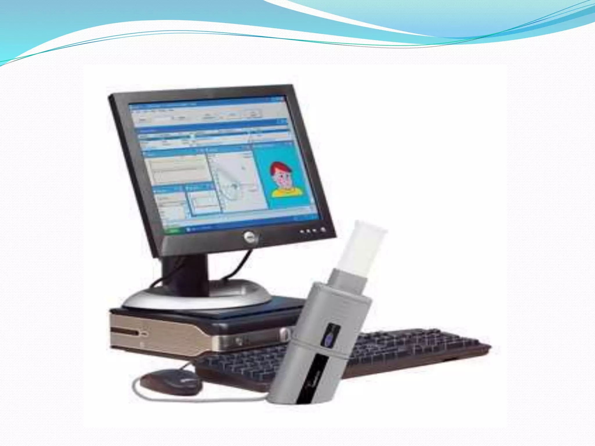
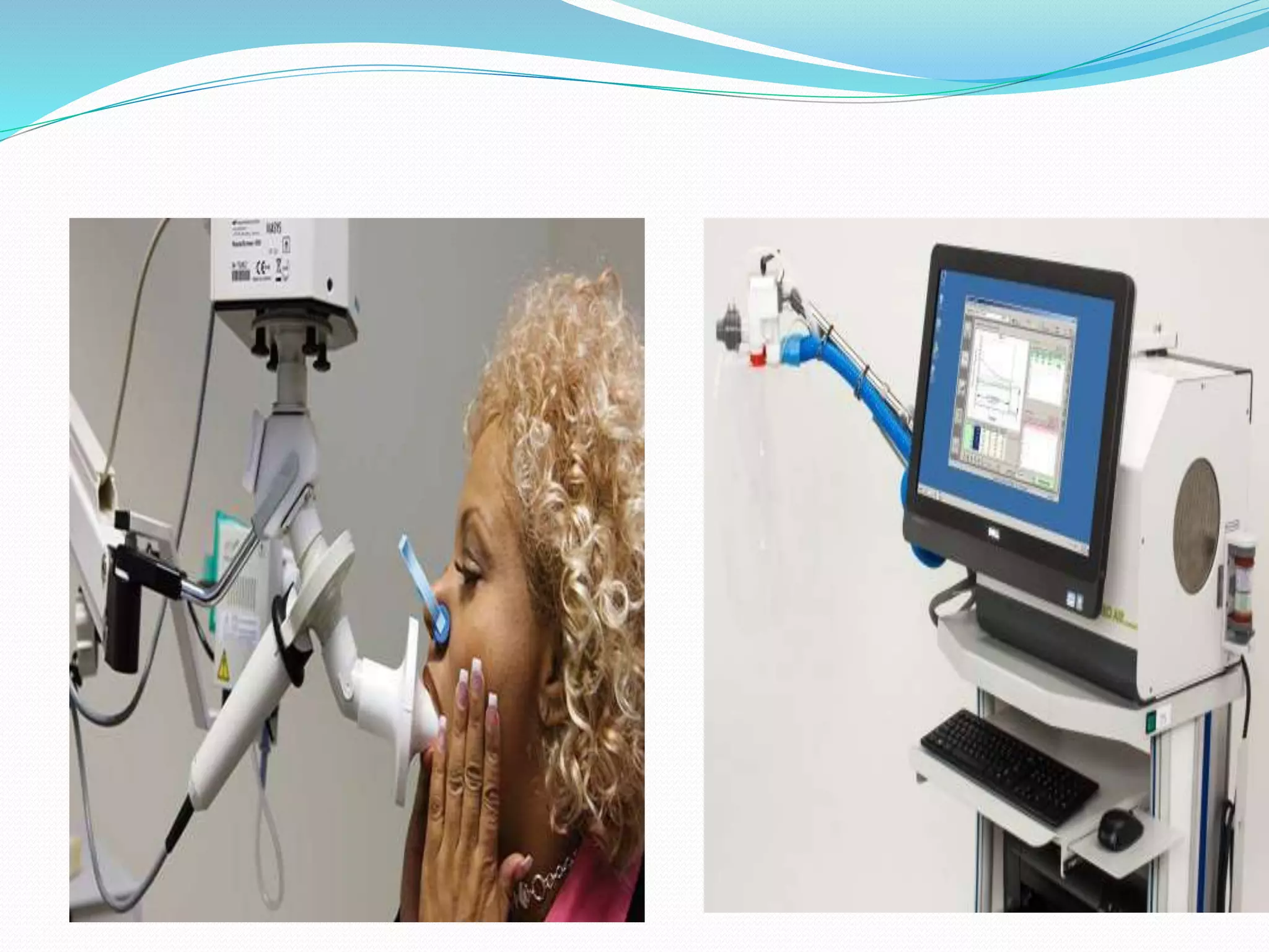
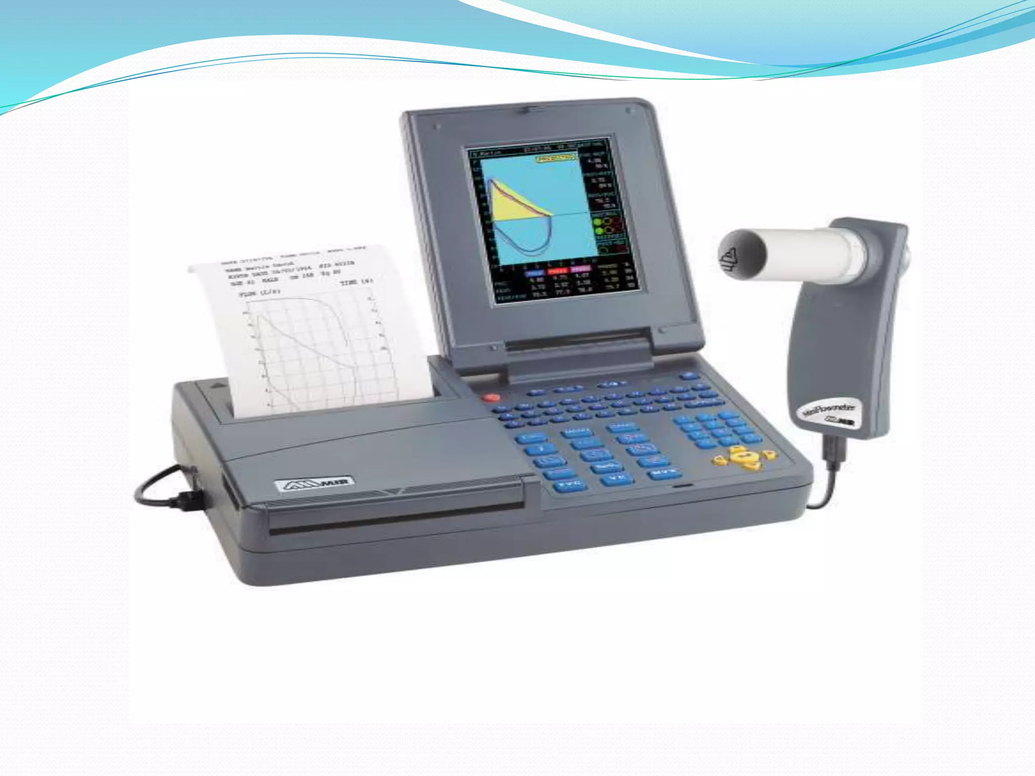
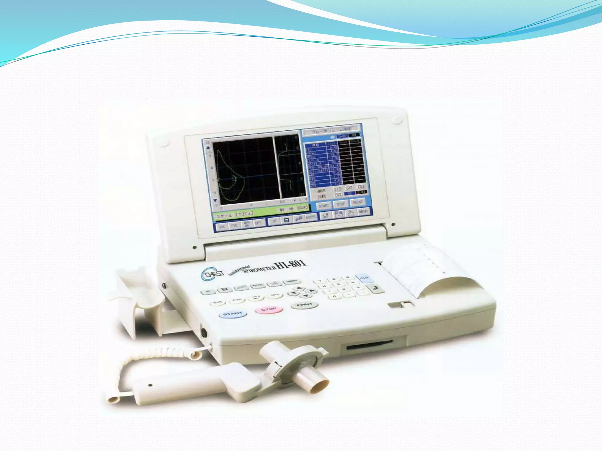
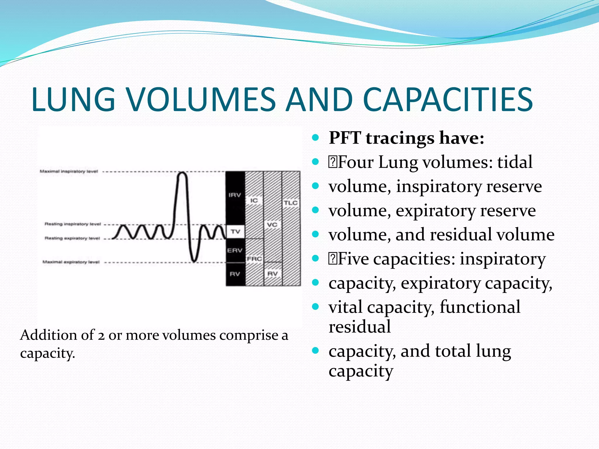
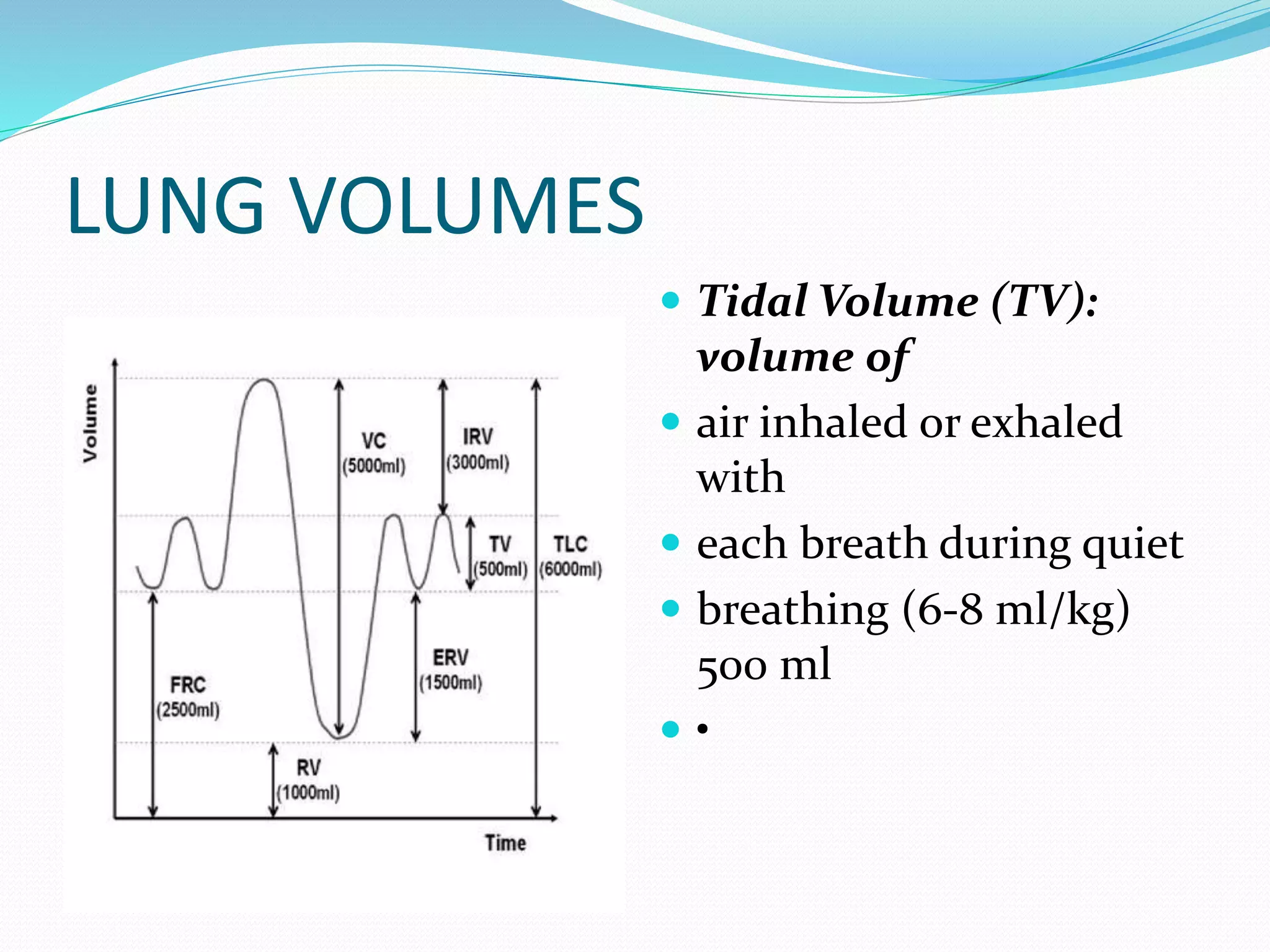
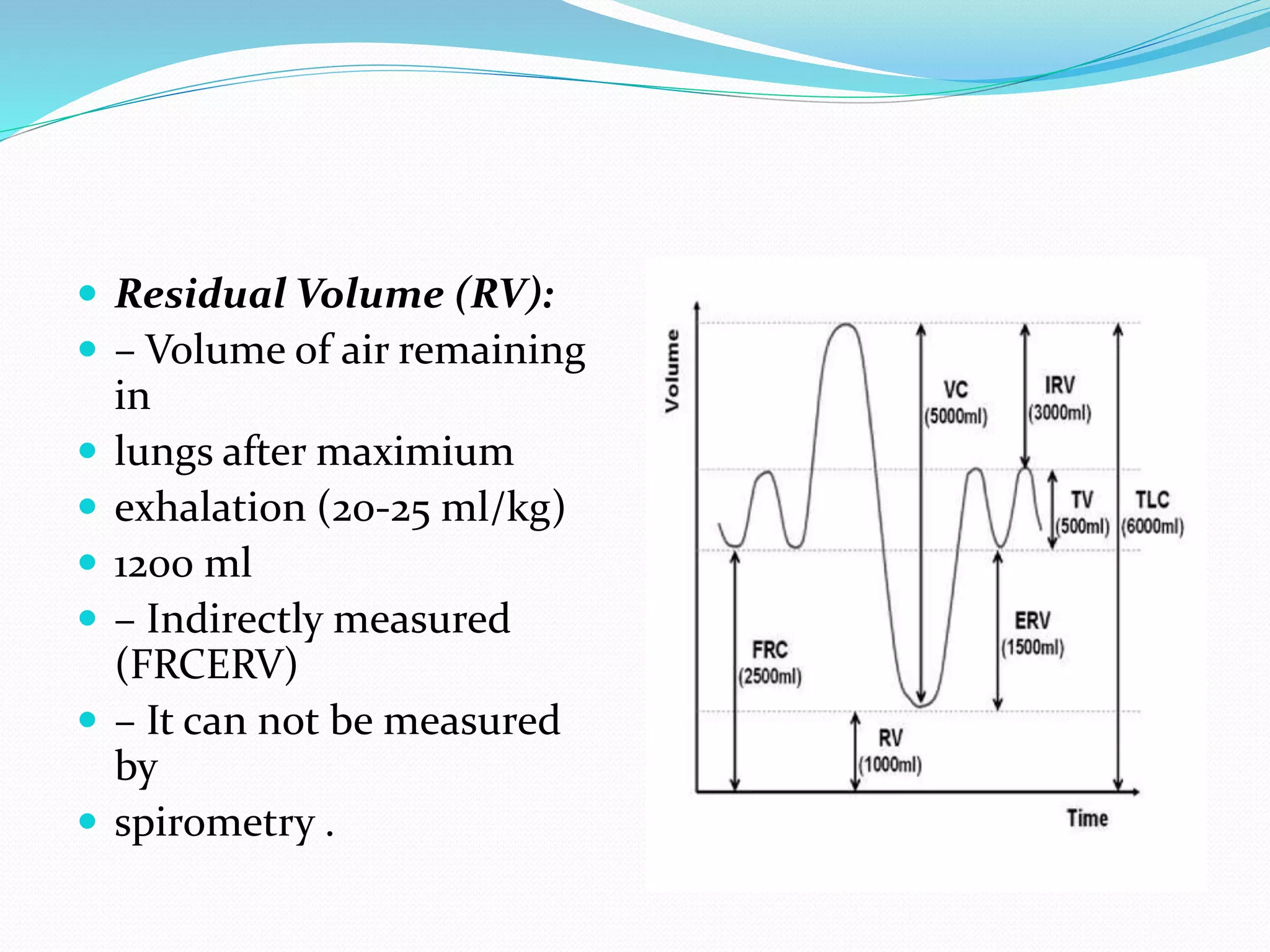
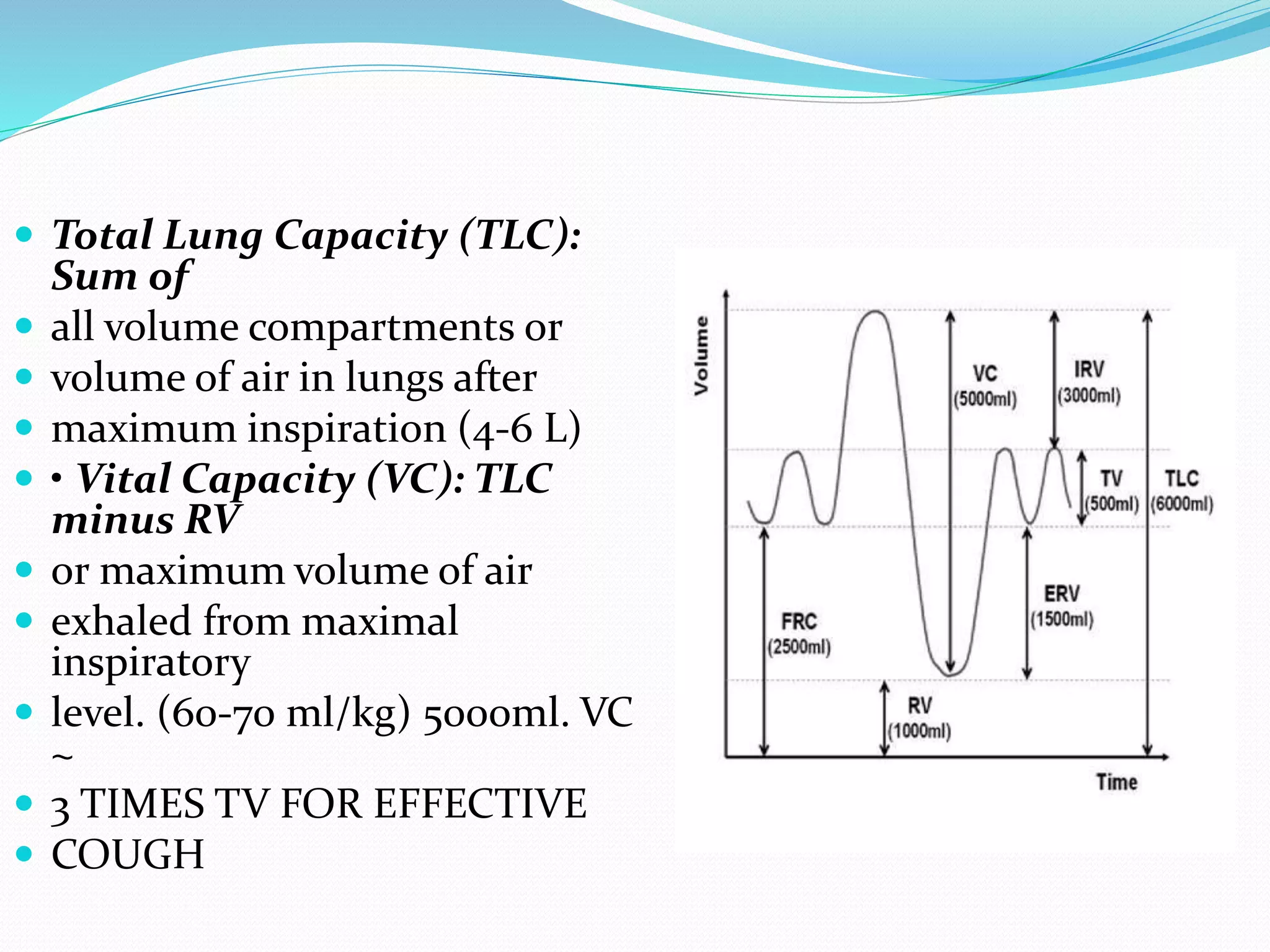
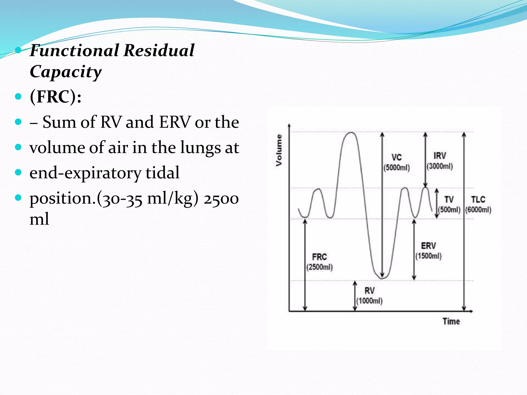
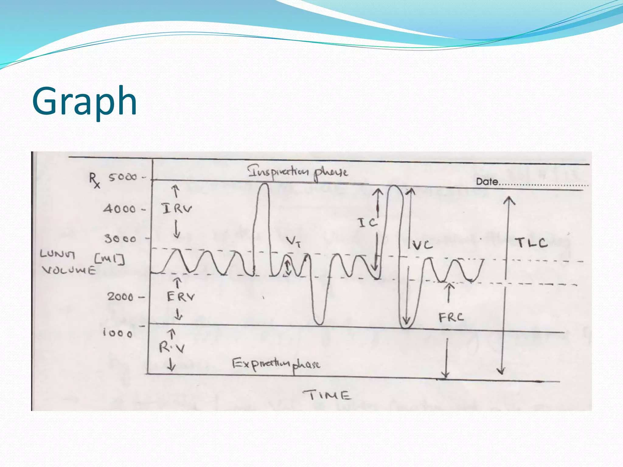
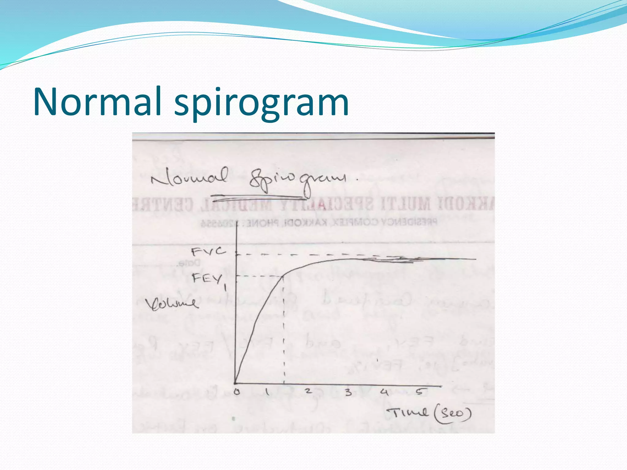
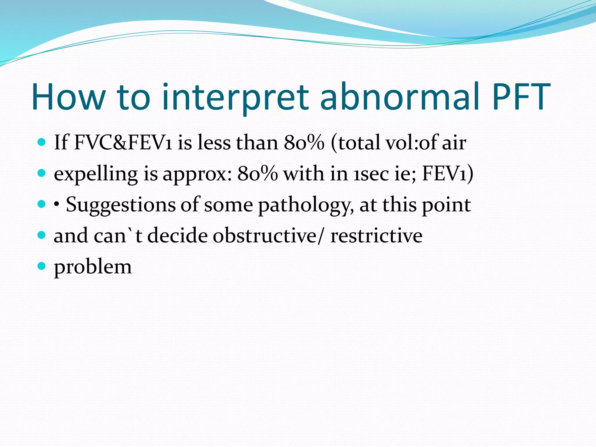
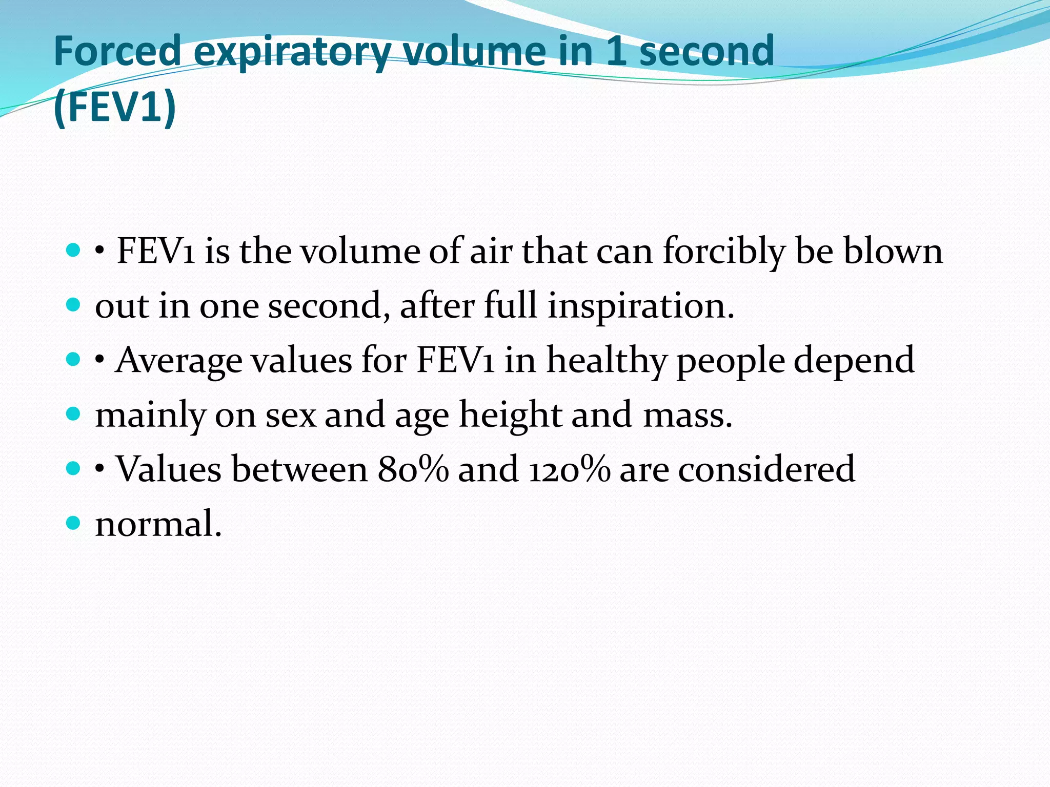
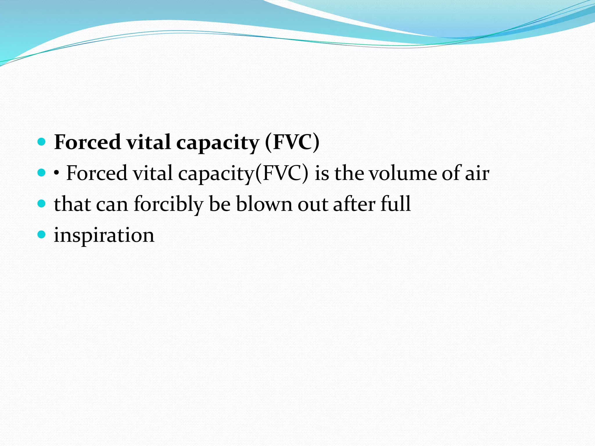
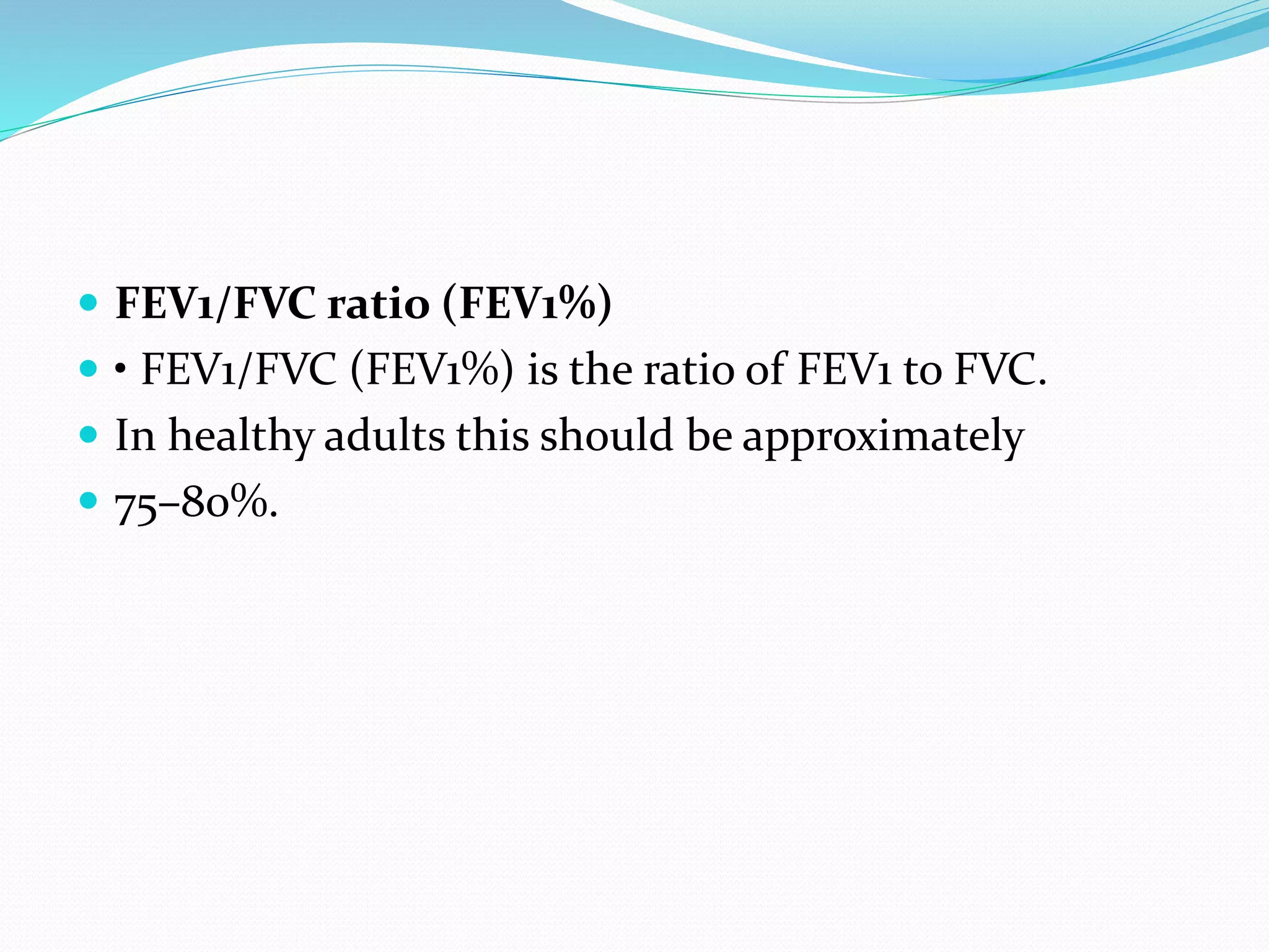
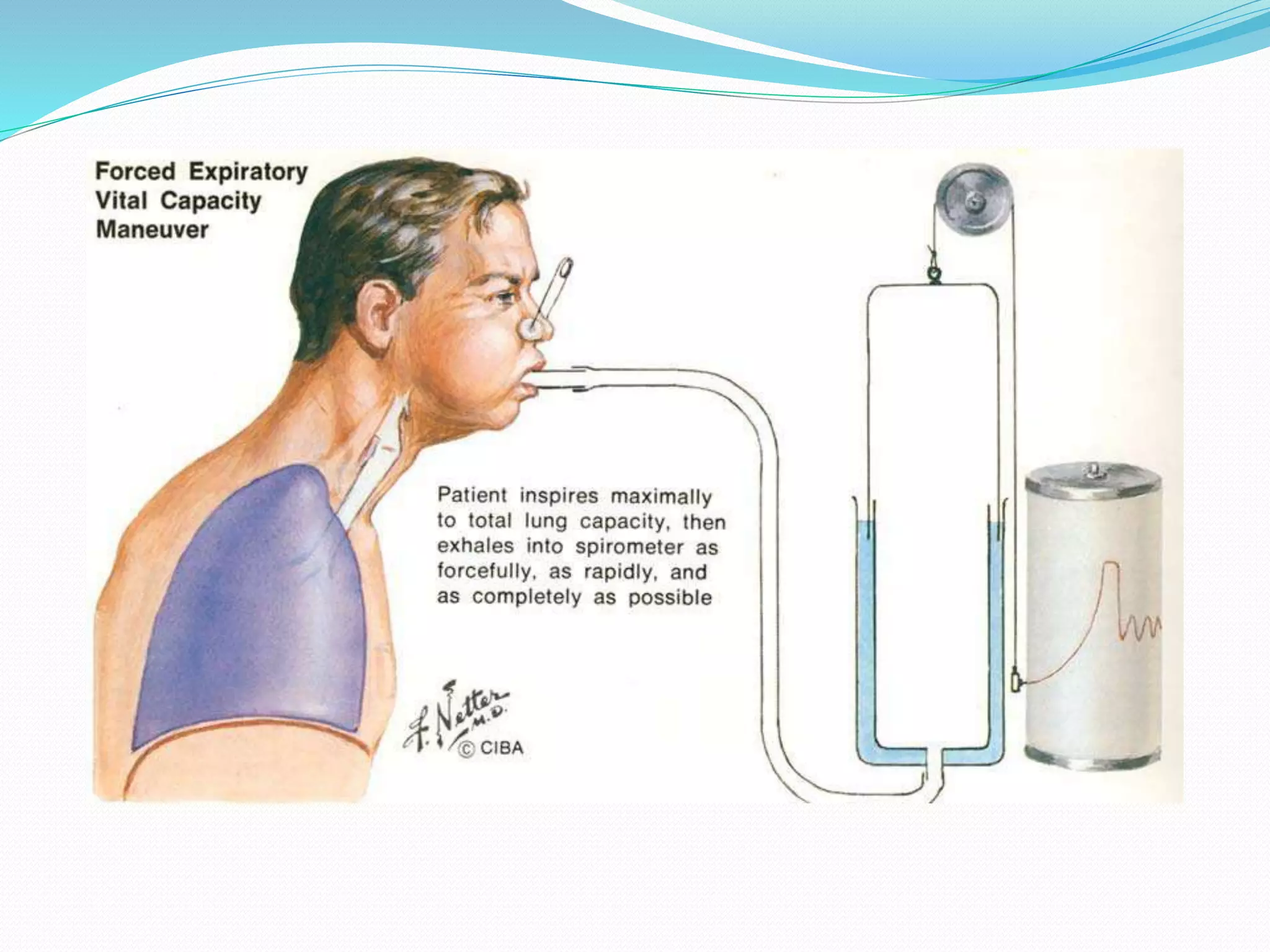
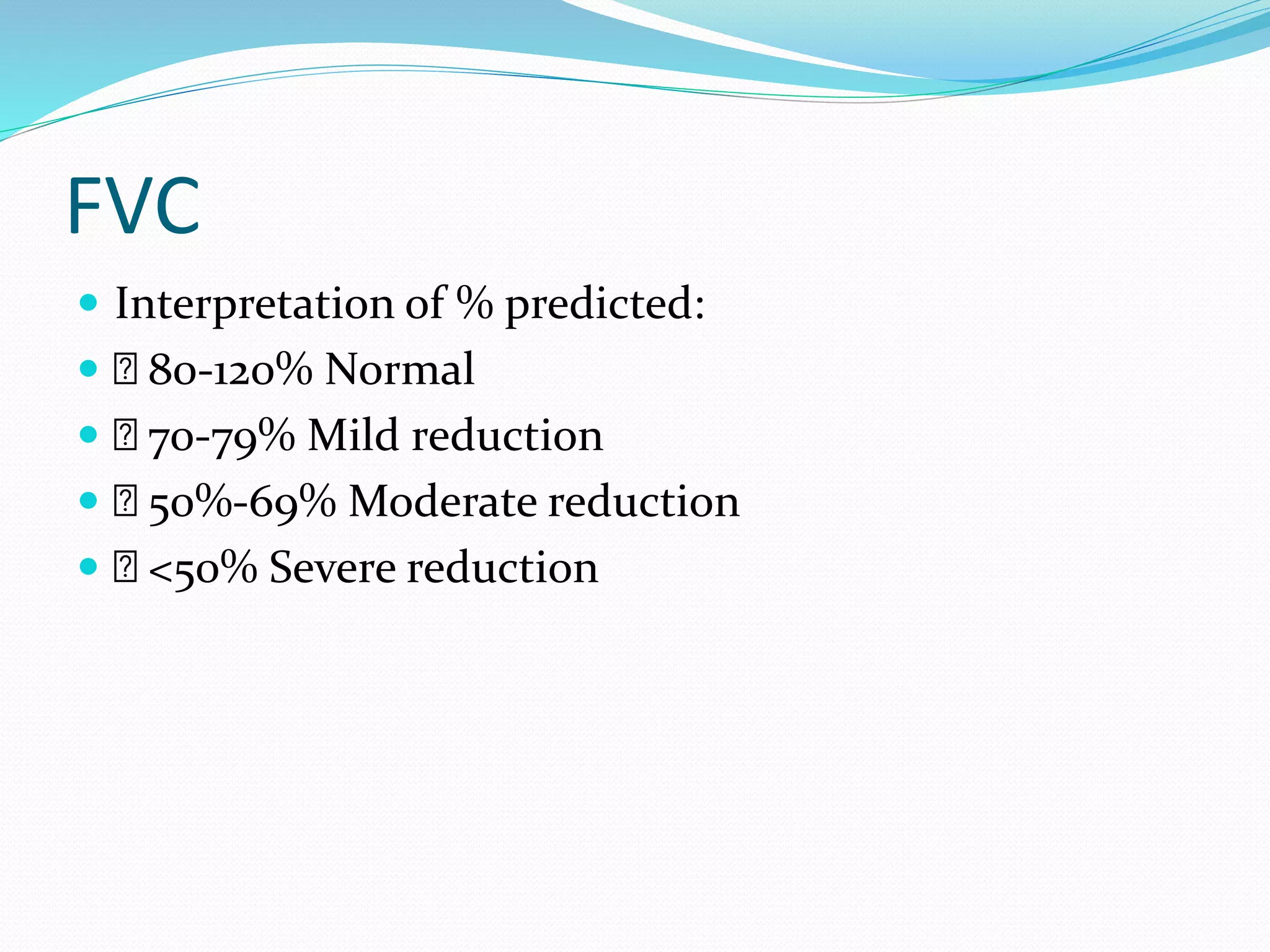
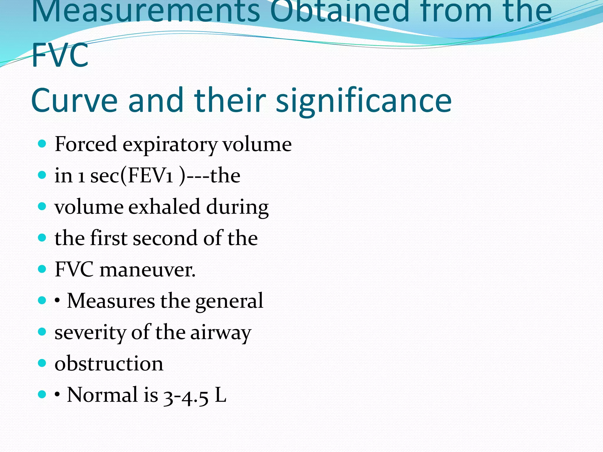
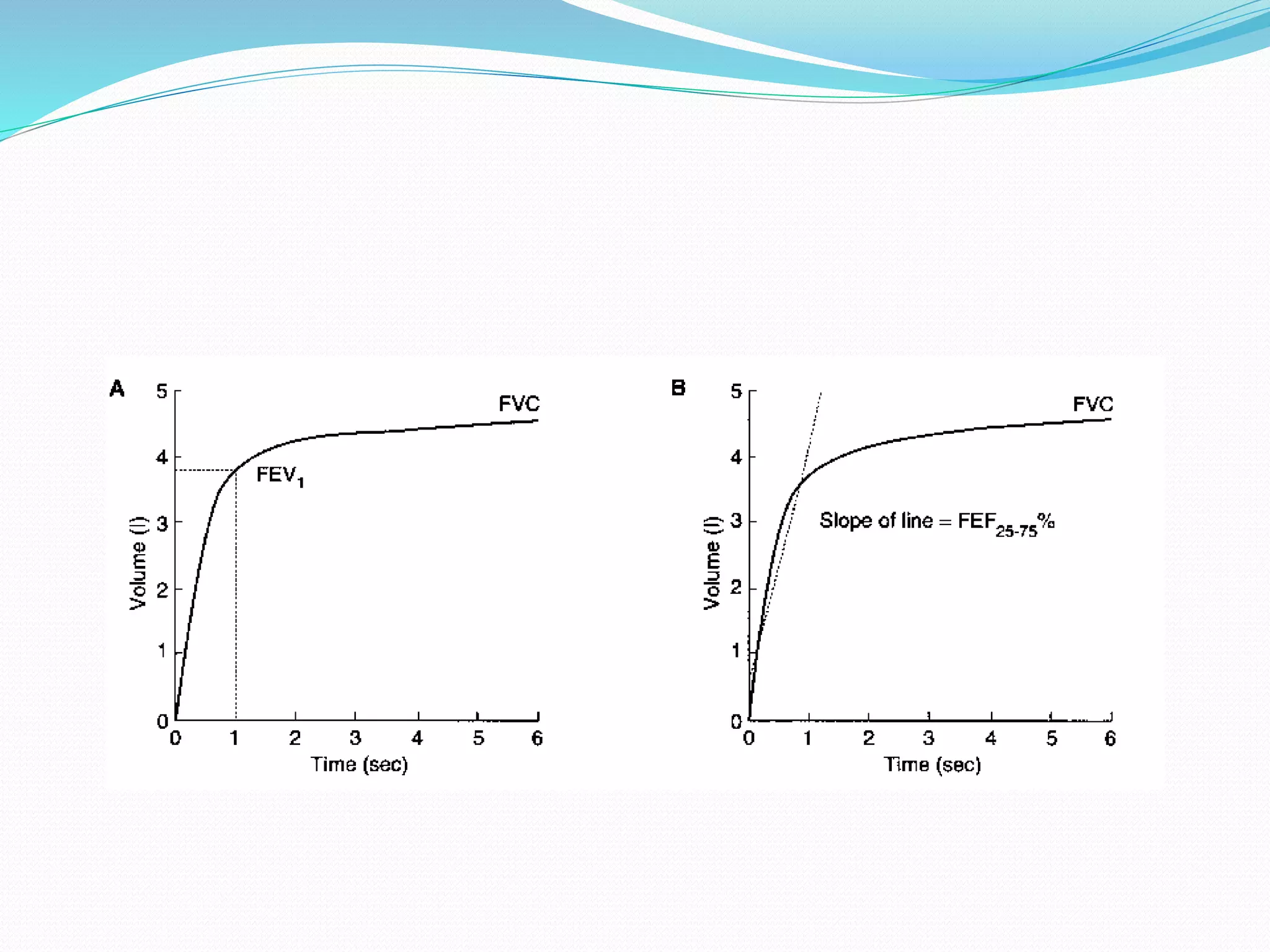
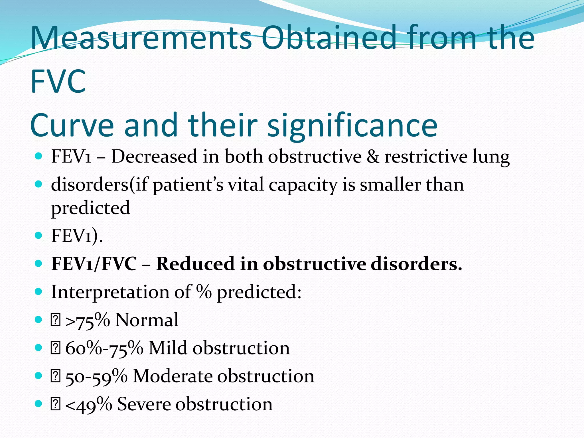
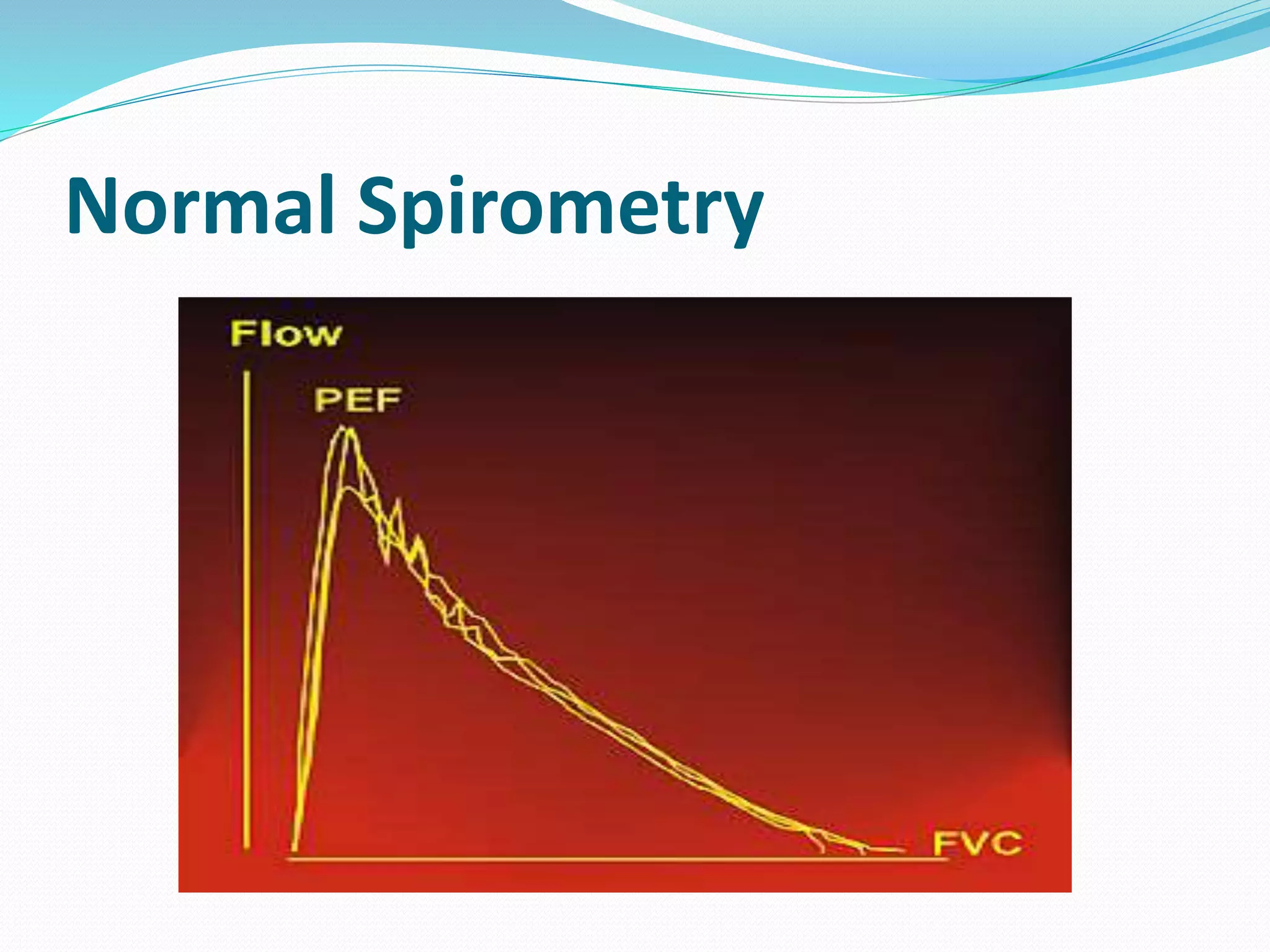
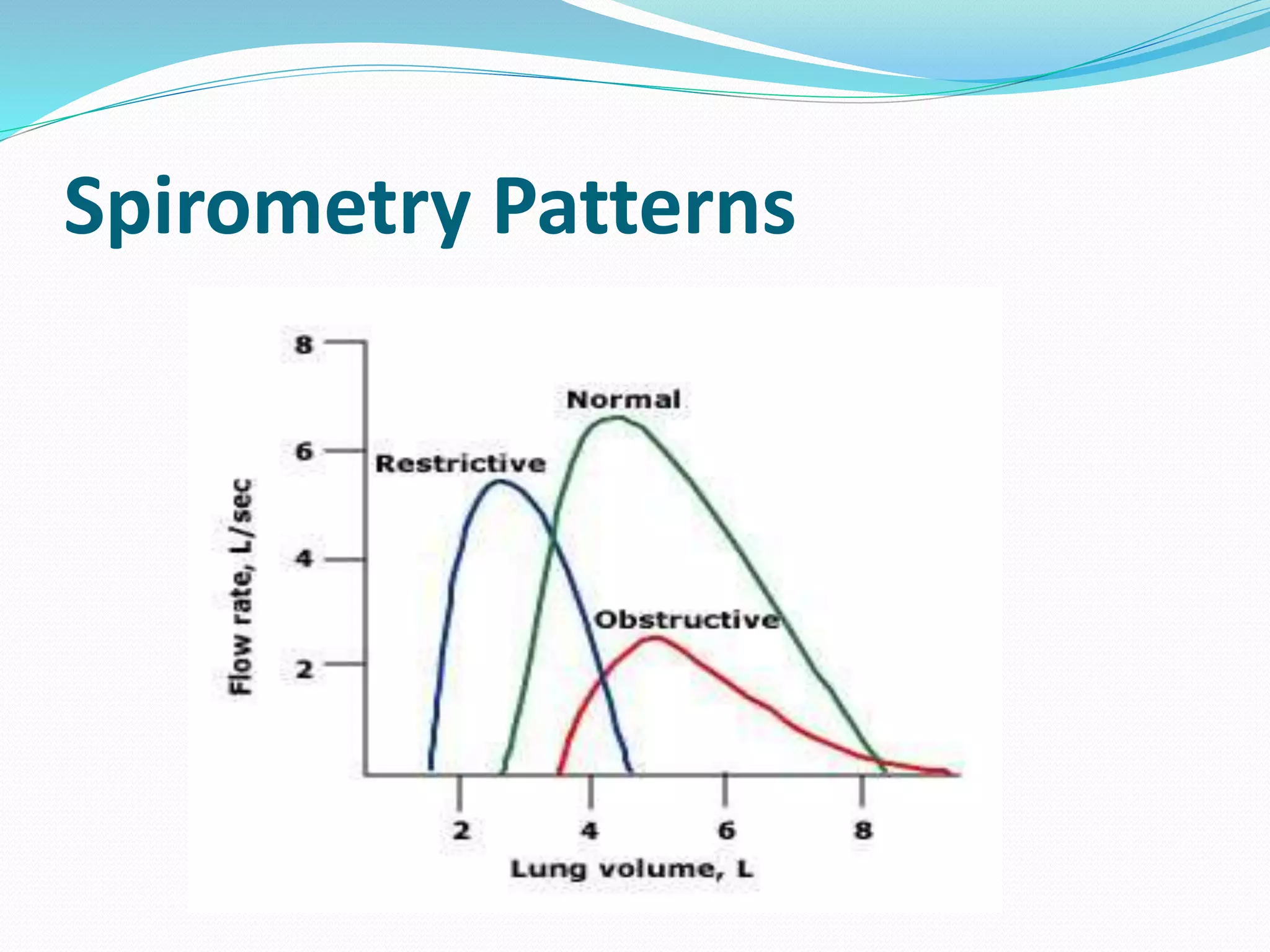
By-Dr.Ranjeet Singha,PT(MPT in Neurology)
HAAD Licensed
Associate Professor,
College of Physiotherapy and Medical Sciences,
Guwahati,Assam.
 Pulmonary function tests is a generic term used to
 indicate a battery of studies or maneuvers that may
 be performed using standardized equipment to
 measure lung function.
 Pulmonary function tests (PFTs) are a group of tests
 that measure how well your lungs works, how well
 the lungs take in and exhale air, and how efficiently
 they transfer oxygen into the blood
 PFT or LFT are useful in assessing the functional
 status of the respiratory system both in
 physiological and pathological condition
 • It is base on the measurement of volumes of air
 breathed in and out in normal breathing and forced
 breathing
 • It is carried out by using a spirometer
 Evaluates one or more aspects of the respiratory
 system
 – Respiratory mechanics
 – Lung parenchymal function/ Gas exchange
 – Cardiopulmonary interaction
INDICATIONS
 DIAGNOSTIC
 Evaluation of signs &
symptoms‐ chronic
cough, exertional
dyspnea
 Screening at risk pts
 Measure the effect of Ds
on pulmonary function
 PROGNOSTIC
 Assess severity
 Follow response to
therapy
 Determine further
treatment goals
 Evaluating degree of
disability
PFTs can help diagnose
 Asthma
 • Chronic bronchitis
 • Respiratory infections
 • Lung fibrosis
 • Bronchiectasis
 • Allergy
 Emphysema
 • Cystic fibrosis
 • Asbestosis which is a
condition caused by
exposure
 to asbestos
 • Sarcoidosis, which is an
inflammation of your
lungs,
 liver, lymph nodes, eyes,
skin, or other tissues
 • Pulmonary tumor
Contraindications
 Recent eye surgery
 • Thoracic , abdominal and cerebral aneurysms
 • Active hemoptysis
 • Pneumothorax
 • Unstable angina/ recent MI within 1 month
STATIC LUNG VOLUMES AND
CAPACITIES
 SPIROMETRY : CORNERSTONE OF ALL PFTs.
 • John hutchinson – invented spirometer.
 • “Spirometry is a medical test that measures the
 volume of air an individual inhales or exhales as a
 function of time.”
 • CAN’T MEASURE – FRC, RV, TLC
Spirometry
 It is an instrument for measuring the air capacity of
 the lungs
 • Measurement of the pattern of air movement in
 and out of the lungs during controlled ventilatory
 maneuvers.
 • spirometre is used to measure the air flow,
 ventilatory regulation, ventilatory mechanics and
 lung volume during a forced expiratory maneuver
 from full inspiration.
 Pft used to evaluate physiological aspect of
 breathing from resp:muscle function to the
 diffusion of gas at the alviolar wall.
 • Pft helps physiotherapist to distinguish between
 obstructive and restrictive lung problem and to
 select appropriate treatment
 • It also measure the effect of the given treatment
SPIROMETRY‐Acceptability Criteria
 Good start of test‐ without any hesitation
 • No coughing / glottic closure
 • No variable flow
 • No early termination(> 6 sec)
 • No air leak
 • Reproducibility‐ The test is without excessive
variability
 The two largest values for FVC and the two largest
values for FEV1 should vary by no more than 0.2L.
Spirometry Interpretation: So what
constitutes normal?
 Normal values vary and depend on:
 I. Height – Directly proportional
 II. Age – Inversely proportional
 III. Gender
 IV. Ethnicity
LUNG VOLUMES AND CAPACITIES
 PFT tracings have:
 Four Lung volumes: tidal
 volume, inspiratory reserve
 volume, expiratory reserve
 volume, and residual volume
 Five capacities: inspiratory
 capacity, expiratory capacity,
 vital capacity, functional
residual
 capacity, and total lung
capacity
Addition of 2 or more volumes comprise a
capacity.
LUNG VOLUMES
 Tidal Volume (TV):
volume of
 air inhaled or exhaled
with
 each breath during quiet
 breathing (6‐8 ml/kg)
500 ml
 •
 Inspiratory Reserve Volume
 (IRV): maximum volume of air
 inhaled from the endinspiratory
 tidal position.3000
 ml
 • Expiratory Reserve Volume
 (ERV): maximum volume of
 air that can be exhaled from
 resting end‐expiratory tidal
 position.1500 ml
 Residual Volume (RV):
 – Volume of air remaining
in
 lungs after maximium
 exhalation (20‐25 ml/kg)
 1200 ml
 – Indirectly measured
(FRCERV)
 – It can not be measured
by
 spirometry .
 Total Lung Capacity (TLC):
Sum of
 all volume compartments or
 volume of air in lungs after
 maximum inspiration (4‐6 L)
 • Vital Capacity (VC): TLC
minus RV
 or maximum volume of air
 exhaled from maximal
inspiratory
 level. (60‐70 ml/kg) 5000ml. VC
~
 3 TIMES TV FOR EFFECTIVE
 COUGH
 • Inspiratory Capacity (IC): Sum of
 IRV and TV or the maximum
 volume of air that can be inhaled
 from the end‐expiratory tidal
 position. (2400‐3800ml).
 • Expiratory Capacity (EC): TV+ ERV
 Functional Residual
Capacity
 (FRC):
 – Sum of RV and ERV or the
 volume of air in the lungs at
 end‐expiratory tidal
 position.(30‐35 ml/kg) 2500
ml
 – Decreases
 1.in supine position(0.5‐1L)
 2.Obese pts
 3.Induction of anesthesia: by 16‐
 20%
Graph
Mechanics of Breathing
 • Inspiration
 Active process
 • Expiration
 Quiet breathing: passive
 Can become active
 Pulmonary Function Tests Evaluates 1 or more
 major aspects of the respiratory system
 • Lung volumes
 • Airway function
 • Gas exchange
 Lung Factors Affecting Spirometry
 • Mechanical properties
 • Resistive elements
Mechanical Properties
 • Compliance
 – Describes the stiffness of the lungs
 – Change in volume over the change in
 pressure
 • Elastic recoil
 – The tendency of the lung to return to it’s
 resting state
 – A lung that is fully stretched has more
 elastic recoil and thus larger/ maximal flows
 of gas
Resistive Properties
 Affected by:
 Lung volume
 Age
 Sex
 Height
 Weight
 Race
 Disease
 Bronchial smooth muscles
PFT procedure
 • Forced expiratory maneuver is the common clinical
 approach
 • Results are found in patients chart/moniter
 • Common spirometric values areFEV1 and FVC
 FEV1/FVC ratio
 • Lung volume and peak expiratory flow rate (PEF or
 PEFR) are measured to differentiate obstructive or
 restrictive problems
 • Forced expiratory flow (FEF)
Procedure
 Sit up straight
 • Get a good seal around the mouth piece
 • Rapid inhale maximally
 • Without any delay blow out as hard as fast as
 possible (blast out)
 • Continue the exhale until the patient can`t
 blow no more
 • Expiration should continue at least 6sec (in
 adult) and 3 sec (children under 10yrs)
 • Repeat at least 3 technically acceptable times
 (without cough, air leak and false start)
Normal spirogram
How to interpret abnormal PFT
 If FVC&FEV1 is less than 80% (total vol:of air
 expelling is approx: 80% with in 1sec ie; FEV1)
 • Suggestions of some pathology, at this point
 and can`t decide obstructive/ restrictive
 problem
Forced expiratory volume in 1 second
(FEV1)
 • FEV1 is the volume of air that can forcibly be blown
 out in one second, after full inspiration.
 • Average values for FEV1 in healthy people depend
 mainly on sex and age height and mass.
 • Values between 80% and 120% are considered
 normal.
 Forced vital capacity (FVC)
 • Forced vital capacity(FVC) is the volume of air
 that can forcibly be blown out after full
 inspiration
 FEV1/FVC ratio (FEV1%)
 • FEV1/FVC (FEV1%) is the ratio of FEV1 to FVC.
 In healthy adults this should be approximately
 75–80%.
 Forced expiratory flow (FEF)
 • Forced expiratory flow (FEF) is the flow (or speed)
 of air coming out of the lung during the middle
 portion of a forced expiration.
 • generally defined by fraction, The usual intervals
 are 25%, 50% and 75% (FEF25, FEF50 and FEF75)
Measuring RV, FRC
 It can be measured by
 – nitrogen washout technique
 – Helium dilution method
 – Body plethysmography
N2 Washout Technique
 The patient breathes 100% oxygen, and all the
 nitrogen in the lungs is washed out.
 • The exhaled volume and the nitrogen
 concentration in that volume are measured.
 • The difference in nitrogen volume at the initial
 concentration and at the final exhaled
 concentration allows a calculation of
 intrathoracic volume, usually FRC.
Helium Dilution technique
 Pt breathes in and out from a reservoir with known
 volume of gas containing trace of helium.
 • Helium gets diluted by gas previously present in
lungs.
Body Plethysmography
 • Plethysmography (derived from greek word meaning
 enlargement).
 • Based on principle of BOYLE’S LAW(P*V=k)
 • Priniciple advantage over other two method is it
 quantifies non‐ communicating gas volumes
 A patient is placed in a sitting
 position in a closed body box
 with a known volume
 • The patient pants with an
 open glottis against a closed
 shutter to produce changes
 in the box pressure
 proportionate to the volume
 of air in the chest.
 • As measurements done at
 end of expiration, it yields
 FRC
FORCED SPIROMETRY/TIMED
EXPIRATORY
SPIROGRAM
 Includes measuring:
 • pulmonary mechanics – to
 assess the ability of the lung to
 move large vol of air quickly
 through the airways to identify
 airway obstruction
 • FVC
 •FEV1
 •Several FEF values
 •Forced inspiratory rates(FIF’s)
 •MVV
FORCED VITAL CAPACITY
 • The FVC is the maximum volume of air that can be
 breathed out as forcefully and rapidly as possible
 following a maximum inspiration.
 • Characterized by full inspiration to TLC followed by
 abrupt onset of expiration to RV
 • Indirectly reflects flow resistance property of
 airways.
FVC
 Interpretation of % predicted:
 80-120% Normal
 70-79% Mild reduction
 50%-69% Moderate reduction
 <50% Severe reduction
Measurements Obtained from the
FVC
Curve and their significance
 Forced expiratory volume
 in 1 sec(FEV1 )‐‐‐the
 volume exhaled during
 the first second of the
 FVC maneuver.
 • Measures the general
 severity of the airway
 obstruction
 • Normal is 3‐4.5 L
Measurements Obtained from the
FVC
Curve and their significance
 FEV1 – Decreased in both obstructive & restrictive lung
 disorders(if patient’s vital capacity is smaller than
predicted
 FEV1).
 FEV1/FVC – Reduced in obstructive disorders.
 Interpretation of % predicted:
 >75% Normal
 60%‐75% Mild obstruction
 50‐59% Moderate obstruction
 <49% Severe obstruction
Normal Spirometry
Spirometry Patterns

Pulmonary function tests

  • 1.
    By-Dr.Ranjeet Singha,PT(MPT inNeurology) HAAD Licensed Associate Professor, College of Physiotherapy and Medical Sciences, Guwahati,Assam.
  • 2.
     Pulmonary functiontests is a generic term used to  indicate a battery of studies or maneuvers that may  be performed using standardized equipment to  measure lung function.  Pulmonary function tests (PFTs) are a group of tests  that measure how well your lungs works, how well  the lungs take in and exhale air, and how efficiently  they transfer oxygen into the blood
  • 3.
     PFT orLFT are useful in assessing the functional  status of the respiratory system both in  physiological and pathological condition  • It is base on the measurement of volumes of air  breathed in and out in normal breathing and forced  breathing  • It is carried out by using a spirometer
  • 6.
     Evaluates oneor more aspects of the respiratory  system  – Respiratory mechanics  – Lung parenchymal function/ Gas exchange  – Cardiopulmonary interaction
  • 7.
    INDICATIONS  DIAGNOSTIC  Evaluationof signs & symptoms‐ chronic cough, exertional dyspnea  Screening at risk pts  Measure the effect of Ds on pulmonary function  PROGNOSTIC  Assess severity  Follow response to therapy  Determine further treatment goals  Evaluating degree of disability
  • 8.
    PFTs can helpdiagnose  Asthma  • Chronic bronchitis  • Respiratory infections  • Lung fibrosis  • Bronchiectasis  • Allergy  Emphysema  • Cystic fibrosis  • Asbestosis which is a condition caused by exposure  to asbestos  • Sarcoidosis, which is an inflammation of your lungs,  liver, lymph nodes, eyes, skin, or other tissues  • Pulmonary tumor
  • 9.
    Contraindications  Recent eyesurgery  • Thoracic , abdominal and cerebral aneurysms  • Active hemoptysis  • Pneumothorax  • Unstable angina/ recent MI within 1 month
  • 10.
    STATIC LUNG VOLUMESAND CAPACITIES  SPIROMETRY : CORNERSTONE OF ALL PFTs.  • John hutchinson – invented spirometer.  • “Spirometry is a medical test that measures the  volume of air an individual inhales or exhales as a  function of time.”  • CAN’T MEASURE – FRC, RV, TLC
  • 11.
    Spirometry  It isan instrument for measuring the air capacity of  the lungs  • Measurement of the pattern of air movement in  and out of the lungs during controlled ventilatory  maneuvers.  • spirometre is used to measure the air flow,  ventilatory regulation, ventilatory mechanics and  lung volume during a forced expiratory maneuver  from full inspiration.
  • 14.
     Pft usedto evaluate physiological aspect of  breathing from resp:muscle function to the  diffusion of gas at the alviolar wall.  • Pft helps physiotherapist to distinguish between  obstructive and restrictive lung problem and to  select appropriate treatment  • It also measure the effect of the given treatment
  • 15.
    SPIROMETRY‐Acceptability Criteria  Goodstart of test‐ without any hesitation  • No coughing / glottic closure  • No variable flow  • No early termination(> 6 sec)  • No air leak  • Reproducibility‐ The test is without excessive variability  The two largest values for FVC and the two largest values for FEV1 should vary by no more than 0.2L.
  • 16.
    Spirometry Interpretation: Sowhat constitutes normal?  Normal values vary and depend on:  I. Height – Directly proportional  II. Age – Inversely proportional  III. Gender  IV. Ethnicity
  • 17.
    LUNG VOLUMES ANDCAPACITIES  PFT tracings have:  Four Lung volumes: tidal  volume, inspiratory reserve  volume, expiratory reserve  volume, and residual volume  Five capacities: inspiratory  capacity, expiratory capacity,  vital capacity, functional residual  capacity, and total lung capacity Addition of 2 or more volumes comprise a capacity.
  • 18.
    LUNG VOLUMES  TidalVolume (TV): volume of  air inhaled or exhaled with  each breath during quiet  breathing (6‐8 ml/kg) 500 ml  •
  • 19.
     Inspiratory ReserveVolume  (IRV): maximum volume of air  inhaled from the endinspiratory  tidal position.3000  ml  • Expiratory Reserve Volume  (ERV): maximum volume of  air that can be exhaled from  resting end‐expiratory tidal  position.1500 ml
  • 20.
     Residual Volume(RV):  – Volume of air remaining in  lungs after maximium  exhalation (20‐25 ml/kg)  1200 ml  – Indirectly measured (FRCERV)  – It can not be measured by  spirometry .
  • 21.
     Total LungCapacity (TLC): Sum of  all volume compartments or  volume of air in lungs after  maximum inspiration (4‐6 L)  • Vital Capacity (VC): TLC minus RV  or maximum volume of air  exhaled from maximal inspiratory  level. (60‐70 ml/kg) 5000ml. VC ~  3 TIMES TV FOR EFFECTIVE  COUGH
  • 22.
     • InspiratoryCapacity (IC): Sum of  IRV and TV or the maximum  volume of air that can be inhaled  from the end‐expiratory tidal  position. (2400‐3800ml).  • Expiratory Capacity (EC): TV+ ERV
  • 23.
     Functional Residual Capacity (FRC):  – Sum of RV and ERV or the  volume of air in the lungs at  end‐expiratory tidal  position.(30‐35 ml/kg) 2500 ml
  • 24.
     – Decreases 1.in supine position(0.5‐1L)  2.Obese pts  3.Induction of anesthesia: by 16‐  20%
  • 25.
  • 26.
    Mechanics of Breathing • Inspiration  Active process  • Expiration  Quiet breathing: passive  Can become active  Pulmonary Function Tests Evaluates 1 or more  major aspects of the respiratory system  • Lung volumes  • Airway function  • Gas exchange
  • 27.
     Lung FactorsAffecting Spirometry  • Mechanical properties  • Resistive elements
  • 28.
    Mechanical Properties  •Compliance  – Describes the stiffness of the lungs  – Change in volume over the change in  pressure  • Elastic recoil  – The tendency of the lung to return to it’s  resting state  – A lung that is fully stretched has more  elastic recoil and thus larger/ maximal flows  of gas
  • 29.
    Resistive Properties  Affectedby:  Lung volume  Age  Sex  Height  Weight  Race  Disease  Bronchial smooth muscles
  • 30.
    PFT procedure  •Forced expiratory maneuver is the common clinical  approach  • Results are found in patients chart/moniter  • Common spirometric values areFEV1 and FVC  FEV1/FVC ratio  • Lung volume and peak expiratory flow rate (PEF or  PEFR) are measured to differentiate obstructive or  restrictive problems  • Forced expiratory flow (FEF)
  • 31.
    Procedure  Sit upstraight  • Get a good seal around the mouth piece  • Rapid inhale maximally  • Without any delay blow out as hard as fast as  possible (blast out)  • Continue the exhale until the patient can`t  blow no more  • Expiration should continue at least 6sec (in  adult) and 3 sec (children under 10yrs)  • Repeat at least 3 technically acceptable times  (without cough, air leak and false start)
  • 32.
  • 33.
    How to interpretabnormal PFT  If FVC&FEV1 is less than 80% (total vol:of air  expelling is approx: 80% with in 1sec ie; FEV1)  • Suggestions of some pathology, at this point  and can`t decide obstructive/ restrictive  problem
  • 34.
    Forced expiratory volumein 1 second (FEV1)  • FEV1 is the volume of air that can forcibly be blown  out in one second, after full inspiration.  • Average values for FEV1 in healthy people depend  mainly on sex and age height and mass.  • Values between 80% and 120% are considered  normal.
  • 35.
     Forced vitalcapacity (FVC)  • Forced vital capacity(FVC) is the volume of air  that can forcibly be blown out after full  inspiration
  • 36.
     FEV1/FVC ratio(FEV1%)  • FEV1/FVC (FEV1%) is the ratio of FEV1 to FVC.  In healthy adults this should be approximately  75–80%.
  • 37.
     Forced expiratoryflow (FEF)  • Forced expiratory flow (FEF) is the flow (or speed)  of air coming out of the lung during the middle  portion of a forced expiration.  • generally defined by fraction, The usual intervals  are 25%, 50% and 75% (FEF25, FEF50 and FEF75)
  • 38.
    Measuring RV, FRC It can be measured by  – nitrogen washout technique  – Helium dilution method  – Body plethysmography
  • 39.
    N2 Washout Technique The patient breathes 100% oxygen, and all the  nitrogen in the lungs is washed out.  • The exhaled volume and the nitrogen  concentration in that volume are measured.  • The difference in nitrogen volume at the initial  concentration and at the final exhaled  concentration allows a calculation of  intrathoracic volume, usually FRC.
  • 40.
    Helium Dilution technique Pt breathes in and out from a reservoir with known  volume of gas containing trace of helium.  • Helium gets diluted by gas previously present in lungs.
  • 41.
    Body Plethysmography  •Plethysmography (derived from greek word meaning  enlargement).  • Based on principle of BOYLE’S LAW(P*V=k)  • Priniciple advantage over other two method is it  quantifies non‐ communicating gas volumes
  • 42.
     A patientis placed in a sitting  position in a closed body box  with a known volume  • The patient pants with an  open glottis against a closed  shutter to produce changes  in the box pressure  proportionate to the volume  of air in the chest.  • As measurements done at  end of expiration, it yields  FRC
  • 43.
    FORCED SPIROMETRY/TIMED EXPIRATORY SPIROGRAM  Includesmeasuring:  • pulmonary mechanics – to  assess the ability of the lung to  move large vol of air quickly  through the airways to identify  airway obstruction  • FVC  •FEV1  •Several FEF values  •Forced inspiratory rates(FIF’s)  •MVV
  • 44.
    FORCED VITAL CAPACITY • The FVC is the maximum volume of air that can be  breathed out as forcefully and rapidly as possible  following a maximum inspiration.  • Characterized by full inspiration to TLC followed by  abrupt onset of expiration to RV  • Indirectly reflects flow resistance property of  airways.
  • 46.
    FVC  Interpretation of% predicted:  80-120% Normal  70-79% Mild reduction  50%-69% Moderate reduction  <50% Severe reduction
  • 47.
    Measurements Obtained fromthe FVC Curve and their significance  Forced expiratory volume  in 1 sec(FEV1 )‐‐‐the  volume exhaled during  the first second of the  FVC maneuver.  • Measures the general  severity of the airway  obstruction  • Normal is 3‐4.5 L
  • 49.
    Measurements Obtained fromthe FVC Curve and their significance  FEV1 – Decreased in both obstructive & restrictive lung  disorders(if patient’s vital capacity is smaller than predicted  FEV1).  FEV1/FVC – Reduced in obstructive disorders.  Interpretation of % predicted:  >75% Normal  60%‐75% Mild obstruction  50‐59% Moderate obstruction  <49% Severe obstruction
  • 50.
  • 51.