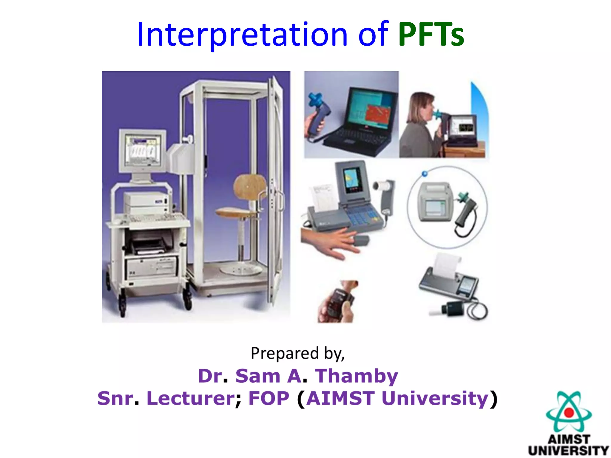
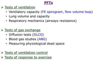
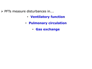
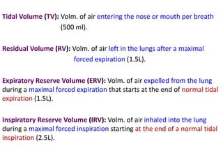
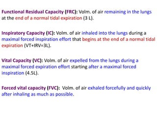
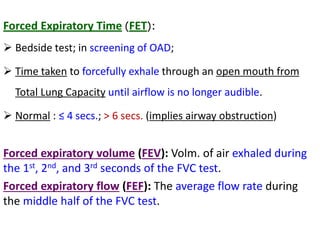
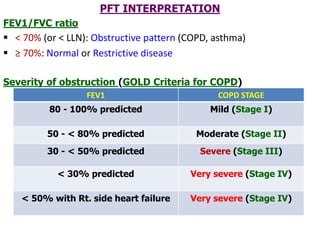
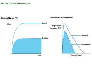
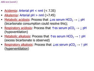
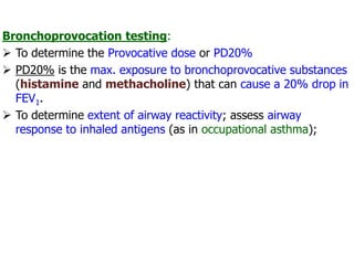
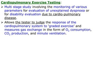
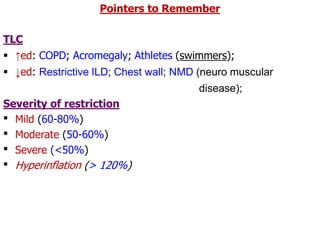
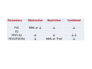
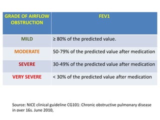
PFTs measure lung function through tests of ventilation, gas exchange, ventilation control, and exercise response. Key measurements include FEV1, FVC, FEV1/FVC ratio, DLCO, and arterial blood gases. PFT results are interpreted to identify obstructive, restrictive, or combined patterns and determine the severity of lung abnormalities.
Dynamic Central Airway Obstruction: Tracheomalacia, Tracheobronchomalacia, An...Bassel Ericsoussi, MD
Dynamic Central Airway Obstruction: Tracheomalacia, Tracheobronchomalacia, And Excessive Dynamic Airway Collapse: Classification, Diagnosis, and Treatment
What are the pulmonary function tests used?
What are the indications?
What are the contraindications?
How to perform each and prepare patients?
How to interpret and reach a diagnosis?
How to clean and calibrate devices?
Pulmonary function testing is the process of having the patient perform specific inspiratory and expiratory maneuvers while breathing in and out of tubing attached to the equipment that measure a variety of variables
Pulmonary function tests (PFT) are series of tests that measure lung function and aid in the management of patients with respiratory disease.
They are performed using standardized equipment and can be used for diagnosis, prognostication, management and follow-up of patients with pulmonary pathology.
Although PFT may not identify the exact pathology, it broadly classifies respiratory disorders as either obstructive or restrictive. In this session , the role of PFT in the measurement of lung mechanics and diagnosis of various diseases will be discussed in detail.
PULMONARY FUNCTION TESTS - LAB DATA INTERPRETATIONLincyAsha
PULMONARY FUNCTION TESTS
LAB DATA INTERPRETATION
CLINICAL PHARMACY PRACTICE
M.PHARMACY
PHARMACY PRACTICE
1ST YEAR
Pulmonary function tests are a series of tests performed to examine a patient’s respiratory system and identify the severity of pulmonary impairment.
These tests are performed to measure a patient’s lung volume, capacity, flow rate and gas exchange.
This allows medical professionals to obtain an accurate diagnosis and determine the best course of medical intervention for the patient.
In general there are two types of lung disorders that these tests can be used to assess
Obstructive lung diseases
Restrictive lung diseases
1.OBSTRUCTIVE LUNG DISEASES
It include conditions that make it difficult to exhale air out of the lungs
This results in shortness of breath that occurs from narrowing and constriction of the airways and causes the patient to have decreased flow rates. Eg. COPD, Asthma
2.RESTRICTIVE LUNG DISEASES
It include conditions that make it difficult to fully fill the lungs with air during inhalation.
When the lungs aren’t fully able to expand it causes the patient to have decreased lung volumes. Eg. Pulmonary fibrosis, interstitial lung disease
Pulmonary function tests would be indicated for the following:
On healthy patients as part of a routine physical exam
Evaluate signs and symptoms of lung disease
Diagnosis of certain medical conditions
Measure current stage of disease and evaluate its progress
Assess how a patient is responding to different treatments
Determine patient’s condition before surgery to assess the risk of respiratory complications
Screen people who are at risk of pulmonary disease
Determine how much a patient’s airways have narrowed due to disorders
In certain types of work environments to assess the health of employees.
Additionally PFTs may be indicated for the following
Chronic lung conditions
Restrictive airway problems
Asthma
COPD
Shortness of breath
Impairment or disability
Early morning wheezing
Chest muscle weakness
Lung cancer
Respiratory infections
STATIC LUNG VOLUMES
Lung volume is the amount of air breathed by an individual under a specific condition.
1.Tidal Volume (TV)
It is the volume of air inspired or expired during normal breathing at rest.
2.Inspiratory Reserve Volume (IRV)
It is the volume of air inspired with maximum effort over and above the normal tidal volume.
3.Expiratory Reserve Volume (ERV)
It is the volume of air expired forcefully after a normal respiration.
4.Residual Volume (RV)
It is the volume of air remaining in the lungs after a forceful expiration
STATIC LUNG CAPACITIES
1.Inspiratory capacity (IC)
It is the amount of air a person can inspire forcefully after a normal respiration.
IC = TV+IRV
2.Functional Residual Capacity (FRC)
It is the amount of air that remains in the lungs at the end of normal respiration.
FRC = ERV+RV
3.Vital Capacity (VC)
It is the maximum volume of air exhaled forcefully from the lungs after a maximum inspiration.
4.Total Lung Capacity
a detailed study on pulmonary function testmartinshaji
this study details about all the aspects of pulmonary function test, lung volumes& capacities , tests such as spirometry , carbon monoxide diffusion capacity, chest x ray, body plethesmography , nitrogen washout etc
please comment
thank u
HOT NEW PRODUCT! BIG SALES FAST SHIPPING NOW FROM CHINA!! EU KU DB BK substit...GL Anaacs
Contact us if you are interested:
Email / Skype : kefaya1771@gmail.com
Threema: PXHY5PDH
New BATCH Ku !!! MUCH IN DEMAND FAST SALE EVERY BATCH HAPPY GOOD EFFECT BIG BATCH !
Contact me on Threema or skype to start big business!!
Hot-sale products:
NEW HOT EUTYLONE WHITE CRYSTAL!!
5cl-adba precursor (semi finished )
5cl-adba raw materials
ADBB precursor (semi finished )
ADBB raw materials
APVP powder
5fadb/4f-adb
Jwh018 / Jwh210
Eutylone crystal
Protonitazene (hydrochloride) CAS: 119276-01-6
Flubrotizolam CAS: 57801-95-3
Metonitazene CAS: 14680-51-4
Payment terms: Western Union,MoneyGram,Bitcoin or USDT.
Deliver Time: Usually 7-15days
Shipping method: FedEx, TNT, DHL,UPS etc.Our deliveries are 100% safe, fast, reliable and discreet.
Samples will be sent for your evaluation!If you are interested in, please contact me, let's talk details.
We specializes in exporting high quality Research chemical, medical intermediate, Pharmaceutical chemicals and so on. Products are exported to USA, Canada, France, Korea, Japan,Russia, Southeast Asia and other countries.
MANAGEMENT OF ATRIOVENTRICULAR CONDUCTION BLOCK.pdfJim Jacob Roy
Cardiac conduction defects can occur due to various causes.
Atrioventricular conduction blocks ( AV blocks ) are classified into 3 types.
This document describes the acute management of AV block.
micro teaching on communication m.sc nursing.pdfAnurag Sharma
Microteaching is a unique model of practice teaching. It is a viable instrument for the. desired change in the teaching behavior or the behavior potential which, in specified types of real. classroom situations, tends to facilitate the achievement of specified types of objectives.
These simplified slides by Dr. Sidra Arshad present an overview of the non-respiratory functions of the respiratory tract.
Learning objectives:
1. Enlist the non-respiratory functions of the respiratory tract
2. Briefly explain how these functions are carried out
3. Discuss the significance of dead space
4. Differentiate between minute ventilation and alveolar ventilation
5. Describe the cough and sneeze reflexes
Study Resources:
1. Chapter 39, Guyton and Hall Textbook of Medical Physiology, 14th edition
2. Chapter 34, Ganong’s Review of Medical Physiology, 26th edition
3. Chapter 17, Human Physiology by Lauralee Sherwood, 9th edition
4. Non-respiratory functions of the lungs https://academic.oup.com/bjaed/article/13/3/98/278874
TEST BANK for Operations Management, 14th Edition by William J. Stevenson, Ve...kevinkariuki227
TEST BANK for Operations Management, 14th Edition by William J. Stevenson, Verified Chapters 1 - 19, Complete Newest Version.pdf
Prix Galien International 2024 Forum ProgramLevi Shapiro
June 20, 2024, Prix Galien International and Jerusalem Ethics Forum in ROME. Detailed agenda including panels:
- ADVANCES IN CARDIOLOGY: A NEW PARADIGM IS COMING
- WOMEN’S HEALTH: FERTILITY PRESERVATION
- WHAT’S NEW IN THE TREATMENT OF INFECTIOUS,
ONCOLOGICAL AND INFLAMMATORY SKIN DISEASES?
- ARTIFICIAL INTELLIGENCE AND ETHICS
- GENE THERAPY
- BEYOND BORDERS: GLOBAL INITIATIVES FOR DEMOCRATIZING LIFE SCIENCE TECHNOLOGIES AND PROMOTING ACCESS TO HEALTHCARE
- ETHICAL CHALLENGES IN LIFE SCIENCES
- Prix Galien International Awards Ceremony
The prostate is an exocrine gland of the male mammalian reproductive system
It is a walnut-sized gland that forms part of the male reproductive system and is located in front of the rectum and just below the urinary bladder
Function is to store and secrete a clear, slightly alkaline fluid that constitutes 10-30% of the volume of the seminal fluid that along with the spermatozoa, constitutes semen
A healthy human prostate measures (4cm-vertical, by 3cm-horizontal, 2cm ant-post ).
It surrounds the urethra just below the urinary bladder. It has anterior, median, posterior and two lateral lobes
It’s work is regulated by androgens which are responsible for male sex characteristics
Generalised disease of the prostate due to hormonal derangement which leads to non malignant enlargement of the gland (increase in the number of epithelial cells and stromal tissue)to cause compression of the urethra leading to symptoms (LUTS
Pulmonary Thromboembolism - etilogy, types, medical- Surgical and nursing man...VarunMahajani
Disruption of blood supply to lung alveoli due to blockage of one or more pulmonary blood vessels is called as Pulmonary thromboembolism. In this presentation we will discuss its causes, types and its management in depth.
Title: Sense of Smell
Presenter: Dr. Faiza, Assistant Professor of Physiology
Qualifications:
MBBS (Best Graduate, AIMC Lahore)
FCPS Physiology
ICMT, CHPE, DHPE (STMU)
MPH (GC University, Faisalabad)
MBA (Virtual University of Pakistan)
Learning Objectives:
Describe the primary categories of smells and the concept of odor blindness.
Explain the structure and location of the olfactory membrane and mucosa, including the types and roles of cells involved in olfaction.
Describe the pathway and mechanisms of olfactory signal transmission from the olfactory receptors to the brain.
Illustrate the biochemical cascade triggered by odorant binding to olfactory receptors, including the role of G-proteins and second messengers in generating an action potential.
Identify different types of olfactory disorders such as anosmia, hyposmia, hyperosmia, and dysosmia, including their potential causes.
Key Topics:
Olfactory Genes:
3% of the human genome accounts for olfactory genes.
400 genes for odorant receptors.
Olfactory Membrane:
Located in the superior part of the nasal cavity.
Medially: Folds downward along the superior septum.
Laterally: Folds over the superior turbinate and upper surface of the middle turbinate.
Total surface area: 5-10 square centimeters.
Olfactory Mucosa:
Olfactory Cells: Bipolar nerve cells derived from the CNS (100 million), with 4-25 olfactory cilia per cell.
Sustentacular Cells: Produce mucus and maintain ionic and molecular environment.
Basal Cells: Replace worn-out olfactory cells with an average lifespan of 1-2 months.
Bowman’s Gland: Secretes mucus.
Stimulation of Olfactory Cells:
Odorant dissolves in mucus and attaches to receptors on olfactory cilia.
Involves a cascade effect through G-proteins and second messengers, leading to depolarization and action potential generation in the olfactory nerve.
Quality of a Good Odorant:
Small (3-20 Carbon atoms), volatile, water-soluble, and lipid-soluble.
Facilitated by odorant-binding proteins in mucus.
Membrane Potential and Action Potential:
Resting membrane potential: -55mV.
Action potential frequency in the olfactory nerve increases with odorant strength.
Adaptation Towards the Sense of Smell:
Rapid adaptation within the first second, with further slow adaptation.
Psychological adaptation greater than receptor adaptation, involving feedback inhibition from the central nervous system.
Primary Sensations of Smell:
Camphoraceous, Musky, Floral, Pepperminty, Ethereal, Pungent, Putrid.
Odor Detection Threshold:
Examples: Hydrogen sulfide (0.0005 ppm), Methyl-mercaptan (0.002 ppm).
Some toxic substances are odorless at lethal concentrations.
Characteristics of Smell:
Odor blindness for single substances due to lack of appropriate receptor protein.
Behavioral and emotional influences of smell.
Transmission of Olfactory Signals:
From olfactory cells to glomeruli in the olfactory bulb, involving lateral inhibition.
Primitive, less old, and new olfactory systems with different path
- Video recording of this lecture in English language: https://youtu.be/lK81BzxMqdo
- Video recording of this lecture in Arabic language: https://youtu.be/Ve4P0COk9OI
- Link to download the book free: https://nephrotube.blogspot.com/p/nephrotube-nephrology-books.html
- Link to NephroTube website: www.NephroTube.com
- Link to NephroTube social media accounts: https://nephrotube.blogspot.com/p/join-nephrotube-on-social-media.html
Couples presenting to the infertility clinic- Do they really have infertility...Sujoy Dasgupta
Dr Sujoy Dasgupta presented the study on "Couples presenting to the infertility clinic- Do they really have infertility? – The unexplored stories of non-consummation" in the 13th Congress of the Asia Pacific Initiative on Reproduction (ASPIRE 2024) at Manila on 24 May, 2024.
New Directions in Targeted Therapeutic Approaches for Older Adults With Mantl...i3 Health
i3 Health is pleased to make the speaker slides from this activity available for use as a non-accredited self-study or teaching resource.
This slide deck presented by Dr. Kami Maddocks, Professor-Clinical in the Division of Hematology and
Associate Division Director for Ambulatory Operations
The Ohio State University Comprehensive Cancer Center, will provide insight into new directions in targeted therapeutic approaches for older adults with mantle cell lymphoma.
STATEMENT OF NEED
Mantle cell lymphoma (MCL) is a rare, aggressive B-cell non-Hodgkin lymphoma (NHL) accounting for 5% to 7% of all lymphomas. Its prognosis ranges from indolent disease that does not require treatment for years to very aggressive disease, which is associated with poor survival (Silkenstedt et al, 2021). Typically, MCL is diagnosed at advanced stage and in older patients who cannot tolerate intensive therapy (NCCN, 2022). Although recent advances have slightly increased remission rates, recurrence and relapse remain very common, leading to a median overall survival between 3 and 6 years (LLS, 2021). Though there are several effective options, progress is still needed towards establishing an accepted frontline approach for MCL (Castellino et al, 2022). Treatment selection and management of MCL are complicated by the heterogeneity of prognosis, advanced age and comorbidities of patients, and lack of an established standard approach for treatment, making it vital that clinicians be familiar with the latest research and advances in this area. In this activity chaired by Michael Wang, MD, Professor in the Department of Lymphoma & Myeloma at MD Anderson Cancer Center, expert faculty will discuss prognostic factors informing treatment, the promising results of recent trials in new therapeutic approaches, and the implications of treatment resistance in therapeutic selection for MCL.
Target Audience
Hematology/oncology fellows, attending faculty, and other health care professionals involved in the treatment of patients with mantle cell lymphoma (MCL).
Learning Objectives
1.) Identify clinical and biological prognostic factors that can guide treatment decision making for older adults with MCL
2.) Evaluate emerging data on targeted therapeutic approaches for treatment-naive and relapsed/refractory MCL and their applicability to older adults
3.) Assess mechanisms of resistance to targeted therapies for MCL and their implications for treatment selection
Report Back from SGO 2024: What’s the Latest in Cervical Cancer?bkling
Are you curious about what’s new in cervical cancer research or unsure what the findings mean? Join Dr. Emily Ko, a gynecologic oncologist at Penn Medicine, to learn about the latest updates from the Society of Gynecologic Oncology (SGO) 2024 Annual Meeting on Women’s Cancer. Dr. Ko will discuss what the research presented at the conference means for you and answer your questions about the new developments.
2. PFTs
Tests of ventilation
• Ventilatory capacity (FE spirogram, flow volume loop)
• Lung volume and capacity
• Respiratory mechanics (airways resistance)
Tests of gas exchange
• Diffusion tests (DLCO)
• Blood gas studies (ABG)
• Measuring physiological dead space
Tests of ventilation control
Tests of response to exercise
3. PFTs measure disturbances in….
• Ventilatory function
• Pulmonary circulation
• Gas exchange
4. Tidal Volume (TV): Volm. of air entering the nose or mouth per breath
(500 ml).
Residual Volume (RV): Volm. of air left in the lungs after a maximal
forced expiration (1.5L).
Expiratory Reserve Volume (ERV): Volm. of air expelled from the lung
during a maximal forced expiration that starts at the end of normal tidal
expiration (1.5L).
Inspiratory Reserve Volume (IRV): Volm. of air inhaled into the lung
during a maximal forced inspiration starting at the end of a normal tidal
inspiration (2.5L).
5. Functional Residual Capacity (FRC): Volm. of air remaining in the lungs
at the end of a normal tidal expiration (3 L).
Inspiratory Capacity (IC): Volm. of air inhaled into the lungs during a
maximal forced inspiration effort that begins at the end of a normal tidal
expiration (VT+IRV=3L).
Vital Capacity (VC): Volm. of air expelled from the lungs during a
maximal forced expiration effort starting after a maximal forced
inspiration (4.5L).
Forced vital capacity (FVC): Volm. of air exhaled forcefully and quickly
after inhaling as much as possible.
6. Total Lung Capacity (TLC):
Volm. of air inhaled into the lungs after a maximal inspiration effort
(5-6 L);
TLC = RV + VC
↑ed: obstructive defects (e.g., emphysema);
↓ed: restrictive abnormalities, chest wall abnormalities and
kyphoscoliosis.
Residual Volume (RV):
Volm. of air left in the lungs after a maximal forced expiration (1.5L);
↑ed: in obstructive lung diseases (incomplete emptying of lungs and
air trapping);
7. Forced Expiratory Time (FET):
Bedside test; in screening of OAD;
Time taken to forcefully exhale through an open mouth from
Total Lung Capacity until airflow is no longer audible.
Normal : ≤ 4 secs.; > 6 secs. (implies airway obstruction)
Forced expiratory volume (FEV): Volm. of air exhaled during
the 1st, 2nd, and 3rd seconds of the FVC test.
Forced expiratory flow (FEF): The average flow rate during
the middle half of the FVC test.
8. Peak Expiratory Flow Rate (PEFR):
Done using the peak flow meter;
It is the largest expiratory flow achieved w/ a
maximally forced expiratory effort from a position of
maximal inspiration.
Best of three successive readings are noted.
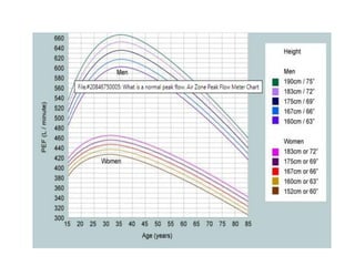
Males (450 – 550 L/min.); Females (350 – 450 L/min.);
PEFR (L/min.) = [Height (in cms.) – 80] x 5
9.
10. Useful in…
• diagnosis of asthma, exercise-induced asthma;
occupational asthma;
• recognizing the severity of asthma;
Peak flow meters are cheap; compact; portable; simple
to use.
Diurnal variability =
PEFR evening – PEFR morning
x 100
½ (PEFR evening + PEFR morning)
11. Diurnal variability of PEFR > 20% for at least three
days in a week for 2 weeks is typical of asthma.
Disadvantages:
• Any condition which reduces the Vital Capacity also
reduces the PEFR.
• It cannot distinguish between obstruction and restriction.
• The calculation is time-consuming when in clinical
practice.
12. Forced Expiratory Spirogram
Is a simple yet highly informative test of lung function;
The recording is obtained as Volume-time tracing.
The patient breathes in maximally and then exhales as
fast and as much as possible.
The forced expiratory spirogram enables the following
to be calculated:
• Forced Vital Capacity (FVC)
• Forced Expiratory Volume (FEV, FEV1)
• Forced Expiratory Flow rates (FEF)at various % of FVC
(e.g., FEF 25-75%);
13. FVC: max. amount of air exhaled after a maximal inhalation;
If normal:
o Normal lung tissue (fairly normal elasticity)
o Normal thoracic cage (configuration of thoracic spine/ribs).
o Respiratory muscles are not too weak.
If low: one or more of the above is an issue (Restrictive disorder)
FEV1: amount of air exhaled in the first second as forcefully as
possible from a full lung;
Measuring the speed of the air being exhaled can provide info.
about the airways;
Unit is litres/second;
↓ed speed indicates ‘obstructed’ or narrowed airways;
14. PFT INTERPRETATION
FEV1/FVC ratio
< 70% (or < LLN): Obstructive pattern (COPD, asthma)
≥ 70%: Normal or Restrictive disease
Severity of obstruction (GOLD Criteria for COPD)
FEV1 COPD STAGE
80 - 100% predicted Mild (Stage I)
50 - < 80% predicted Moderate (Stage II)
30 - < 50% predicted Severe (Stage III)
< 30% predicted Very severe (Stage IV)
< 50% with Rt. side heart failure Very severe (Stage IV)
15. Classification of airflow limitation severity in COPD (based
on post-bronchodilator FEV1):
FEV1 Airflow
obstruction
CATEGORY
≥ 80% predicted MILD GOLD-1
50 to < 80%
predicted
MODERATE GOLD-2
50 - 60% predicted MODERATELY
SEVERE
30 to < 50%
predicted
SEVERE GOLD-3
< 30% predicted VERY SEVERE GOLD-4
In patients with FEV1/FVC < 0.70
16. Bronchodilator response:
≥ 12% ↑ and an absolute improvement of at least 200 ml after
inhaling a beta agonist (usually neb. Salbutamol)
FEV1 or FVC is considered a significant response.
Total Lung Capacity (TLC)
Normal range: 80 - 120% of predicted
> 120% = Hyperinflation
< 80% = Restrictive disease
Use the ATS criteria for severity.
RV/TLC ratio
Normal range: < 35% or < predicted
> 35% or > predicted indicates Air trapping
17. Flow Volume Loop
It is a graphical analysis of the flow generated during
the FEV manoeuvre (plotted against the volume
change) followed by a FIV manoeuvre (plotted against
the volume change).
Volume (X-axis); Flow rates (Y-axis)
Gives additional info. about airflow at various lung
volumes from RV (reserve volume) to TLC.
Ascending part of expiratory component is patient-
effort dependent.
Descending part of expiratory component is mostly
effort independent; represents the elasticity and recoil
of the alveoli and smaller airways;
The peaks represent PEFR and PIFR.
Observing the loop indicates either ‘Obstruction’ or
‘Restriction’.
18.
19.
20. Spirometry Patterns:
These depend on how much air is breathed out and
what proportion is exhaled in the first second.
Usually shows one of three main patterns: Normal;
Obstructive; Restrictive patterns
Normal pattern:
21. Obstructive pattern:
When lung conditions (COPD and asthma) narrow the
airways.
Inference: Air flows out of the lungs more slowly than
it should (low FEV1) < 70% of the total amount in the
first second.
22. Restrictive Pattern:
The total amount of air breathed in is reduced but the
speed to breathe out is preserved.
Both FEV1 and FVC will be lower than predicted.
Causes:
• by various conditions that affect the lung tissues or the
capacity of the lungs to expand and hold a normal
amount of air, (pulmonary fibrosis, sarcoidosis,
pneumoconiosis, pleural effusion, pneumothorax,
pleural thickening).
• also seen in people who are significantly overweight,
have an abnormal curvature of the spine or weak
respiratory muscles.
24. Combined patterns:
Sometimes these two processes combine (obstruction
and restriction).
Both the total amount of air (inspired and expired), and
how fast a patient can blow out are reduced. This
happens in severe emphysema, cystic fibrosis, in
obesity.
25. Common Spirometry Patterns
Normal:
Normal spirometry (normal FVC, FEV1/FVC ratio)
Obstruction to airflow:
Normal FVC + ↓ FEV1 + low FEV1/FVC%; must assess BD
response
Restrictive changes:
↓ FVC + ↓ FEV1 + ‘normal’ FEV1/FVC%; must assess lung
volumes and DLCO
Combined obstructive and restrictive:
↓ FVC + ↓ FEV1 + ↓ FEV1/FVC%; must assess lung volumes
and DLCO
26. Reversibility testing:
Helps to assess if inhaled medication can open up the
airways.
Reversibility testing helps to grade the severity of the
conditions according to the FEV1 measurement after
taking the bronchodilator (usually by using Neb.
Salbutamol).
If there is obstructive abnormality, then bronchodilators
are given to assess the degree of reversibility. Post-
obstruction bronchodilator study is done, and the
graphs (FE Spirogram and Flow volume) are assessed
to determine if reversal had occurred, and to what
extent.
27. RV/TLC Ratio:
Ratio of residual volume to the total lung capacity.
Indicates the extent of air trapping in the lungs after
complete expiration.
Normal values upto 35%
Can be ↑ed in bullous disease, emphysema, AEBA
DLCO:
Measures the diffusing capacity of the lungs for carbon
monoxide.
The measure of the amount of gas transferred across
the interstitium per unit time as a function of the mean
pressure gradient of the gas across the interstitium.
Units: ml/min./mm of Hg
Is a diagnostic indicator of Interstitial lung disease.
Values < 80% of predicted are abnormal.
28. DLCO is ↑ed in….
• Alveolar haemorrhage
• Polycythemia
• L to R cardiac shunt
• Exercise
• Supine position
• Pregnancy
DLCO is ↓ed in….
• ILD
• Emphysema
• Anaemia
• Pneumonectomy
29. Pre and Post Arterial Blood Gas Analysis:
Helps to determine the blood gas status pre and post exercise and
post-administration of 100% O2;
Provides important information on gas exchange and oxygen
delivery to the tissues;
Post-exercise: ↑ of upto 10 mm Hg in PaO2;
PaCO2 should be normal or lower;
Abnormality can be inferred if post-exercise…
• PaO2 does not rise or is ↓ed;
• ↑ed PaCO2
Post 100% O2: There should be a normal rise of upto atleast 5x in
PaO2 levels. If this rise in PaO2 levels is lower, it is indicative of an
abnormality.
30. ABG test (contd’.)
Type-1 Respiratory Failure
PaO2 < 8 kPa with normal PaCO2;
Causes: Pneumonia and Pulmonary embolism;
Type-2 Respiratory Failure
When hypoxia is accompanied by hypercapnia (PaCO2 > 6.5 kPa);
Seen in ventilatory failure;
Causes: Respiratory muscle weakness and COPD;
Type-2 respiratory failure may also occur in patients with advanced
type-1 respiratory failure as they tire and develop ventilatory failure.
31. ABG test (contd’.)
Acidemia: Arterial pH < nml (< 7.35)
Alkalemia: Arterial pH > nml (>7.45)
Metabolic acidosis: Process that ↓es serum HCO3 → ↓ pH
(bicarbonate consumption could resolve this);
Respiratory acidosis: Process that ↑es serum pCO2 → ↓ pH
(hypoventilation)
Metabolic alkalosis: Process that ↑es serum HCO3 → ↑ pH
(excess bicarbonate is observed)
Respiratory alkalosis: Process that ↓es serum pCO2 → ↑ pH
(hyperventilation)
32. Bronchoprovocation testing:
To determine the Provocative dose or PD20%
PD20% is the max. exposure to bronchoprovocative substances
(histamine and methacholine) that can cause a 20% drop in
FEV1.
To determine extent of airway reactivity; assess airway
response to inhaled antigens (as in occupational asthma);
33. Cardiopulmonary Exercise Testing:
Multi stage study involving the monitoring of various
parameters for evaluation of unexplained dyspnoea or
for disability evaluation due to cardio-pulmonary
disease.
Allows the tester to judge the response of the
cardiopulmonary system to ‘graded exercise’ and
measures gas exchange in the form of O2 consumption,
CO2 production, and minute ventilation.
35. Residual Volume (RV)
↑ed: Air trapping (Obstructive disorders such as COPD and
asthma);
↓ed: Parenchymal restriction;
Functional Residual Capacity (FRC)
The volume of air left in the lungs after a normal, passive
exhalation;
Is used to evaluate the lung function;
↑ed: Age; Emphysema; Loss of elastic recoil
↓ed: Lung fibrosis; Obesity;
41. Source: NICE clinical guideline CG101: Chronic obstructive pulmonary disease
in over 16s. June 2010,
GRADE OF AIRFLOW
OBSTRUCTION
FEV1
MILD ≥ 80% of the predicted value.
MODERATE 50-79% of the predicted value after medication
SEVERE 30-49% of the predicted value after medication
VERY SEVERE < 30% of the predicted value after medication