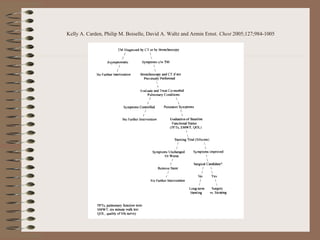
SlideShare a Scribd company logo
Clinical Case Conference

Gagan Kumar MD
Fellow
Pulmonary & Critical Care
GL 95yo M
• C/O progressive shortness of breath x 1
month
• Associated with dry cough
• C/O Wheezing on lying down – better with
sitting up.
• No fever/chills/night sweats/weight loss
• No PND/orthopnea
• Treated for CAP with levaquin
Past Medical History
•

•
•
•
•
•
•
•
•
•
•

MAI (progressive pulmonary infilterates on CT scan + per
culture reports from BAL in 2007)– started on Azithro + Rifampin
+ Ethambutol but patient took it x 1 month and stopped. No
follow ups.
Tracheomalacia per bronchoscopy in 08/2008, - focal on the
right side.
Tachycardia-bradycardia syndrome, status post pacemaker
placement.
Atrial fibrillation, status post AV ablation.
Diastolic dysfunction.
Hypertension.
Hypothyroidism.
GERD.
History of previous GI bleeding.
Osteoarthritis.
Benign positional vertigo.
Allergies - NKDA
• Family history: Non contributory
• Social history:
–
–
–
–

Non smoker
Occasional EtOH – quit 10 years back
Retired steel worker.
Lives at NH
Medications
•
•
•
•
•
•
•
•
•
•
•

Combivent 2 puffs 4 times/day
Duoneb TIS
Prednisone 60 mg daily.
Azithromycin 500 mg daily.
Rocephin 1 gram daily.
Cordarone 200 mg daily.
Calcium with Vitamin D 600 mg 1 tab t.i.d.
Lovenox 40 mg subQ at bedtime.
Pepcid 20 mg b.i.d.
Synthroid 150 mcg daily.
Multivitamin with mineral 1 daily.
Examination
•

BP 116/62, HR 70. Pulse ox is 99% on 2L/min via NC.

•

GENERAL: AO x 3 pleasant and in no distress.

•

HEENT: No Oral/Nasal lesions or exudates noted.

•

NECK: No JVD, lymphadenopathy or thyromegaly.

•

RESPIRATORY: Chest expansion is equal and bilateral with good effort. Faint bilateral
wheezing throughout.

•

HEART: RRR , normal S1 and S2, No GMR.

•

ABDOMEN: Slightly protuberant, soft, NT/ND , BS + No organomegaly.

•

EXTREMITIES: Warm. No cyanosis, clubbing or edema.

•

NEUROLOGIC: Gait normal. Cranial nerves 2-12 are grossly intact.

•

SKIN: Warm, dry and intact.
Labs
14.4
22.5

197
40.8

Blood cultures –ve

124

87

15

3.9

28

1.0

120
Radiology
CXR 10/6/10: no focal infiltrates
CT chest 10/12/10:
• Diffuse emphysematous changes
• Patchy opacities in RLL and LLL concerning for
pneumonia
• Multiple linear nodular opacities at apices
• Bilateral pleural effusions with compressive
atelectasis
• Stable RUL nodule, LLL nodule not visualized
PFTs
• 10/28/05
– FEV1/FVC – normal
– FEV1 – normal
–
–
–
–

No significant changes with bronchodilator
Flow volume loop – unremarkable
TLC – elevated (shows hyperinflation)
DLco - normal
Bronchoscopy
• 8/28/2007 by Dr Hubley
– VC normal
– Trachea revealed significant tracheomalacia seen on
right proximal trachea to cause an approximate
collapse down to 30% of the original diameter with
inspiration.
– No lesions
– Carina – sharp/symmetric
– Mild erythema throughout the airways
– No endobronchial lesions
Tracheomalacia
•
•

•

•

Malacia = “softness”
Normal intrathoracic trachea
dilates somewhat with
inspiration and narrows with
expiration
Narrowing is most
prominent when
intrathoracic pressure is
substantially greater than
intraluminal pressure, as it
is during forced expiration,
cough, or the Valsalva
maneuver
Extrathoracic or cervical TM
upper airway collapses
during inspiration
Types
• Tracheomalacia - trachea
• Bronchomalacia - one or both of the
main-stem bronchi
• Tracheobronchomalacia - both
Are Tracheomalacia & Dynamic
airway collapse the same?
Excessive dynamic
airway collapse?

1.
2.
3.
4.

Tracheobronchial collapse,
Expiratory tracheobronchial collapse,
Expiratory tracheobronchial stenosis,
Tacheobronchial dyskinesia

Tracheobronchial lumen during
coughing is 18–39% narrower than the
maximal inspiratory lumen observed during
restful respiration

Invagination of the posterior membrane

softening of the supporting cartilage and hypotonia of myoelastic
elements
Airway lumen during inspiration (A). During expiration there is inward bulging of the posterior membrane.
This process is physiological and is called dynamic airway collapse (DAC) (B). The pathologic
exaggeration of this process results in a reduction in cross sectional area of 50% or more and is called
excessive dynamic airway collapse (EDAC) (C). The pathological collapse of the cartilaginous rings
represents tracheobronchomalacia (TBM). The crescent type TBM occurs when the anterior cartilaginous
wall is softened and results in excessive narrowing of the sagittal airway diameter (D).The saber-sheath
type TBM is due to softening of the lateral walls and excessive narrowing of the transverse airway diameter
(E). Circumferential (combined) type TBM is characterized by anterior and lateral airway walls collapse and
is usually associated with significant airway wall inflammation
Tracheobronchomalacia and excessive dynamic airway collapse Septimiu D. MURGU AND Henri G. COLTRespirology (2006) 11, 388–406
History
•

1897 – Czyhlarz : was the first to describe the postmortem finding of
an unusually large trachea and bronchi.

•

1949 - Lemoine : was the first to use bronchoscopy to document
acquired tracheal enlargement in the adult.

•

1950 – Ferraris : described two patients with acquired TM who both
reported “expiratory dyspnea,” the inability to clear secretions, and
recurrent respiratory infections. Both had been labeled and treated as
asthmatic patients

•

1954 - Herzog and Nissen: “Relaxation and expiratory invagination of
the membranous portion of the intrathoracic trachea and the main
bronchi as cause of asphyxial attacks in bronchial asthma and the
chronic asthmoid bronchitis of pulmonary emphysema”. Bone graft in
the membranous trachea to prevent collapse
Embryology

1. Stomodeum
2. Pharyngeal gut
3. Thyroglossal duct
4. Tracheobronchial diverticulum
4th week : endodermal lining
of the respiratory diverticulum
gives rise to the epithelial
lining of the larynx, trachea,
bronchi and alveoli.
The cartilaginous and
muscular components of the
trachea and lungs are derived
from the surrounding
splanchnic mesoderm.

www.chronolab.com/embryo/respiratory.htm
Classification
•

•

Congenital disease (also called primary): consequence of the
inadequate maturity of tracheobronchial cartilage
– Polychondritis
– Chondromalacia
– Mucopolysaccharidoses: Hunter syndrome and Hurler
syndrome
– Idiopathic “giant trachea” or Mounier- Kuhn
syndrome
– most common associated disease is tracheoesophageal
fistula
• trachea receiving too much tissue during embryologic
separation
Acquired disease (also called secondary).
Acquired Tracheomalacia
•

Posttraumatic

•
•
•

Emphysema
Chronic infection/bronchitis
Chronic inflammation

•

Chronic external compression of the trachea

•

Vascular rings, previously undiagnosed in childhood

–
–
–
–

Post-intubation
Post-tracheostomy
External chest trauma
Post-lung transplantation

–

Relapsing polychondritis

–
–
–
–
–

Malignancy
Benign tumors
Cysts
Abscesses
Aortic aneurysm
Mounier- Kuhn syndrome
• Third or fourth decade of life.
•

Atrophy of longitudinal elastic fibers and thinning of the
muscularis mucosa

•

Diagnostic criteria: if

•

Secretions are poorly mobilized, leading to the chronic
accumulation of secretions
– Recurrent infections,
– Bronchiectasis
– Rarely pulmonary fibrosis

– Right mainstem > 2.4cm
– Left mainstem > 2.3cm
– Trachea exceed > 3.0 cm
Tracheostomy
•

Degeneration of normal cartilaginous support
– Prolonged intubation
– Tracheotomy
– Severe tracheobronchitis

•

Post-intubation ‘malacia’ is most commonly 3 cm in length
and is segmental in nature

•

Predisposing factors
– Recurrent intubation,
– Duration of intubation
– Use of high-dose steroids
– Chronic inflammation
– Irritants, such as cigarette smoke
Tracheostomy/Intubation
• SITES

– Stoma
– cuff site
– impingement point

• Mechanism

– Pressure necrosis,
– Impairment of the blood
supply
– Infection
– Mucosal damage caused
by friction

Where does blood supply
to cartilage comes from?
Relapsing Polychondritis
•

“recurrent episodes of
inflammation of the cartilage
of various tissues of the
body”

•

Involves tracheal rings in
56% of cases, but the
respiratory symptoms are
found on presentation in
only 14% of cases

•

Worse prognosis and
poorer response to
corticosteroids

World J Radiol. 2010 July 28; 2(7): 237-24

Characteristic thickening of the anterior cartilaginous wall of
the trachea . The posterior membranous wall is uninvolved
Histology
•

Pars membranacea is
dilated and flaccid.

•

Anterio-posterior narrowing
of the bronchial lumen

•

Atrophy of the longitudinal
elastic fibers of the pars
membranacea

•

The normal tracheal
cartilage-to-soft tissue ratio
is approximately 4.5 : 1. In
patients with TBM, this ratio
is often as low as 2 : 1.
Prevalence
•

1958 - Herzog : reported TBM in 16 of 1,500 patients (1%)
undergoing bronchoscopy for various respiratory symptoms

•

1977 - Jokinen et al: reported bronchoscopic findings for
2,150 patients with a range of symptoms and found that 94
patients (4.5%) had some form of malacia.
– TM -22%, TBM - 62%, isolated BM in15%

•

1992 - Ikeda S, Hanawa T, Konishi T, et al..
– Rate of airway collapse was 50% in 542 of 4,283 patients
(12.7%) with from pulmonary disease who underwent
bronchoscopy
Symptoms
• Dyspnea*
• Cough
•
•

Sputum production
Hemoptysis

•
•
•
•

More symptoms during forced exhalation
Inspiratory wheezing or stridor
Barking cough, which has been likened to a barking seal
Syncope associated with forced exhalation or cough

•

Differentials: “emphysema, chronic bronchitis,
cigarette smoking,or asthma”

*Nuutinen J. Acquired tracheobronchomalacia. Eur J RespirDis 1982; 63:380–387
http://www.youtube.com/watch?v=j2-61pPx-ZE&feature=related
Diagnosis
•In intubated patients,
– positive-pressure ventilatory support
keeps the airway open.
– “experience respiratory distress,
wheezing, and apparent stridor” on
extubation

“Unexplained extubation failure
should prompt evaluation for TM.”
Radiology
• Plain films: not good
– compression from other structures may be
occasionally seen
Radiology
• 1970s: Tracheograms and
Bronchograms:
– radiopaque material into the trachea, to
outline the bronchial tree and to evaluate
the size of the structures

• Cinetracheograms were used in the
hopes of seeing “tracheal flutter,”
• Fluoroscopy
“Gold Standard”
• Direct visualization by bronchoscopy to
document a narrowing of at least 50% in the
sagittal diameter in expiration*
– Mild : obstruction during expiration is to one half
of the lumen
– Moderate : reaches three quarters of the lumen
– Severe : the posterior wall touches the anterior
wall

• Straining/Coughing/Valsalva :

– to elicit airway wall collapse,
– the expiratory effort to achieve collapse has
never been standardized

* Nuutinen J. Acquired tracheobronchomalacia: a clinical study with bronchological correlations. Ann Clin Res 1977;9:350–355
Bronchoscopy
Dynamic CT scan
• Dynamic CT scan images, although not the
reference standard, are useful in diagnosing TM
• End-expiratory imaging rather than dynamic
expiratory imaging may require a lower threshold
criterion for diagnosing TBM.

Frown face

http://imaging.consult.com/imageSearch?query=lumen&thes=false&resultOffset=11
Multi-detector CT
• Permit imaging of the entire central airways in only a
few seconds
• Gilkeson et al (2001): reported agreement between
dynamic expiratory CT scan findings and
collapsibility seen during bronchoscopy
• Zhang et al (2005):
– low-dose CT scan technique is comparable to a standarddose technique for measuring the tracheal lumen
– Air trapping was seen at a higher frequency (TM patients,
100%; control subjects, 60%) and was more severe in the
patients with TM
Excessive narrowing of bronchi (black arrows)
Areas of geographically marginated radiolucency (white arrows) within lungs, = air trapping

Zhang J, Hasegawa I, Hatabu H, et al. Frequency and severity of air trapping at dynamic expiratory CT in patients with
tracheobronchomalacia. AJR Am J Roentgenol 2004; 182:81–85
Saggital reconstruction
Dynamic MRI
• Suto and Tanabe (1998)

– forced expiration and cough to compare
the collapsibility of the trachea in patients
with TM to that of healthy subjects by
using a “collapsibility index”
– CI = (Maxcsa– Mincsa)/Maxcsa
– lack of ionizing radiation
Multiplanar CT
• Three-dimensional CT scan reconstructions,
• Virtual bronchoscopy
• WHY multiplanar?

– images are less than ideal for evaluating airways that
course obliquely (eg., the mainstem bronchi)

• In patients who had relative contraindications to
bronchoscopy
Shaded-surface
display image of
central airways in
postero-lateral
projection shows
diffuse narrowing
of trachea and
bronchi (arrows ).

Virtual bronchoscopic image obtained at level of bronchus
intermedius during full inspiration shows mildly narrowed
but patent right middle (M) and lower (L) lobe bronchi.
Virtual bronchoscopic image obtained during dynamic
expiration shows marked narrowing of right middle lobe
bronchus (straight arrow ) with complete collapse of lower
lobe orifice (curved arrow ).
Pulmonary function studies
• Useful but not diagnostic
• Spirometry is not in proportion to the severity of
malacia
Decreased FEV1 and
a low PFR with a
rapid decrease in
flow

Near complete absence
of the usual sloping
phase of the midportion of the curve
“Break” or notch in
the expiratory phase
of the flow-volume
loop*

*May be seen in moderate-to-severe emphysema
Flow oscillations
•

Sequence of alternating decelerations and accelerations of flow,
are often seen on the expiratory curve

•

Also seen in
– redundant pharyngeal tissue, as in obstructive sleep apnea
syndrome,
– structural or functional disorders of the larynx,
– neuromuscular diseases

Vincken W, Cosio MG. Flow oscillations on the flow-volume loop: a nonspecific indicator of
upper airway dysfunction. Bull Eur Physiopathol Respir 1985; 21:559–567
Treatment
•

•
•

•

Supportive –
– unless the situation is emergent or progressively
worsening.
TM frequently occurs in patients who also have COPD:
– the obstructive disorder optimally should be treated first.
Bronchospasm must be controlled
– large pressure swings in the thorax
– worsening the degree of collapse of the malacic tracheal
segments
In relapsing polychondritis
– NSAIDs
– Steroids
• If the patient is in critical condition:
– noninvasive, positive-pressure ventilation
– ‘short term’ to keep the airway open and
to facilitate secretion drainage
Bronchoscopy + Stenting
Metal stents:
• Easily placed by flexible bronchoscopy,
• Are visible on plain radiographs,
• Expand dynamically
• Preserve mucociliary function
Problems:
• Formation of granulation tissue,
• Breakage over time,
• Airway obstruction, airway perforation
• Make future options such as surgical
interventions difficult or impossible
• Can not be removed easily

Not the first choice for patients with TM.
Stents . . .
• Silicone stents
– easily inserted, repositioned,
and removed

• Problems
– rigid bronchoscopy and
general anesthesia
– stent migration (new cough)
– direct visualization and
repositioning
• Dynamic features of TBM are quite different
from stenosis
• Constant change in size and shape of the airway
predisposes to stent migration and fracture.
• Long-term safety and efficacy data are sparse
• Ernst et al. 2007 : silicone stent placement.
– But they encountered high rate of stent-related
complications in 3 months (n=75):
• 21 partial stent obstructions, 14 infections, and 10 stent
migrations

• Thornton et al. : metallic stents.
– N=40
– Survival at 1, 2, 3, 4, 5 and 6 years as 79, 76, 51, 47,
38 and 23%, respectively. (died of comorbid causes )
Surgical Options
• Tracheostomy
– either bypass the malacic segment
– might splint the airway open
• If generalized and extensive TM,
– a longer tube may be necessary,
• Tracheostomy may aggravate the underlying
disorder and is, therefore, not a first-line treatment
Surgery
• Bone graft.
• Tracheal implantation of from one to three biocompatible
ceramic rings (Amedee et al) n=16. follow up 6.5 years.
• *TRACHEOPLASTY ( with Prosthetic and autologous
materials):
– Surgical placation of the posterior wall of the trachea with
crystalline polypropylene and high density polyethylene mesh.

• Conventional resection and reconstruction can be
considered for treatment of focal malacia of the trachea
Measure of success !!
1.
2.
3.
4.
5.

Improvement of respiratory symptoms,
Clearing of infectious processes,
Lack of stent complications
Bronchoscopy
Imaging technique

If airway stenting does not improve symptoms or the functional baseline
of the patient, the stents should be removed to avoid any stent-related
complications.
Gotway MB, Golden JA, LaBerge JM, et al. Benign tracheobronchial stenoses: changes in short-term
and long-term pulmonary function testing after expandable metallic stent placement. J Comput Assist
Tomogr 2002; 26:564–572

Twenty-two patients underwent 34 tracheal and/or bronchial stent placement
procedures for benign airway stenoses and had the results of pulmonary
function tests available. Stent placement indications included bronchomalacia
after lung transplantation (n11), postintubation stenoses (n6), relapsing
polychondritis (n2), and 1 each of tracheomalacia, tracheal compression, and
histoplasmosis.
Kelly A. Carden, Philip M. Boiselle, David A. Waltz and Armin Ernst. Chest 2005;127;984-1005
Thanks

More Related Content

What's hot

Pneumothorax case based
Pneumothorax case basedPneumothorax case based
Pneumothorax case based
Siruhan Ali
 
Dyspnea
DyspneaDyspnea
Stridor
StridorStridor
Stridor
yuyuricci
 
Stridor In Children
Stridor In ChildrenStridor In Children
Aortic stenosis
Aortic stenosis Aortic stenosis
Aortic stenosis
Pratap Tiwari
 
Aortic regurgitation
Aortic regurgitationAortic regurgitation
Aortic regurgitation
Pratap Tiwari
 
Massive hemoptysis / Nahid Sherbini
Massive hemoptysis / Nahid SherbiniMassive hemoptysis / Nahid Sherbini
Massive hemoptysis / Nahid Sherbini
Nahid Sherbini
 
Complications of pulmonary tb
Complications of pulmonary tbComplications of pulmonary tb
Complications of pulmonary tb
Ankur Gupta
 
Kartagener Syndrome ( USMLE Step 1)
Kartagener Syndrome ( USMLE Step 1)Kartagener Syndrome ( USMLE Step 1)
Kartagener Syndrome ( USMLE Step 1)
Greta Valadez
 
ENT (EAR NOSE THROAT ) Surgical instruments with their uses
ENT (EAR NOSE THROAT ) Surgical instruments with their usesENT (EAR NOSE THROAT ) Surgical instruments with their uses
ENT (EAR NOSE THROAT ) Surgical instruments with their uses
bannu medical college bannu KPK pakistan
 
Adenoids Hypertrophy
Adenoids HypertrophyAdenoids Hypertrophy
Adenoids Hypertrophy
Asst.Prof.Dr.Terdsak Rojsurakitti
 
Obesity Hypoventilation Syndrome
Obesity Hypoventilation Syndrome Obesity Hypoventilation Syndrome
Obesity Hypoventilation Syndrome
Ade Wijaya
 
HYPERSENSITIVITY PNEUMONITIS
HYPERSENSITIVITY PNEUMONITISHYPERSENSITIVITY PNEUMONITIS
HYPERSENSITIVITY PNEUMONITIS
DR.SHARIF AHSAN
 
Pneumothorax and pneumomediastinum
Pneumothorax and pneumomediastinum Pneumothorax and pneumomediastinum
Pneumothorax and pneumomediastinum
Dr Emad efat
 
dyspnea approach
dyspnea approachdyspnea approach
dyspnea approach
Prateek Singh
 
Medical Thoracoscopy
Medical ThoracoscopyMedical Thoracoscopy
Medical Thoracoscopy
Dr Subin Ahmed
 
Bronchiectasis.
Bronchiectasis.Bronchiectasis.
Bronchiectasis.
Davis Kurian
 
Chylothorax
ChylothoraxChylothorax
Chylothorax
Vijay Anand
 
Ards
ArdsArds
Bronchopleural fistula
Bronchopleural fistulaBronchopleural fistula
Bronchopleural fistula
Chiranjib Bhattacharyya
 

What's hot (20)

Pneumothorax case based
Pneumothorax case basedPneumothorax case based
Pneumothorax case based
 
Dyspnea
DyspneaDyspnea
Dyspnea
 
Stridor
StridorStridor
Stridor
 
Stridor In Children
Stridor In ChildrenStridor In Children
Stridor In Children
 
Aortic stenosis
Aortic stenosis Aortic stenosis
Aortic stenosis
 
Aortic regurgitation
Aortic regurgitationAortic regurgitation
Aortic regurgitation
 
Massive hemoptysis / Nahid Sherbini
Massive hemoptysis / Nahid SherbiniMassive hemoptysis / Nahid Sherbini
Massive hemoptysis / Nahid Sherbini
 
Complications of pulmonary tb
Complications of pulmonary tbComplications of pulmonary tb
Complications of pulmonary tb
 
Kartagener Syndrome ( USMLE Step 1)
Kartagener Syndrome ( USMLE Step 1)Kartagener Syndrome ( USMLE Step 1)
Kartagener Syndrome ( USMLE Step 1)
 
ENT (EAR NOSE THROAT ) Surgical instruments with their uses
ENT (EAR NOSE THROAT ) Surgical instruments with their usesENT (EAR NOSE THROAT ) Surgical instruments with their uses
ENT (EAR NOSE THROAT ) Surgical instruments with their uses
 
Adenoids Hypertrophy
Adenoids HypertrophyAdenoids Hypertrophy
Adenoids Hypertrophy
 
Obesity Hypoventilation Syndrome
Obesity Hypoventilation Syndrome Obesity Hypoventilation Syndrome
Obesity Hypoventilation Syndrome
 
HYPERSENSITIVITY PNEUMONITIS
HYPERSENSITIVITY PNEUMONITISHYPERSENSITIVITY PNEUMONITIS
HYPERSENSITIVITY PNEUMONITIS
 
Pneumothorax and pneumomediastinum
Pneumothorax and pneumomediastinum Pneumothorax and pneumomediastinum
Pneumothorax and pneumomediastinum
 
dyspnea approach
dyspnea approachdyspnea approach
dyspnea approach
 
Medical Thoracoscopy
Medical ThoracoscopyMedical Thoracoscopy
Medical Thoracoscopy
 
Bronchiectasis.
Bronchiectasis.Bronchiectasis.
Bronchiectasis.
 
Chylothorax
ChylothoraxChylothorax
Chylothorax
 
Ards
ArdsArds
Ards
 
Bronchopleural fistula
Bronchopleural fistulaBronchopleural fistula
Bronchopleural fistula
 

Viewers also liked

Dynamic Central Airway Obstruction: Tracheomalacia, Tracheobronchomalacia, An...
Dynamic Central Airway Obstruction: Tracheomalacia, Tracheobronchomalacia, An...Dynamic Central Airway Obstruction: Tracheomalacia, Tracheobronchomalacia, An...
Dynamic Central Airway Obstruction: Tracheomalacia, Tracheobronchomalacia, An...
Bassel Ericsoussi, MD
 
E.N.T 5th year, 4th lecture (Dr. Hiwa)
E.N.T 5th year, 4th lecture (Dr. Hiwa)E.N.T 5th year, 4th lecture (Dr. Hiwa)
E.N.T 5th year, 4th lecture (Dr. Hiwa)
College of Medicine, Sulaymaniyah
 
Silicosis
SilicosisSilicosis
Silicosis
maaferhc
 
Silicosis
SilicosisSilicosis
Silicosis
font Fawn
 
Pedrespiemergencyupper 110315115727-phpapp02 (1)
Pedrespiemergencyupper 110315115727-phpapp02 (1)Pedrespiemergencyupper 110315115727-phpapp02 (1)
Pedrespiemergencyupper 110315115727-phpapp02 (1)
Dimitrije123
 
Tracheal pathologies
Tracheal pathologiesTracheal pathologies
Tracheal pathologies
Jino Justin
 
Targeted Temperature Management (Therapeutic Hypothermia) in Critical Care: ...
Targeted Temperature Management  (Therapeutic Hypothermia) in Critical Care: ...Targeted Temperature Management  (Therapeutic Hypothermia) in Critical Care: ...
Targeted Temperature Management (Therapeutic Hypothermia) in Critical Care: ...
Bassel Ericsoussi, MD
 
Pft lecture for residents mine
Pft lecture for residents minePft lecture for residents mine
Pft lecture for residents mine
katejohnpunag
 
Sarcoidosis
SarcoidosisSarcoidosis
Sarcoidosis
Dr.Manish Kumar
 
sudden infant death syndrome sids
 sudden infant death syndrome  sids sudden infant death syndrome  sids
sudden infant death syndrome sids
Mohamed Abunada
 
Tuberculosis Treatment Symposia - The CRUDEM Foundation
Tuberculosis Treatment Symposia - The CRUDEM FoundationTuberculosis Treatment Symposia - The CRUDEM Foundation
Tuberculosis Treatment Symposia - The CRUDEM Foundation
The CRUDEM Foundation
 
Endometrial polyps
Endometrial polypsEndometrial polyps
Endometrial polyps
raj kumar
 
CCAM Congenital Cystic Adenomatoid Malformation
CCAM Congenital Cystic Adenomatoid MalformationCCAM Congenital Cystic Adenomatoid Malformation
CCAM Congenital Cystic Adenomatoid Malformation
陳 立珣
 
Neonatal Emergencies
Neonatal EmergenciesNeonatal Emergencies
Neonatal Emergencies
Dang Thanh Tuan
 
Basic concepts in treatment of Pulmonary Tuberculosis - By Dr.Tinku Joseph
Basic concepts in treatment of Pulmonary Tuberculosis - By Dr.Tinku JosephBasic concepts in treatment of Pulmonary Tuberculosis - By Dr.Tinku Joseph
Basic concepts in treatment of Pulmonary Tuberculosis - By Dr.Tinku Joseph
Dr.Tinku Joseph
 
NERRS Oct 2013 Thoracic Radiology Case Answers
NERRS Oct 2013 Thoracic Radiology Case AnswersNERRS Oct 2013 Thoracic Radiology Case Answers
NERRS Oct 2013 Thoracic Radiology Case Answers
NERRS
 
Silicosis bengali
Silicosis bengaliSilicosis bengali
Silicosis bengali
drdduttaM
 
Congenital cystic adenomatoid malformation
Congenital cystic adenomatoid malformationCongenital cystic adenomatoid malformation
Congenital cystic adenomatoid malformation
Sneha Volvoikar
 
Presentation1.pptx, radiological imaging of congenital abnormalities of the l...
Presentation1.pptx, radiological imaging of congenital abnormalities of the l...Presentation1.pptx, radiological imaging of congenital abnormalities of the l...
Presentation1.pptx, radiological imaging of congenital abnormalities of the l...
Abdellah Nazeer
 
Chest Xray
Chest XrayChest Xray
Chest Xray
Hassan Tarig
 

Viewers also liked (20)

Dynamic Central Airway Obstruction: Tracheomalacia, Tracheobronchomalacia, An...
Dynamic Central Airway Obstruction: Tracheomalacia, Tracheobronchomalacia, An...Dynamic Central Airway Obstruction: Tracheomalacia, Tracheobronchomalacia, An...
Dynamic Central Airway Obstruction: Tracheomalacia, Tracheobronchomalacia, An...
 
E.N.T 5th year, 4th lecture (Dr. Hiwa)
E.N.T 5th year, 4th lecture (Dr. Hiwa)E.N.T 5th year, 4th lecture (Dr. Hiwa)
E.N.T 5th year, 4th lecture (Dr. Hiwa)
 
Silicosis
SilicosisSilicosis
Silicosis
 
Silicosis
SilicosisSilicosis
Silicosis
 
Pedrespiemergencyupper 110315115727-phpapp02 (1)
Pedrespiemergencyupper 110315115727-phpapp02 (1)Pedrespiemergencyupper 110315115727-phpapp02 (1)
Pedrespiemergencyupper 110315115727-phpapp02 (1)
 
Tracheal pathologies
Tracheal pathologiesTracheal pathologies
Tracheal pathologies
 
Targeted Temperature Management (Therapeutic Hypothermia) in Critical Care: ...
Targeted Temperature Management  (Therapeutic Hypothermia) in Critical Care: ...Targeted Temperature Management  (Therapeutic Hypothermia) in Critical Care: ...
Targeted Temperature Management (Therapeutic Hypothermia) in Critical Care: ...
 
Pft lecture for residents mine
Pft lecture for residents minePft lecture for residents mine
Pft lecture for residents mine
 
Sarcoidosis
SarcoidosisSarcoidosis
Sarcoidosis
 
sudden infant death syndrome sids
 sudden infant death syndrome  sids sudden infant death syndrome  sids
sudden infant death syndrome sids
 
Tuberculosis Treatment Symposia - The CRUDEM Foundation
Tuberculosis Treatment Symposia - The CRUDEM FoundationTuberculosis Treatment Symposia - The CRUDEM Foundation
Tuberculosis Treatment Symposia - The CRUDEM Foundation
 
Endometrial polyps
Endometrial polypsEndometrial polyps
Endometrial polyps
 
CCAM Congenital Cystic Adenomatoid Malformation
CCAM Congenital Cystic Adenomatoid MalformationCCAM Congenital Cystic Adenomatoid Malformation
CCAM Congenital Cystic Adenomatoid Malformation
 
Neonatal Emergencies
Neonatal EmergenciesNeonatal Emergencies
Neonatal Emergencies
 
Basic concepts in treatment of Pulmonary Tuberculosis - By Dr.Tinku Joseph
Basic concepts in treatment of Pulmonary Tuberculosis - By Dr.Tinku JosephBasic concepts in treatment of Pulmonary Tuberculosis - By Dr.Tinku Joseph
Basic concepts in treatment of Pulmonary Tuberculosis - By Dr.Tinku Joseph
 
NERRS Oct 2013 Thoracic Radiology Case Answers
NERRS Oct 2013 Thoracic Radiology Case AnswersNERRS Oct 2013 Thoracic Radiology Case Answers
NERRS Oct 2013 Thoracic Radiology Case Answers
 
Silicosis bengali
Silicosis bengaliSilicosis bengali
Silicosis bengali
 
Congenital cystic adenomatoid malformation
Congenital cystic adenomatoid malformationCongenital cystic adenomatoid malformation
Congenital cystic adenomatoid malformation
 
Presentation1.pptx, radiological imaging of congenital abnormalities of the l...
Presentation1.pptx, radiological imaging of congenital abnormalities of the l...Presentation1.pptx, radiological imaging of congenital abnormalities of the l...
Presentation1.pptx, radiological imaging of congenital abnormalities of the l...
 
Chest Xray
Chest XrayChest Xray
Chest Xray
 

Similar to Tracheomalacia

Pathology of Acute Lungi Injury- Recent advances
Pathology of Acute Lungi Injury- Recent advancesPathology of Acute Lungi Injury- Recent advances
Pathology of Acute Lungi Injury- Recent advances
Dr Snehal Kosale
 
Pediatric chest infection imaging considerations
Pediatric chest infection imaging considerationsPediatric chest infection imaging considerations
Pediatric chest infection imaging considerations
Ahmed Bahnassy
 
COPD.pptx
COPD.pptxCOPD.pptx
COPD.pptx
Wale Ogunlade
 
Lymphangioleiomyomatosis
LymphangioleiomyomatosisLymphangioleiomyomatosis
Lymphangioleiomyomatosis
Ashraf ElAdawy
 
COPD
COPDCOPD
Aime tuberculosis
Aime tuberculosisAime tuberculosis
Aime tuberculosis
aimeAbraham
 
copd 2012.ppt
copd 2012.pptcopd 2012.ppt
copd 2012.ppt
nYnNP
 
Lung malformation part 1
Lung malformation part 1Lung malformation part 1
Lung malformation part 1
Faheem Andrabi
 
Bronchiactasis - Lecture by Dr. Nasir Farooq Butt.pptx
Bronchiactasis - Lecture by Dr. Nasir Farooq Butt.pptxBronchiactasis - Lecture by Dr. Nasir Farooq Butt.pptx
Bronchiactasis - Lecture by Dr. Nasir Farooq Butt.pptx
MukhtarJamac3
 
Chronic obstructive pulmonary disease (copd)
Chronic obstructive pulmonary disease (copd)Chronic obstructive pulmonary disease (copd)
Chronic obstructive pulmonary disease (copd)
Rahil Dalal
 
Murali bronchiectasis.pptx
Murali bronchiectasis.pptxMurali bronchiectasis.pptx
Murali bronchiectasis.pptx
Murali Krishna
 
ppt ild final.pptx
ppt ild final.pptxppt ild final.pptx
ppt ild final.pptx
Mirazul Haque
 
Emphysema
EmphysemaEmphysema
Emphysema
nancygalaly
 
Practical approach to Idiopathic Pulmonary Fibrosis.
Practical approach to Idiopathic Pulmonary Fibrosis.Practical approach to Idiopathic Pulmonary Fibrosis.
Practical approach to Idiopathic Pulmonary Fibrosis.
Hiba Ashibany
 
Paediatric chest 031220
Paediatric chest 031220Paediatric chest 031220
Paediatric chest 031220
FaizahMohdZakiPPUKM
 
COPD
COPDCOPD
Pneumomediastinum
PneumomediastinumPneumomediastinum
Pneumomediastinum
SCGH ED CME
 
Approach to uawo
Approach to uawoApproach to uawo
Approach to uawo
Raddaa
 
Copd
CopdCopd
Pleural diseases
Pleural diseasesPleural diseases
Pleural diseases
Oriba Dan Langoya
 

Similar to Tracheomalacia (20)

Pathology of Acute Lungi Injury- Recent advances
Pathology of Acute Lungi Injury- Recent advancesPathology of Acute Lungi Injury- Recent advances
Pathology of Acute Lungi Injury- Recent advances
 
Pediatric chest infection imaging considerations
Pediatric chest infection imaging considerationsPediatric chest infection imaging considerations
Pediatric chest infection imaging considerations
 
COPD.pptx
COPD.pptxCOPD.pptx
COPD.pptx
 
Lymphangioleiomyomatosis
LymphangioleiomyomatosisLymphangioleiomyomatosis
Lymphangioleiomyomatosis
 
COPD
COPDCOPD
COPD
 
Aime tuberculosis
Aime tuberculosisAime tuberculosis
Aime tuberculosis
 
copd 2012.ppt
copd 2012.pptcopd 2012.ppt
copd 2012.ppt
 
Lung malformation part 1
Lung malformation part 1Lung malformation part 1
Lung malformation part 1
 
Bronchiactasis - Lecture by Dr. Nasir Farooq Butt.pptx
Bronchiactasis - Lecture by Dr. Nasir Farooq Butt.pptxBronchiactasis - Lecture by Dr. Nasir Farooq Butt.pptx
Bronchiactasis - Lecture by Dr. Nasir Farooq Butt.pptx
 
Chronic obstructive pulmonary disease (copd)
Chronic obstructive pulmonary disease (copd)Chronic obstructive pulmonary disease (copd)
Chronic obstructive pulmonary disease (copd)
 
Murali bronchiectasis.pptx
Murali bronchiectasis.pptxMurali bronchiectasis.pptx
Murali bronchiectasis.pptx
 
ppt ild final.pptx
ppt ild final.pptxppt ild final.pptx
ppt ild final.pptx
 
Emphysema
EmphysemaEmphysema
Emphysema
 
Practical approach to Idiopathic Pulmonary Fibrosis.
Practical approach to Idiopathic Pulmonary Fibrosis.Practical approach to Idiopathic Pulmonary Fibrosis.
Practical approach to Idiopathic Pulmonary Fibrosis.
 
Paediatric chest 031220
Paediatric chest 031220Paediatric chest 031220
Paediatric chest 031220
 
COPD
COPDCOPD
COPD
 
Pneumomediastinum
PneumomediastinumPneumomediastinum
Pneumomediastinum
 
Approach to uawo
Approach to uawoApproach to uawo
Approach to uawo
 
Copd
CopdCopd
Copd
 
Pleural diseases
Pleural diseasesPleural diseases
Pleural diseases
 

Recently uploaded

What are the different types of Dental implants.
What are the different types of Dental implants.What are the different types of Dental implants.
What are the different types of Dental implants.
Gokuldas Hospital
 
LOW BIRTH WEIGHT. PRETERM BABIES OR SMALL FOR DATES BABIES
LOW BIRTH WEIGHT. PRETERM BABIES OR SMALL FOR DATES BABIESLOW BIRTH WEIGHT. PRETERM BABIES OR SMALL FOR DATES BABIES
LOW BIRTH WEIGHT. PRETERM BABIES OR SMALL FOR DATES BABIES
ShraddhaTamshettiwar
 
Breast cancer: Post menopausal endocrine therapy
Breast cancer: Post menopausal endocrine therapyBreast cancer: Post menopausal endocrine therapy
Breast cancer: Post menopausal endocrine therapy
Dr. Sumit KUMAR
 
CHEMOTHERAPY_RDP_CHAPTER 1_ANTI TB DRUGS.pdf
CHEMOTHERAPY_RDP_CHAPTER 1_ANTI TB DRUGS.pdfCHEMOTHERAPY_RDP_CHAPTER 1_ANTI TB DRUGS.pdf
CHEMOTHERAPY_RDP_CHAPTER 1_ANTI TB DRUGS.pdf
rishi2789
 
Promoting Wellbeing - Applied Social Psychology - Psychology SuperNotes
Promoting Wellbeing - Applied Social Psychology - Psychology SuperNotesPromoting Wellbeing - Applied Social Psychology - Psychology SuperNotes
Promoting Wellbeing - Applied Social Psychology - Psychology SuperNotes
PsychoTech Services
 
Nano-gold for Cancer Therapy chemistry investigatory project
Nano-gold for Cancer Therapy chemistry investigatory projectNano-gold for Cancer Therapy chemistry investigatory project
Nano-gold for Cancer Therapy chemistry investigatory project
SIVAVINAYAKPK
 
June 2024 Oncology Cartoons By Dr Kanhu Charan Patro
June 2024 Oncology Cartoons By Dr Kanhu Charan PatroJune 2024 Oncology Cartoons By Dr Kanhu Charan Patro
June 2024 Oncology Cartoons By Dr Kanhu Charan Patro
Kanhu Charan
 
10 Benefits an EPCR Software should Bring to EMS Organizations
10 Benefits an EPCR Software should Bring to EMS Organizations   10 Benefits an EPCR Software should Bring to EMS Organizations
10 Benefits an EPCR Software should Bring to EMS Organizations
Traumasoft LLC
 
Alzheimer’s Disease Case Conference: Gearing Up for the Expanding Role of Neu...
Alzheimer’s Disease Case Conference: Gearing Up for the Expanding Role of Neu...Alzheimer’s Disease Case Conference: Gearing Up for the Expanding Role of Neu...
Alzheimer’s Disease Case Conference: Gearing Up for the Expanding Role of Neu...
PVI, PeerView Institute for Medical Education
 
NARCOTICS- POLICY AND PROCEDURES FOR ITS USE
NARCOTICS- POLICY AND PROCEDURES FOR ITS USENARCOTICS- POLICY AND PROCEDURES FOR ITS USE
NARCOTICS- POLICY AND PROCEDURES FOR ITS USE
Dr. Ahana Haroon
 
How to choose the best dermatologists in Indore.
How to choose the best dermatologists in Indore.How to choose the best dermatologists in Indore.
How to choose the best dermatologists in Indore.
Gokuldas Hospital
 
CHEMOTHERAPY_RDP_CHAPTER 2 _LEPROSY.pdf1
CHEMOTHERAPY_RDP_CHAPTER 2 _LEPROSY.pdf1CHEMOTHERAPY_RDP_CHAPTER 2 _LEPROSY.pdf1
CHEMOTHERAPY_RDP_CHAPTER 2 _LEPROSY.pdf1
rishi2789
 
Travel Clinic Cardiff: Health Advice for International Travelers
Travel Clinic Cardiff: Health Advice for International TravelersTravel Clinic Cardiff: Health Advice for International Travelers
Travel Clinic Cardiff: Health Advice for International Travelers
NX Healthcare
 
Medical Quiz ( Online Quiz for API Meet 2024 ).pdf
Medical Quiz ( Online Quiz for API Meet 2024 ).pdfMedical Quiz ( Online Quiz for API Meet 2024 ).pdf
Medical Quiz ( Online Quiz for API Meet 2024 ).pdf
Jim Jacob Roy
 
Cosmetology and Trichology Courses at Kosmoderma Academy PRP (Hair), DR Growt...
Cosmetology and Trichology Courses at Kosmoderma Academy PRP (Hair), DR Growt...Cosmetology and Trichology Courses at Kosmoderma Academy PRP (Hair), DR Growt...
Cosmetology and Trichology Courses at Kosmoderma Academy PRP (Hair), DR Growt...
Kosmoderma Academy Of Aesthetic Medicine
 
Cell Therapy Expansion and Challenges in Autoimmune Disease
Cell Therapy Expansion and Challenges in Autoimmune DiseaseCell Therapy Expansion and Challenges in Autoimmune Disease
Cell Therapy Expansion and Challenges in Autoimmune Disease
Health Advances
 
Ophthalmic drugs latest. Xxxxxxzxxxxxx.pdf
Ophthalmic drugs latest. Xxxxxxzxxxxxx.pdfOphthalmic drugs latest. Xxxxxxzxxxxxx.pdf
Ophthalmic drugs latest. Xxxxxxzxxxxxx.pdf
MuhammadMuneer49
 
Test bank for karp s cell and molecular biology 9th edition by gerald karp.pdf
Test bank for karp s cell and molecular biology 9th edition by gerald karp.pdfTest bank for karp s cell and molecular biology 9th edition by gerald karp.pdf
Test bank for karp s cell and molecular biology 9th edition by gerald karp.pdf
rightmanforbloodline
 
K CỔ TỬ CUNG.pdf tự ghi chép, chữ hơi xấu
K CỔ TỬ CUNG.pdf tự ghi chép, chữ hơi xấuK CỔ TỬ CUNG.pdf tự ghi chép, chữ hơi xấu
K CỔ TỬ CUNG.pdf tự ghi chép, chữ hơi xấu
HongBiThi1
 
CHEMOTHERAPY_RDP_CHAPTER 4_ANTI VIRAL DRUGS.pdf
CHEMOTHERAPY_RDP_CHAPTER 4_ANTI VIRAL DRUGS.pdfCHEMOTHERAPY_RDP_CHAPTER 4_ANTI VIRAL DRUGS.pdf
CHEMOTHERAPY_RDP_CHAPTER 4_ANTI VIRAL DRUGS.pdf
rishi2789
 

Recently uploaded (20)

What are the different types of Dental implants.
What are the different types of Dental implants.What are the different types of Dental implants.
What are the different types of Dental implants.
 
LOW BIRTH WEIGHT. PRETERM BABIES OR SMALL FOR DATES BABIES
LOW BIRTH WEIGHT. PRETERM BABIES OR SMALL FOR DATES BABIESLOW BIRTH WEIGHT. PRETERM BABIES OR SMALL FOR DATES BABIES
LOW BIRTH WEIGHT. PRETERM BABIES OR SMALL FOR DATES BABIES
 
Breast cancer: Post menopausal endocrine therapy
Breast cancer: Post menopausal endocrine therapyBreast cancer: Post menopausal endocrine therapy
Breast cancer: Post menopausal endocrine therapy
 
CHEMOTHERAPY_RDP_CHAPTER 1_ANTI TB DRUGS.pdf
CHEMOTHERAPY_RDP_CHAPTER 1_ANTI TB DRUGS.pdfCHEMOTHERAPY_RDP_CHAPTER 1_ANTI TB DRUGS.pdf
CHEMOTHERAPY_RDP_CHAPTER 1_ANTI TB DRUGS.pdf
 
Promoting Wellbeing - Applied Social Psychology - Psychology SuperNotes
Promoting Wellbeing - Applied Social Psychology - Psychology SuperNotesPromoting Wellbeing - Applied Social Psychology - Psychology SuperNotes
Promoting Wellbeing - Applied Social Psychology - Psychology SuperNotes
 
Nano-gold for Cancer Therapy chemistry investigatory project
Nano-gold for Cancer Therapy chemistry investigatory projectNano-gold for Cancer Therapy chemistry investigatory project
Nano-gold for Cancer Therapy chemistry investigatory project
 
June 2024 Oncology Cartoons By Dr Kanhu Charan Patro
June 2024 Oncology Cartoons By Dr Kanhu Charan PatroJune 2024 Oncology Cartoons By Dr Kanhu Charan Patro
June 2024 Oncology Cartoons By Dr Kanhu Charan Patro
 
10 Benefits an EPCR Software should Bring to EMS Organizations
10 Benefits an EPCR Software should Bring to EMS Organizations   10 Benefits an EPCR Software should Bring to EMS Organizations
10 Benefits an EPCR Software should Bring to EMS Organizations
 
Alzheimer’s Disease Case Conference: Gearing Up for the Expanding Role of Neu...
Alzheimer’s Disease Case Conference: Gearing Up for the Expanding Role of Neu...Alzheimer’s Disease Case Conference: Gearing Up for the Expanding Role of Neu...
Alzheimer’s Disease Case Conference: Gearing Up for the Expanding Role of Neu...
 
NARCOTICS- POLICY AND PROCEDURES FOR ITS USE
NARCOTICS- POLICY AND PROCEDURES FOR ITS USENARCOTICS- POLICY AND PROCEDURES FOR ITS USE
NARCOTICS- POLICY AND PROCEDURES FOR ITS USE
 
How to choose the best dermatologists in Indore.
How to choose the best dermatologists in Indore.How to choose the best dermatologists in Indore.
How to choose the best dermatologists in Indore.
 
CHEMOTHERAPY_RDP_CHAPTER 2 _LEPROSY.pdf1
CHEMOTHERAPY_RDP_CHAPTER 2 _LEPROSY.pdf1CHEMOTHERAPY_RDP_CHAPTER 2 _LEPROSY.pdf1
CHEMOTHERAPY_RDP_CHAPTER 2 _LEPROSY.pdf1
 
Travel Clinic Cardiff: Health Advice for International Travelers
Travel Clinic Cardiff: Health Advice for International TravelersTravel Clinic Cardiff: Health Advice for International Travelers
Travel Clinic Cardiff: Health Advice for International Travelers
 
Medical Quiz ( Online Quiz for API Meet 2024 ).pdf
Medical Quiz ( Online Quiz for API Meet 2024 ).pdfMedical Quiz ( Online Quiz for API Meet 2024 ).pdf
Medical Quiz ( Online Quiz for API Meet 2024 ).pdf
 
Cosmetology and Trichology Courses at Kosmoderma Academy PRP (Hair), DR Growt...
Cosmetology and Trichology Courses at Kosmoderma Academy PRP (Hair), DR Growt...Cosmetology and Trichology Courses at Kosmoderma Academy PRP (Hair), DR Growt...
Cosmetology and Trichology Courses at Kosmoderma Academy PRP (Hair), DR Growt...
 
Cell Therapy Expansion and Challenges in Autoimmune Disease
Cell Therapy Expansion and Challenges in Autoimmune DiseaseCell Therapy Expansion and Challenges in Autoimmune Disease
Cell Therapy Expansion and Challenges in Autoimmune Disease
 
Ophthalmic drugs latest. Xxxxxxzxxxxxx.pdf
Ophthalmic drugs latest. Xxxxxxzxxxxxx.pdfOphthalmic drugs latest. Xxxxxxzxxxxxx.pdf
Ophthalmic drugs latest. Xxxxxxzxxxxxx.pdf
 
Test bank for karp s cell and molecular biology 9th edition by gerald karp.pdf
Test bank for karp s cell and molecular biology 9th edition by gerald karp.pdfTest bank for karp s cell and molecular biology 9th edition by gerald karp.pdf
Test bank for karp s cell and molecular biology 9th edition by gerald karp.pdf
 
K CỔ TỬ CUNG.pdf tự ghi chép, chữ hơi xấu
K CỔ TỬ CUNG.pdf tự ghi chép, chữ hơi xấuK CỔ TỬ CUNG.pdf tự ghi chép, chữ hơi xấu
K CỔ TỬ CUNG.pdf tự ghi chép, chữ hơi xấu
 
CHEMOTHERAPY_RDP_CHAPTER 4_ANTI VIRAL DRUGS.pdf
CHEMOTHERAPY_RDP_CHAPTER 4_ANTI VIRAL DRUGS.pdfCHEMOTHERAPY_RDP_CHAPTER 4_ANTI VIRAL DRUGS.pdf
CHEMOTHERAPY_RDP_CHAPTER 4_ANTI VIRAL DRUGS.pdf
 

Tracheomalacia

  • 1. Clinical Case Conference Gagan Kumar MD Fellow Pulmonary & Critical Care
  • 2. GL 95yo M • C/O progressive shortness of breath x 1 month • Associated with dry cough • C/O Wheezing on lying down – better with sitting up. • No fever/chills/night sweats/weight loss • No PND/orthopnea • Treated for CAP with levaquin
  • 3. Past Medical History • • • • • • • • • • • MAI (progressive pulmonary infilterates on CT scan + per culture reports from BAL in 2007)– started on Azithro + Rifampin + Ethambutol but patient took it x 1 month and stopped. No follow ups. Tracheomalacia per bronchoscopy in 08/2008, - focal on the right side. Tachycardia-bradycardia syndrome, status post pacemaker placement. Atrial fibrillation, status post AV ablation. Diastolic dysfunction. Hypertension. Hypothyroidism. GERD. History of previous GI bleeding. Osteoarthritis. Benign positional vertigo.
  • 4. Allergies - NKDA • Family history: Non contributory • Social history: – – – – Non smoker Occasional EtOH – quit 10 years back Retired steel worker. Lives at NH
  • 5. Medications • • • • • • • • • • • Combivent 2 puffs 4 times/day Duoneb TIS Prednisone 60 mg daily. Azithromycin 500 mg daily. Rocephin 1 gram daily. Cordarone 200 mg daily. Calcium with Vitamin D 600 mg 1 tab t.i.d. Lovenox 40 mg subQ at bedtime. Pepcid 20 mg b.i.d. Synthroid 150 mcg daily. Multivitamin with mineral 1 daily.
  • 6. Examination • BP 116/62, HR 70. Pulse ox is 99% on 2L/min via NC. • GENERAL: AO x 3 pleasant and in no distress. • HEENT: No Oral/Nasal lesions or exudates noted. • NECK: No JVD, lymphadenopathy or thyromegaly. • RESPIRATORY: Chest expansion is equal and bilateral with good effort. Faint bilateral wheezing throughout. • HEART: RRR , normal S1 and S2, No GMR. • ABDOMEN: Slightly protuberant, soft, NT/ND , BS + No organomegaly. • EXTREMITIES: Warm. No cyanosis, clubbing or edema. • NEUROLOGIC: Gait normal. Cranial nerves 2-12 are grossly intact. • SKIN: Warm, dry and intact.
  • 8. Radiology CXR 10/6/10: no focal infiltrates CT chest 10/12/10: • Diffuse emphysematous changes • Patchy opacities in RLL and LLL concerning for pneumonia • Multiple linear nodular opacities at apices • Bilateral pleural effusions with compressive atelectasis • Stable RUL nodule, LLL nodule not visualized
  • 9. PFTs • 10/28/05 – FEV1/FVC – normal – FEV1 – normal – – – – No significant changes with bronchodilator Flow volume loop – unremarkable TLC – elevated (shows hyperinflation) DLco - normal
  • 10. Bronchoscopy • 8/28/2007 by Dr Hubley – VC normal – Trachea revealed significant tracheomalacia seen on right proximal trachea to cause an approximate collapse down to 30% of the original diameter with inspiration. – No lesions – Carina – sharp/symmetric – Mild erythema throughout the airways – No endobronchial lesions
  • 11. Tracheomalacia • • • • Malacia = “softness” Normal intrathoracic trachea dilates somewhat with inspiration and narrows with expiration Narrowing is most prominent when intrathoracic pressure is substantially greater than intraluminal pressure, as it is during forced expiration, cough, or the Valsalva maneuver Extrathoracic or cervical TM upper airway collapses during inspiration
  • 12. Types • Tracheomalacia - trachea • Bronchomalacia - one or both of the main-stem bronchi • Tracheobronchomalacia - both
  • 13. Are Tracheomalacia & Dynamic airway collapse the same? Excessive dynamic airway collapse? 1. 2. 3. 4. Tracheobronchial collapse, Expiratory tracheobronchial collapse, Expiratory tracheobronchial stenosis, Tacheobronchial dyskinesia Tracheobronchial lumen during coughing is 18–39% narrower than the maximal inspiratory lumen observed during restful respiration Invagination of the posterior membrane softening of the supporting cartilage and hypotonia of myoelastic elements
  • 14. Airway lumen during inspiration (A). During expiration there is inward bulging of the posterior membrane. This process is physiological and is called dynamic airway collapse (DAC) (B). The pathologic exaggeration of this process results in a reduction in cross sectional area of 50% or more and is called excessive dynamic airway collapse (EDAC) (C). The pathological collapse of the cartilaginous rings represents tracheobronchomalacia (TBM). The crescent type TBM occurs when the anterior cartilaginous wall is softened and results in excessive narrowing of the sagittal airway diameter (D).The saber-sheath type TBM is due to softening of the lateral walls and excessive narrowing of the transverse airway diameter (E). Circumferential (combined) type TBM is characterized by anterior and lateral airway walls collapse and is usually associated with significant airway wall inflammation Tracheobronchomalacia and excessive dynamic airway collapse Septimiu D. MURGU AND Henri G. COLTRespirology (2006) 11, 388–406
  • 15. History • 1897 – Czyhlarz : was the first to describe the postmortem finding of an unusually large trachea and bronchi. • 1949 - Lemoine : was the first to use bronchoscopy to document acquired tracheal enlargement in the adult. • 1950 – Ferraris : described two patients with acquired TM who both reported “expiratory dyspnea,” the inability to clear secretions, and recurrent respiratory infections. Both had been labeled and treated as asthmatic patients • 1954 - Herzog and Nissen: “Relaxation and expiratory invagination of the membranous portion of the intrathoracic trachea and the main bronchi as cause of asphyxial attacks in bronchial asthma and the chronic asthmoid bronchitis of pulmonary emphysema”. Bone graft in the membranous trachea to prevent collapse
  • 16. Embryology 1. Stomodeum 2. Pharyngeal gut 3. Thyroglossal duct 4. Tracheobronchial diverticulum 4th week : endodermal lining of the respiratory diverticulum gives rise to the epithelial lining of the larynx, trachea, bronchi and alveoli. The cartilaginous and muscular components of the trachea and lungs are derived from the surrounding splanchnic mesoderm. www.chronolab.com/embryo/respiratory.htm
  • 17. Classification • • Congenital disease (also called primary): consequence of the inadequate maturity of tracheobronchial cartilage – Polychondritis – Chondromalacia – Mucopolysaccharidoses: Hunter syndrome and Hurler syndrome – Idiopathic “giant trachea” or Mounier- Kuhn syndrome – most common associated disease is tracheoesophageal fistula • trachea receiving too much tissue during embryologic separation Acquired disease (also called secondary).
  • 18. Acquired Tracheomalacia • Posttraumatic • • • Emphysema Chronic infection/bronchitis Chronic inflammation • Chronic external compression of the trachea • Vascular rings, previously undiagnosed in childhood – – – – Post-intubation Post-tracheostomy External chest trauma Post-lung transplantation – Relapsing polychondritis – – – – – Malignancy Benign tumors Cysts Abscesses Aortic aneurysm
  • 19. Mounier- Kuhn syndrome • Third or fourth decade of life. • Atrophy of longitudinal elastic fibers and thinning of the muscularis mucosa • Diagnostic criteria: if • Secretions are poorly mobilized, leading to the chronic accumulation of secretions – Recurrent infections, – Bronchiectasis – Rarely pulmonary fibrosis – Right mainstem > 2.4cm – Left mainstem > 2.3cm – Trachea exceed > 3.0 cm
  • 20. Tracheostomy • Degeneration of normal cartilaginous support – Prolonged intubation – Tracheotomy – Severe tracheobronchitis • Post-intubation ‘malacia’ is most commonly 3 cm in length and is segmental in nature • Predisposing factors – Recurrent intubation, – Duration of intubation – Use of high-dose steroids – Chronic inflammation – Irritants, such as cigarette smoke
  • 21. Tracheostomy/Intubation • SITES – Stoma – cuff site – impingement point • Mechanism – Pressure necrosis, – Impairment of the blood supply – Infection – Mucosal damage caused by friction Where does blood supply to cartilage comes from?
  • 22. Relapsing Polychondritis • “recurrent episodes of inflammation of the cartilage of various tissues of the body” • Involves tracheal rings in 56% of cases, but the respiratory symptoms are found on presentation in only 14% of cases • Worse prognosis and poorer response to corticosteroids World J Radiol. 2010 July 28; 2(7): 237-24 Characteristic thickening of the anterior cartilaginous wall of the trachea . The posterior membranous wall is uninvolved
  • 23. Histology • Pars membranacea is dilated and flaccid. • Anterio-posterior narrowing of the bronchial lumen • Atrophy of the longitudinal elastic fibers of the pars membranacea • The normal tracheal cartilage-to-soft tissue ratio is approximately 4.5 : 1. In patients with TBM, this ratio is often as low as 2 : 1.
  • 24. Prevalence • 1958 - Herzog : reported TBM in 16 of 1,500 patients (1%) undergoing bronchoscopy for various respiratory symptoms • 1977 - Jokinen et al: reported bronchoscopic findings for 2,150 patients with a range of symptoms and found that 94 patients (4.5%) had some form of malacia. – TM -22%, TBM - 62%, isolated BM in15% • 1992 - Ikeda S, Hanawa T, Konishi T, et al.. – Rate of airway collapse was 50% in 542 of 4,283 patients (12.7%) with from pulmonary disease who underwent bronchoscopy
  • 25. Symptoms • Dyspnea* • Cough • • Sputum production Hemoptysis • • • • More symptoms during forced exhalation Inspiratory wheezing or stridor Barking cough, which has been likened to a barking seal Syncope associated with forced exhalation or cough • Differentials: “emphysema, chronic bronchitis, cigarette smoking,or asthma” *Nuutinen J. Acquired tracheobronchomalacia. Eur J RespirDis 1982; 63:380–387
  • 27. Diagnosis •In intubated patients, – positive-pressure ventilatory support keeps the airway open. – “experience respiratory distress, wheezing, and apparent stridor” on extubation “Unexplained extubation failure should prompt evaluation for TM.”
  • 28. Radiology • Plain films: not good – compression from other structures may be occasionally seen
  • 29. Radiology • 1970s: Tracheograms and Bronchograms: – radiopaque material into the trachea, to outline the bronchial tree and to evaluate the size of the structures • Cinetracheograms were used in the hopes of seeing “tracheal flutter,” • Fluoroscopy
  • 30. “Gold Standard” • Direct visualization by bronchoscopy to document a narrowing of at least 50% in the sagittal diameter in expiration* – Mild : obstruction during expiration is to one half of the lumen – Moderate : reaches three quarters of the lumen – Severe : the posterior wall touches the anterior wall • Straining/Coughing/Valsalva : – to elicit airway wall collapse, – the expiratory effort to achieve collapse has never been standardized * Nuutinen J. Acquired tracheobronchomalacia: a clinical study with bronchological correlations. Ann Clin Res 1977;9:350–355
  • 32. Dynamic CT scan • Dynamic CT scan images, although not the reference standard, are useful in diagnosing TM • End-expiratory imaging rather than dynamic expiratory imaging may require a lower threshold criterion for diagnosing TBM. Frown face http://imaging.consult.com/imageSearch?query=lumen&thes=false&resultOffset=11
  • 33. Multi-detector CT • Permit imaging of the entire central airways in only a few seconds • Gilkeson et al (2001): reported agreement between dynamic expiratory CT scan findings and collapsibility seen during bronchoscopy • Zhang et al (2005): – low-dose CT scan technique is comparable to a standarddose technique for measuring the tracheal lumen – Air trapping was seen at a higher frequency (TM patients, 100%; control subjects, 60%) and was more severe in the patients with TM
  • 34. Excessive narrowing of bronchi (black arrows) Areas of geographically marginated radiolucency (white arrows) within lungs, = air trapping Zhang J, Hasegawa I, Hatabu H, et al. Frequency and severity of air trapping at dynamic expiratory CT in patients with tracheobronchomalacia. AJR Am J Roentgenol 2004; 182:81–85
  • 36. Dynamic MRI • Suto and Tanabe (1998) – forced expiration and cough to compare the collapsibility of the trachea in patients with TM to that of healthy subjects by using a “collapsibility index” – CI = (Maxcsa– Mincsa)/Maxcsa – lack of ionizing radiation
  • 37. Multiplanar CT • Three-dimensional CT scan reconstructions, • Virtual bronchoscopy • WHY multiplanar? – images are less than ideal for evaluating airways that course obliquely (eg., the mainstem bronchi) • In patients who had relative contraindications to bronchoscopy
  • 38. Shaded-surface display image of central airways in postero-lateral projection shows diffuse narrowing of trachea and bronchi (arrows ). Virtual bronchoscopic image obtained at level of bronchus intermedius during full inspiration shows mildly narrowed but patent right middle (M) and lower (L) lobe bronchi. Virtual bronchoscopic image obtained during dynamic expiration shows marked narrowing of right middle lobe bronchus (straight arrow ) with complete collapse of lower lobe orifice (curved arrow ).
  • 39. Pulmonary function studies • Useful but not diagnostic • Spirometry is not in proportion to the severity of malacia Decreased FEV1 and a low PFR with a rapid decrease in flow Near complete absence of the usual sloping phase of the midportion of the curve “Break” or notch in the expiratory phase of the flow-volume loop* *May be seen in moderate-to-severe emphysema
  • 40. Flow oscillations • Sequence of alternating decelerations and accelerations of flow, are often seen on the expiratory curve • Also seen in – redundant pharyngeal tissue, as in obstructive sleep apnea syndrome, – structural or functional disorders of the larynx, – neuromuscular diseases Vincken W, Cosio MG. Flow oscillations on the flow-volume loop: a nonspecific indicator of upper airway dysfunction. Bull Eur Physiopathol Respir 1985; 21:559–567
  • 41. Treatment • • • • Supportive – – unless the situation is emergent or progressively worsening. TM frequently occurs in patients who also have COPD: – the obstructive disorder optimally should be treated first. Bronchospasm must be controlled – large pressure swings in the thorax – worsening the degree of collapse of the malacic tracheal segments In relapsing polychondritis – NSAIDs – Steroids
  • 42. • If the patient is in critical condition: – noninvasive, positive-pressure ventilation – ‘short term’ to keep the airway open and to facilitate secretion drainage
  • 43. Bronchoscopy + Stenting Metal stents: • Easily placed by flexible bronchoscopy, • Are visible on plain radiographs, • Expand dynamically • Preserve mucociliary function Problems: • Formation of granulation tissue, • Breakage over time, • Airway obstruction, airway perforation • Make future options such as surgical interventions difficult or impossible • Can not be removed easily Not the first choice for patients with TM.
  • 44.
  • 45. Stents . . . • Silicone stents – easily inserted, repositioned, and removed • Problems – rigid bronchoscopy and general anesthesia – stent migration (new cough) – direct visualization and repositioning
  • 46. • Dynamic features of TBM are quite different from stenosis • Constant change in size and shape of the airway predisposes to stent migration and fracture. • Long-term safety and efficacy data are sparse • Ernst et al. 2007 : silicone stent placement. – But they encountered high rate of stent-related complications in 3 months (n=75): • 21 partial stent obstructions, 14 infections, and 10 stent migrations • Thornton et al. : metallic stents. – N=40 – Survival at 1, 2, 3, 4, 5 and 6 years as 79, 76, 51, 47, 38 and 23%, respectively. (died of comorbid causes )
  • 47. Surgical Options • Tracheostomy – either bypass the malacic segment – might splint the airway open • If generalized and extensive TM, – a longer tube may be necessary, • Tracheostomy may aggravate the underlying disorder and is, therefore, not a first-line treatment
  • 48. Surgery • Bone graft. • Tracheal implantation of from one to three biocompatible ceramic rings (Amedee et al) n=16. follow up 6.5 years. • *TRACHEOPLASTY ( with Prosthetic and autologous materials): – Surgical placation of the posterior wall of the trachea with crystalline polypropylene and high density polyethylene mesh. • Conventional resection and reconstruction can be considered for treatment of focal malacia of the trachea
  • 49. Measure of success !! 1. 2. 3. 4. 5. Improvement of respiratory symptoms, Clearing of infectious processes, Lack of stent complications Bronchoscopy Imaging technique If airway stenting does not improve symptoms or the functional baseline of the patient, the stents should be removed to avoid any stent-related complications.
  • 50. Gotway MB, Golden JA, LaBerge JM, et al. Benign tracheobronchial stenoses: changes in short-term and long-term pulmonary function testing after expandable metallic stent placement. J Comput Assist Tomogr 2002; 26:564–572 Twenty-two patients underwent 34 tracheal and/or bronchial stent placement procedures for benign airway stenoses and had the results of pulmonary function tests available. Stent placement indications included bronchomalacia after lung transplantation (n11), postintubation stenoses (n6), relapsing polychondritis (n2), and 1 each of tracheomalacia, tracheal compression, and histoplasmosis.
  • 51. Kelly A. Carden, Philip M. Boiselle, David A. Waltz and Armin Ernst. Chest 2005;127;984-1005

Editor's Notes

  1. {}