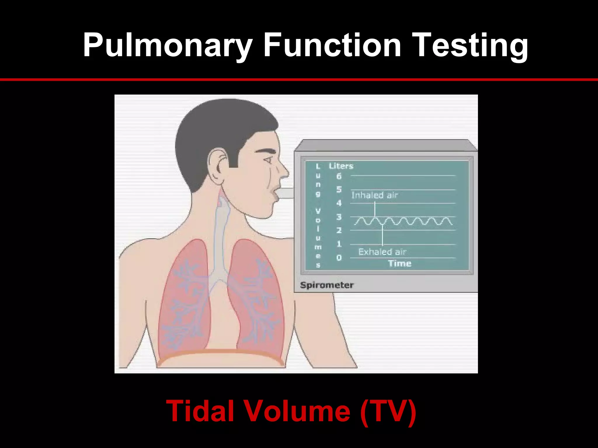
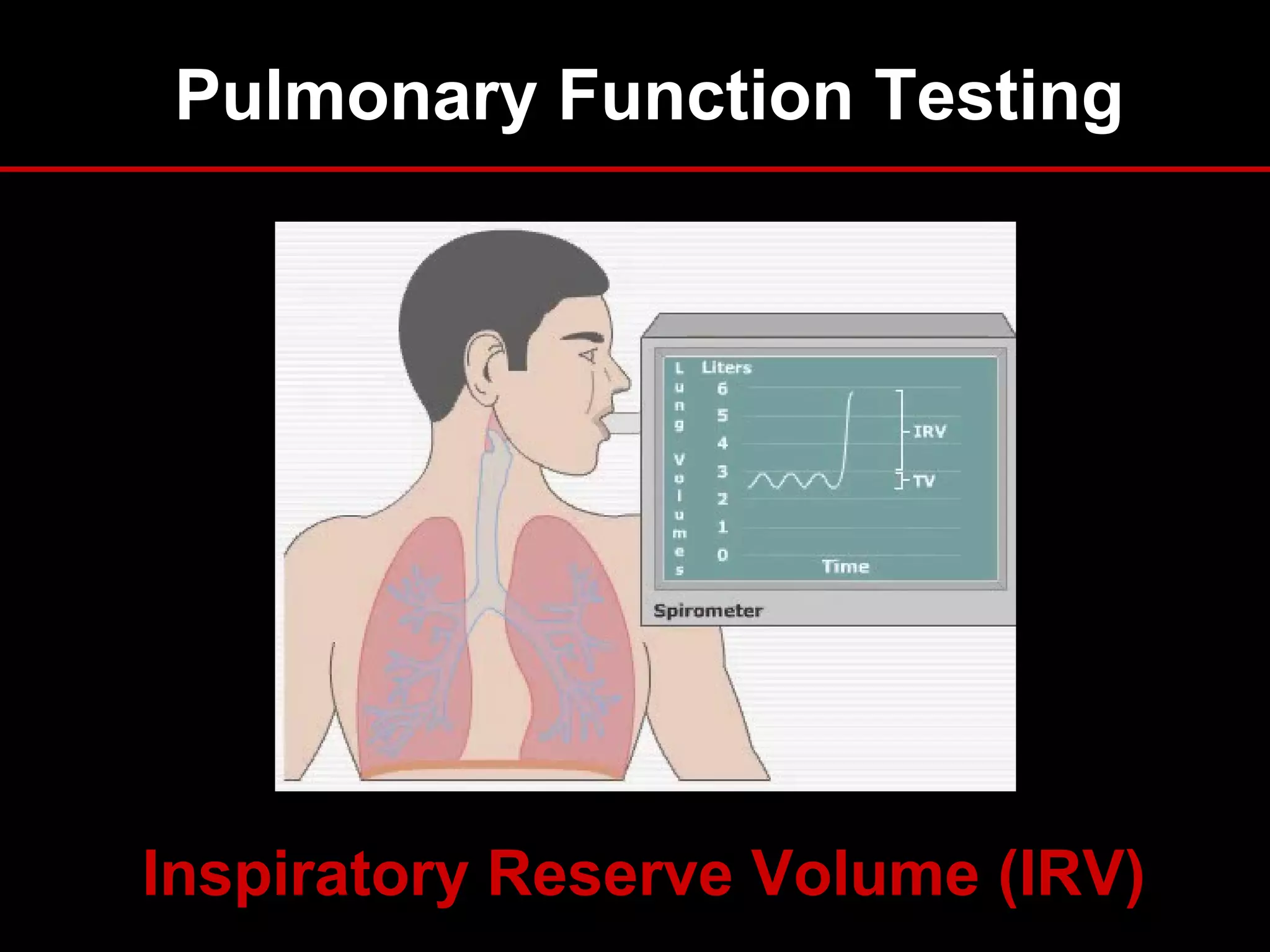
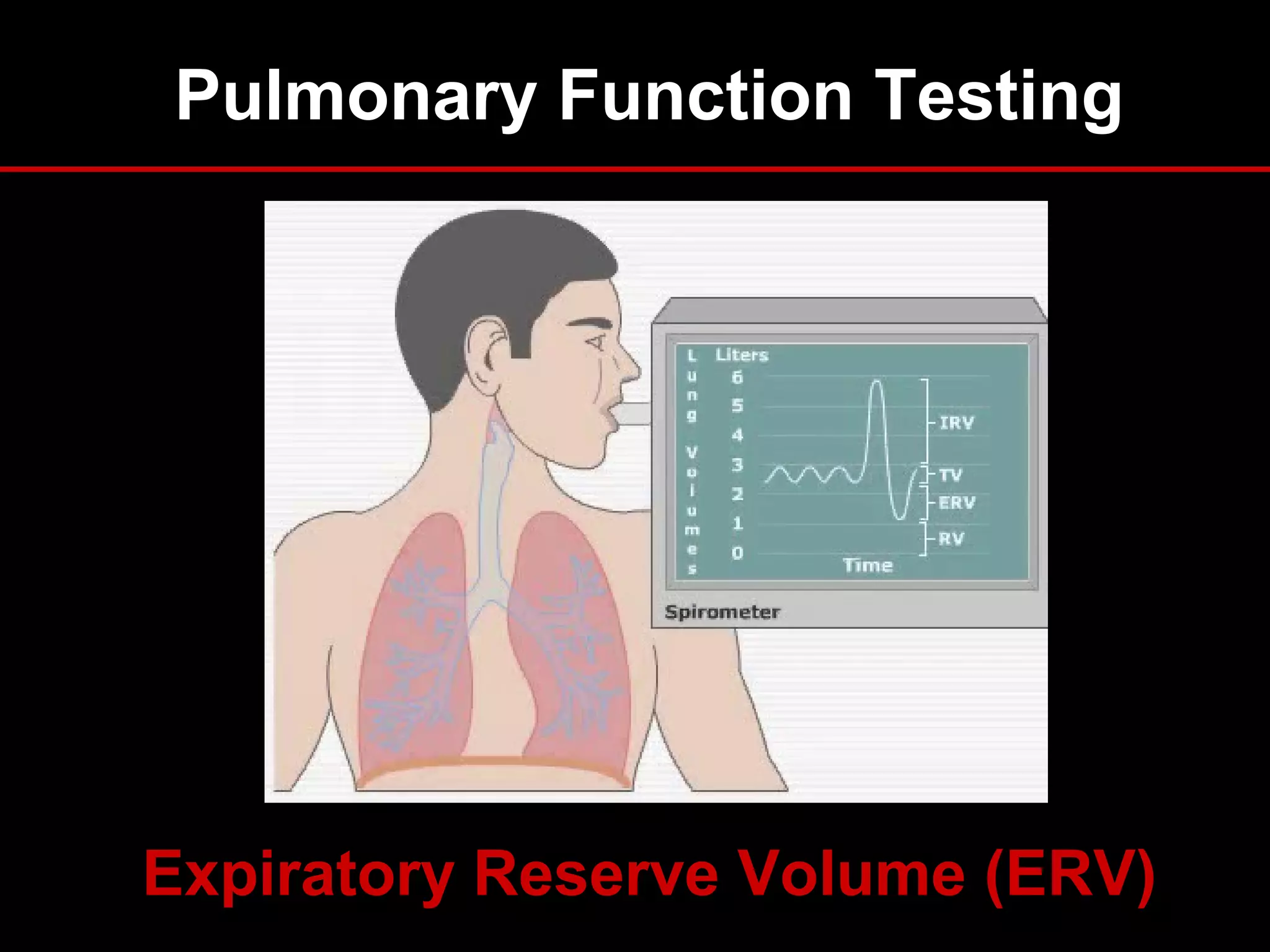
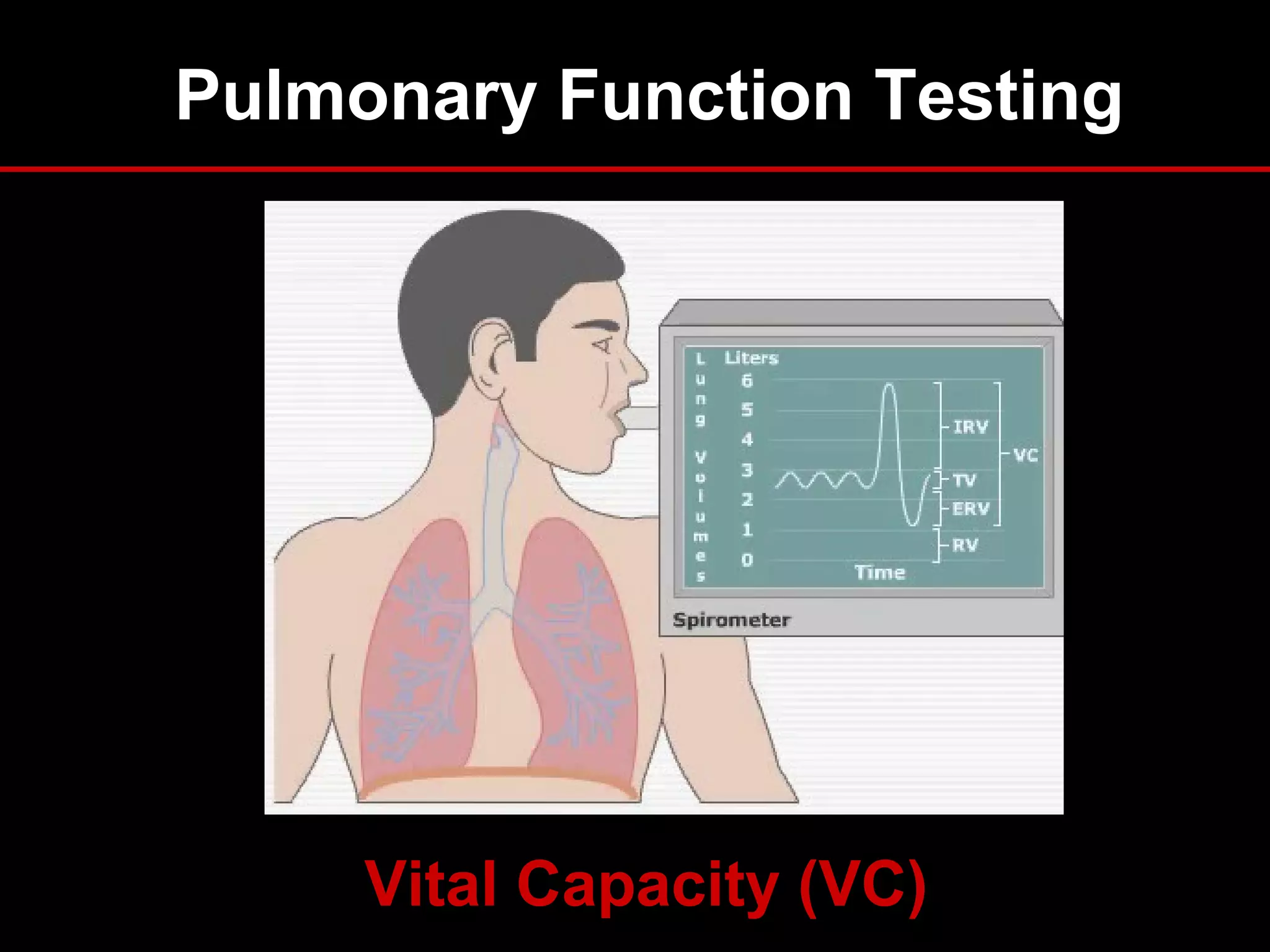
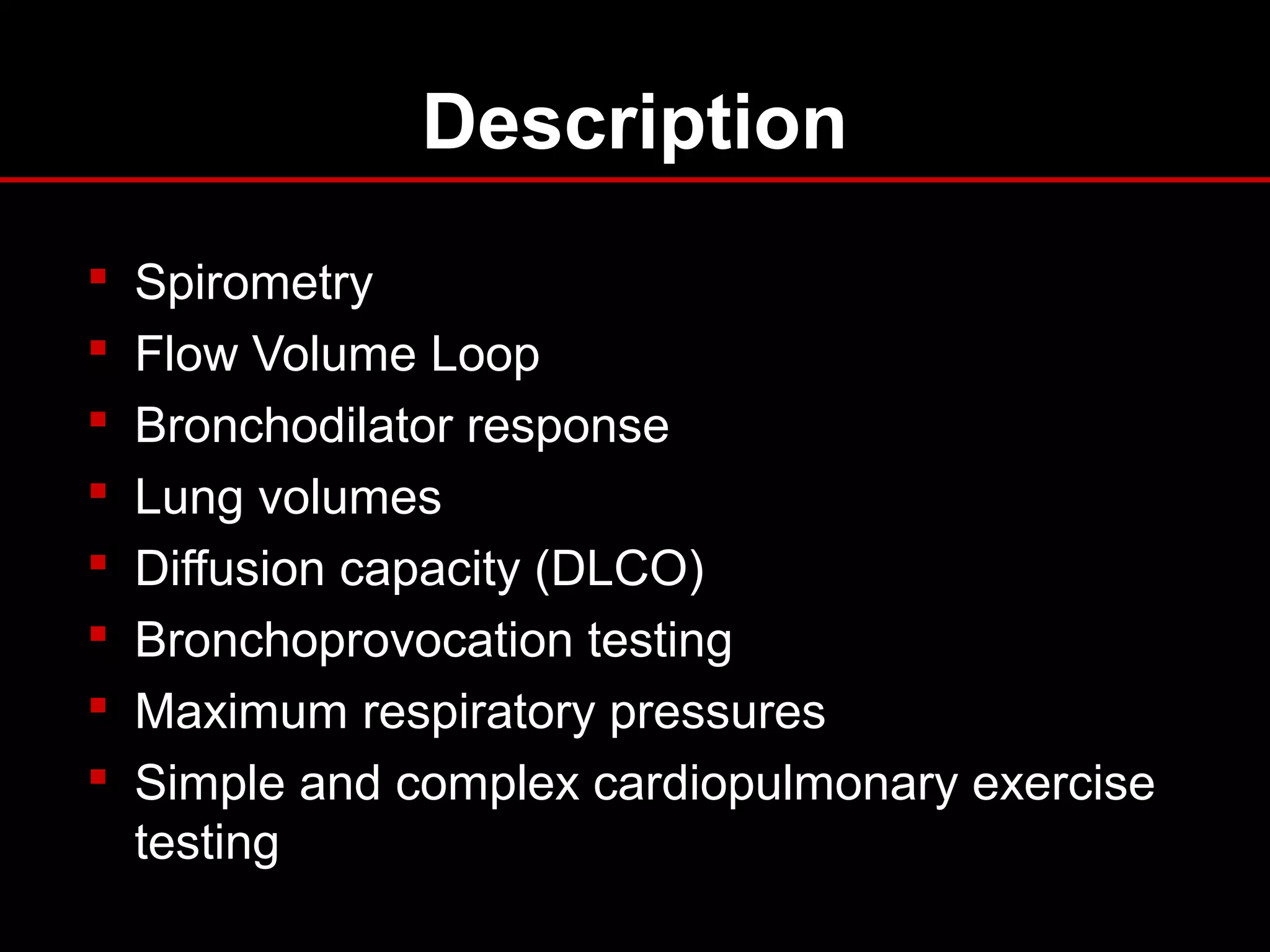
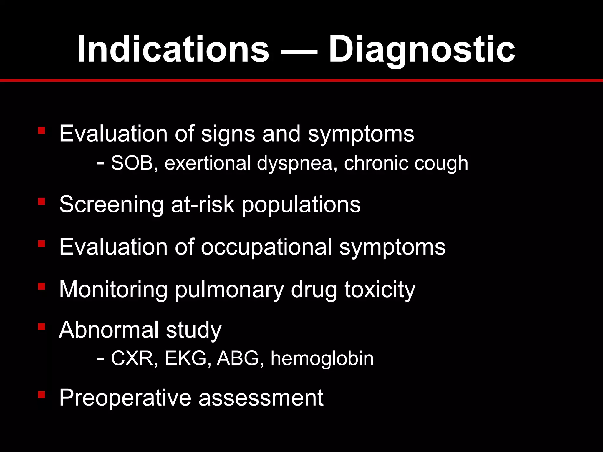
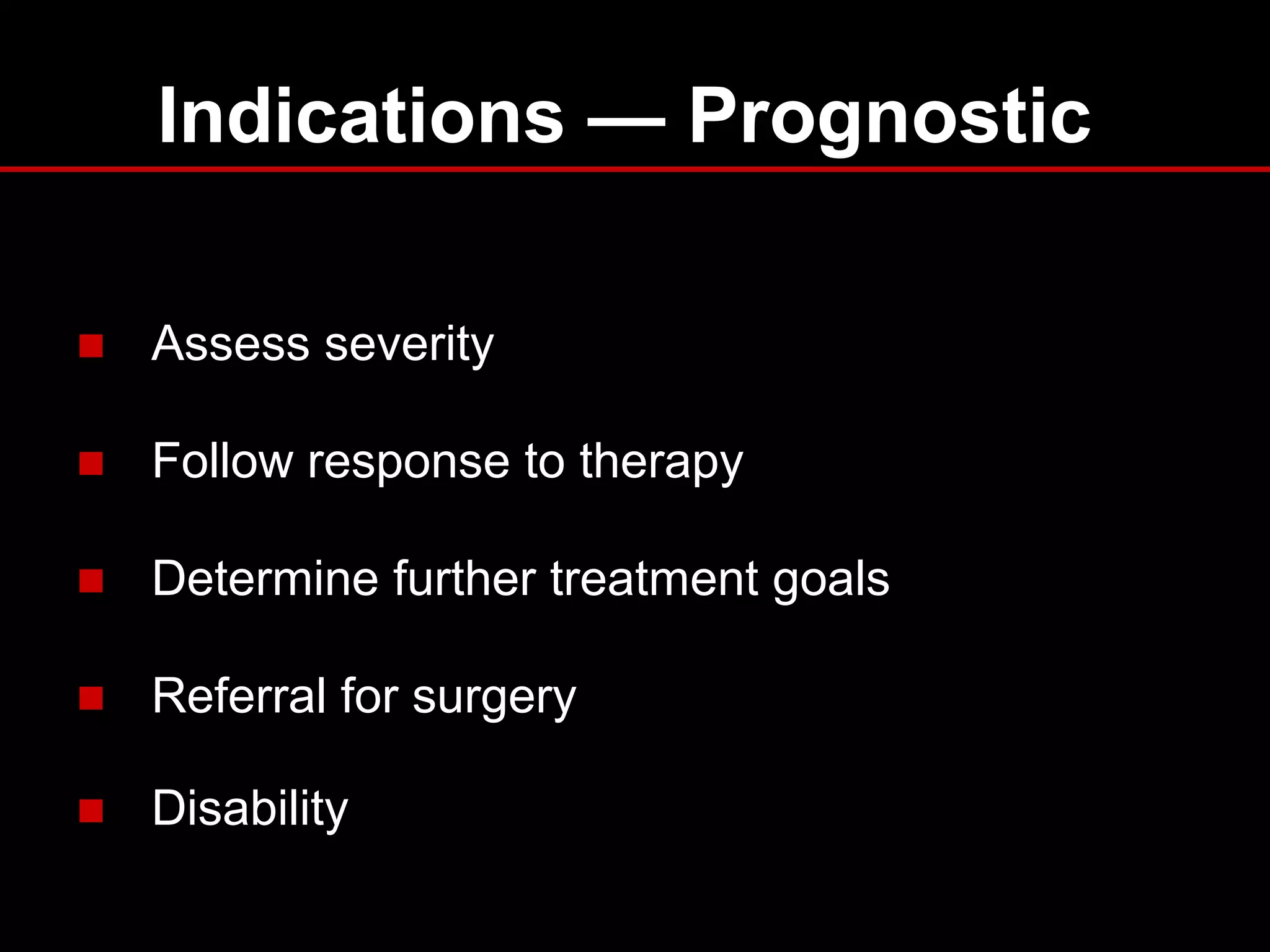
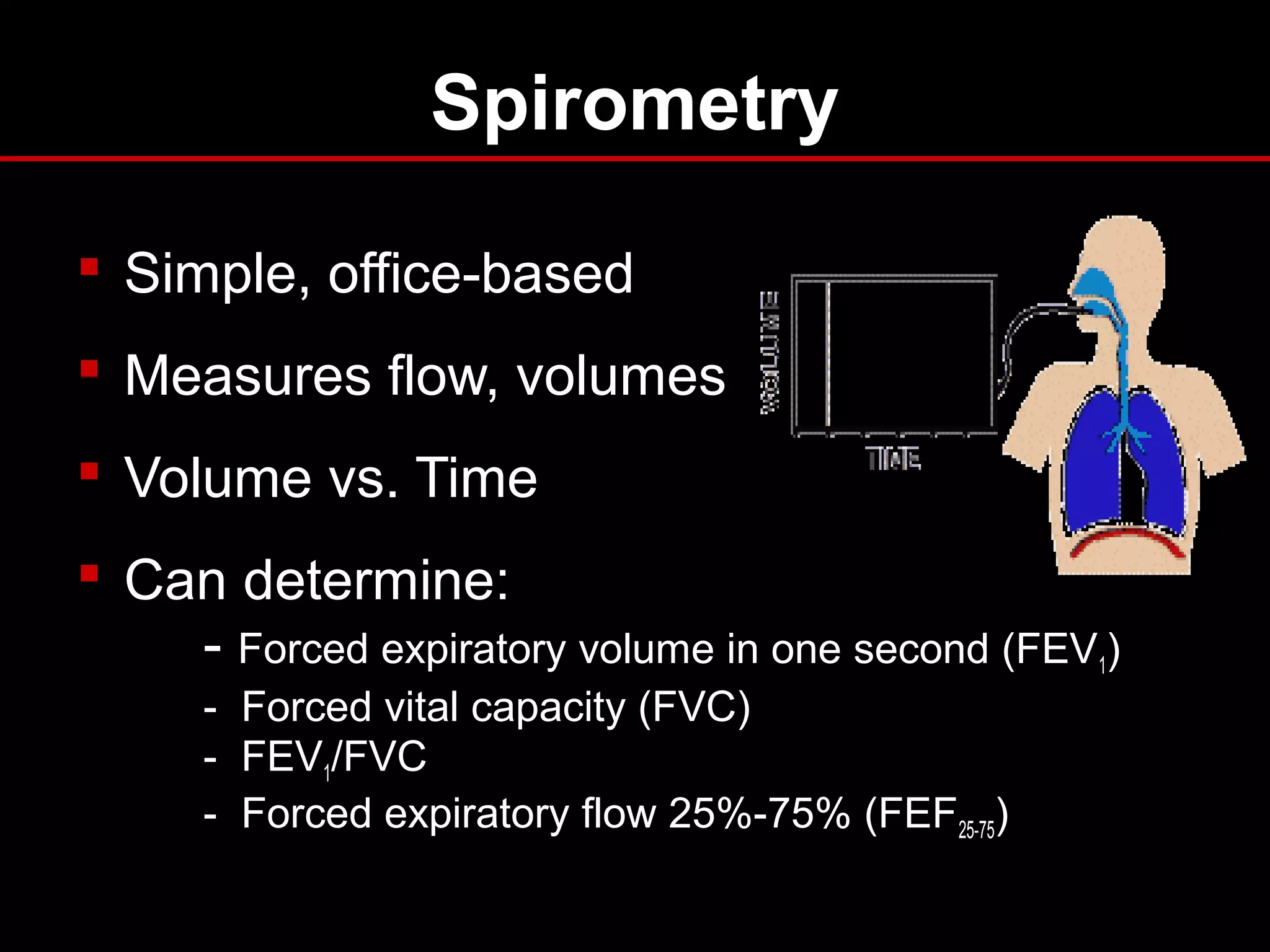
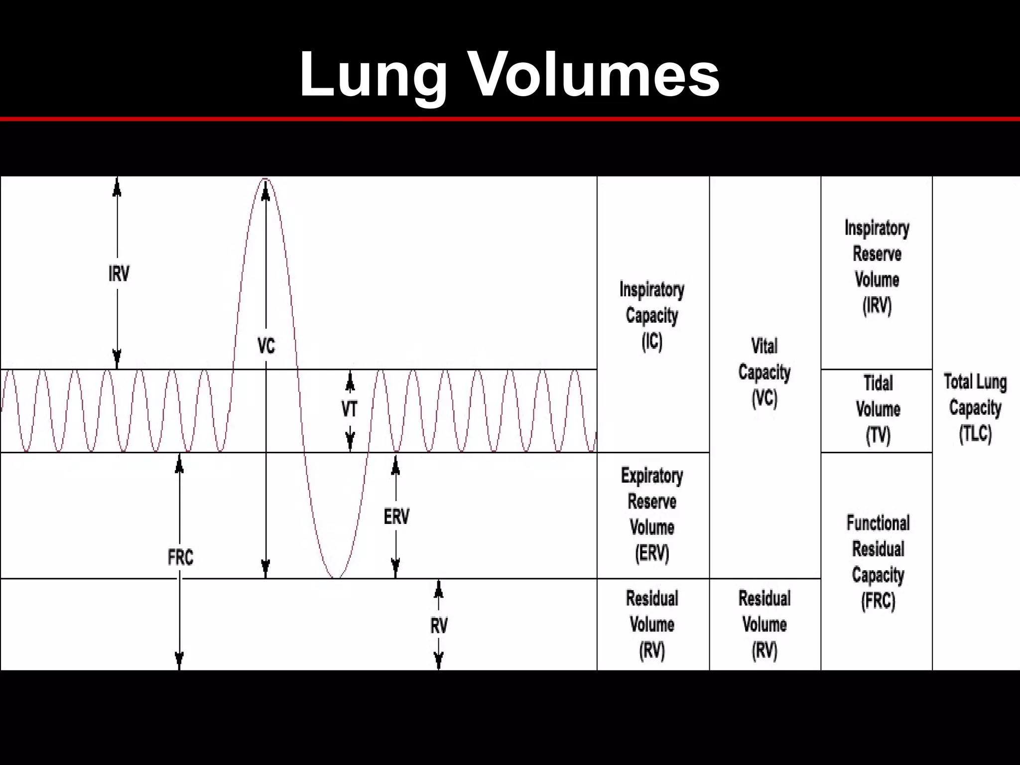
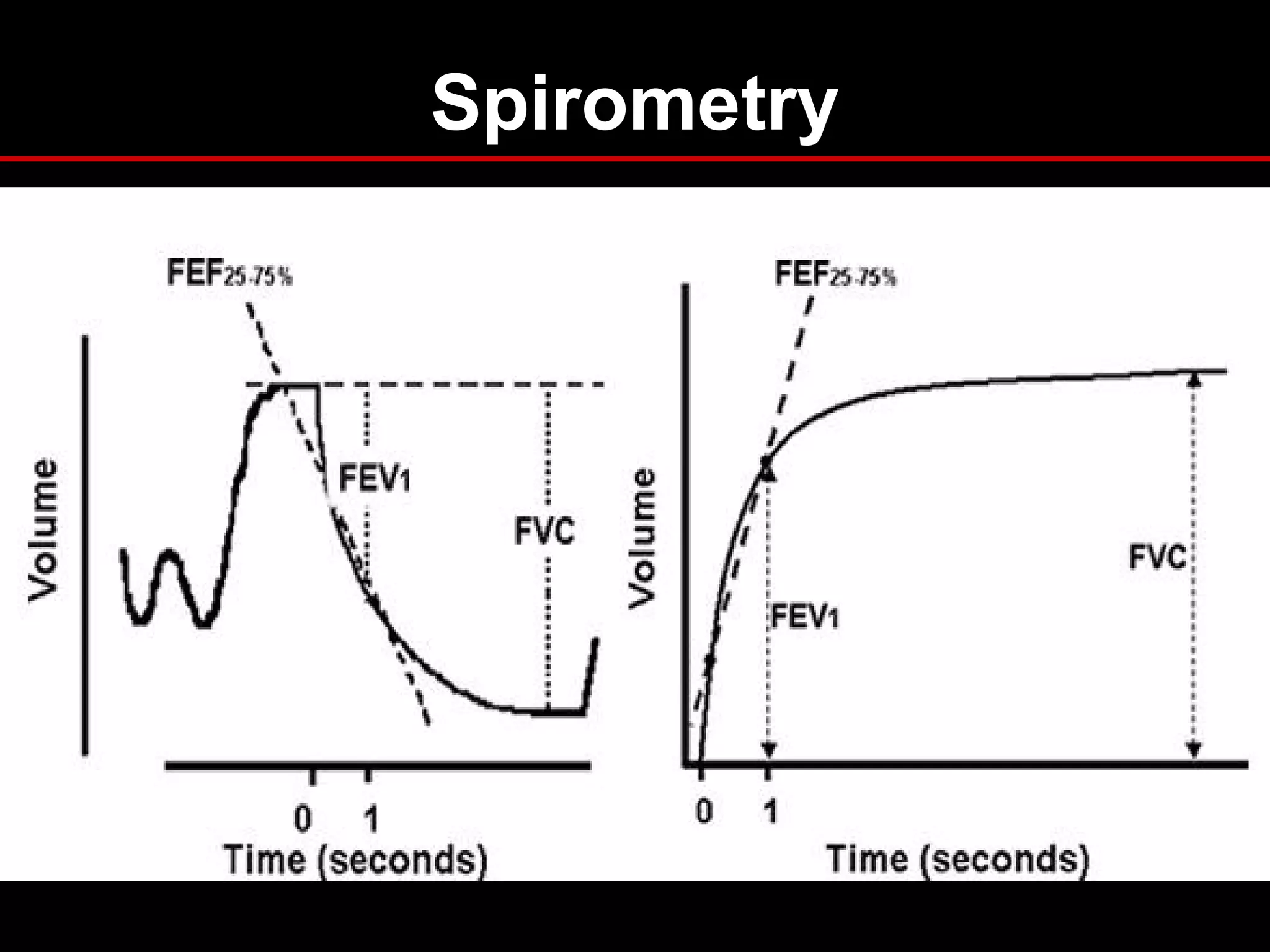
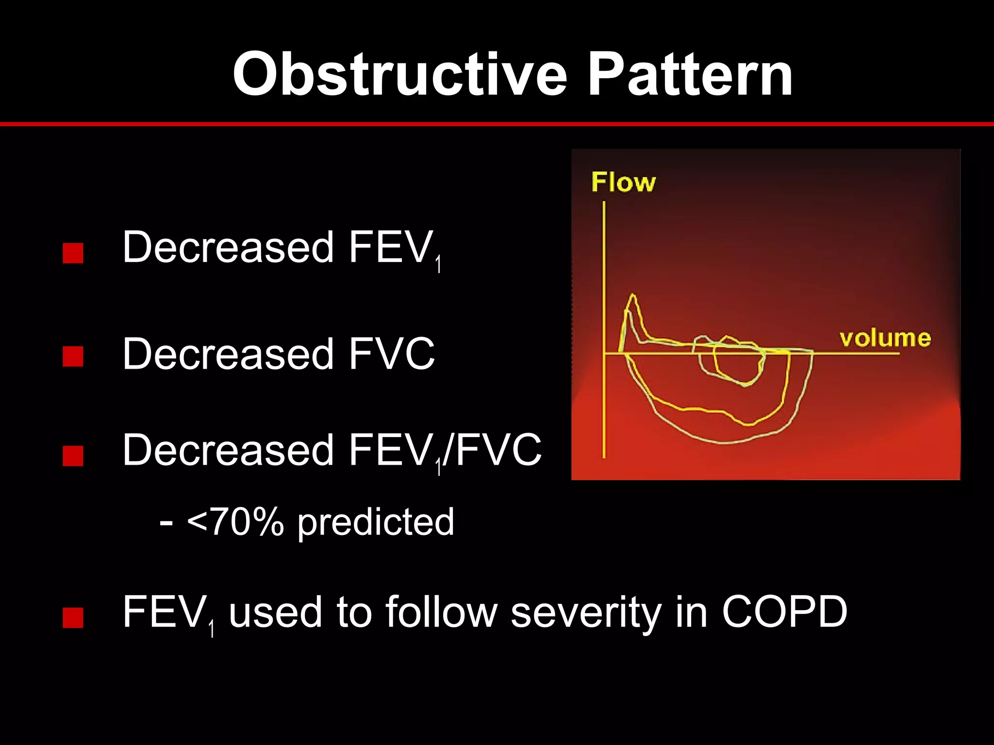
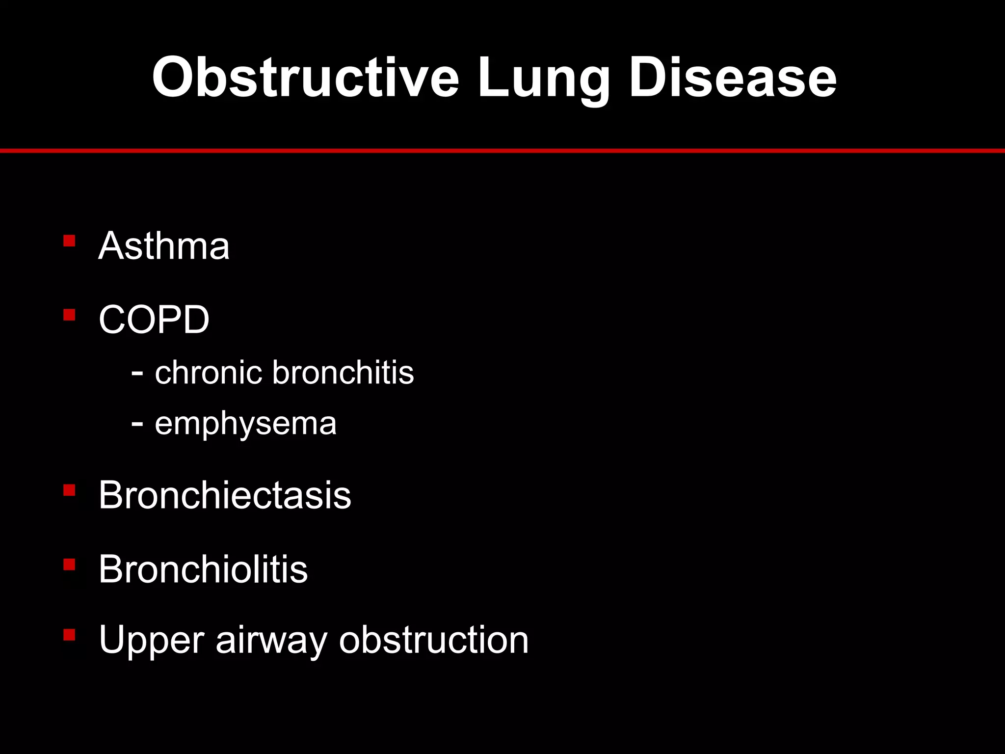
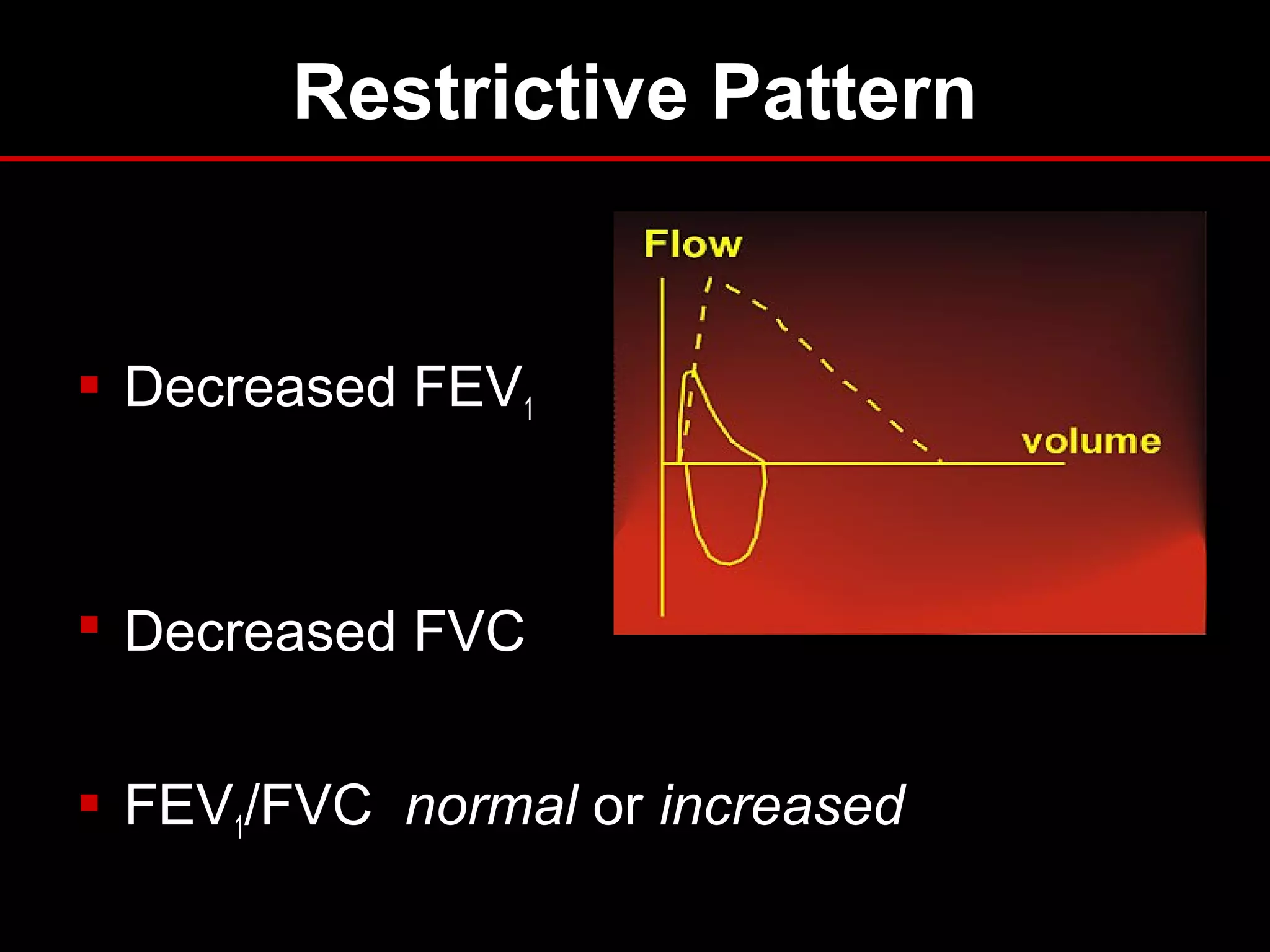
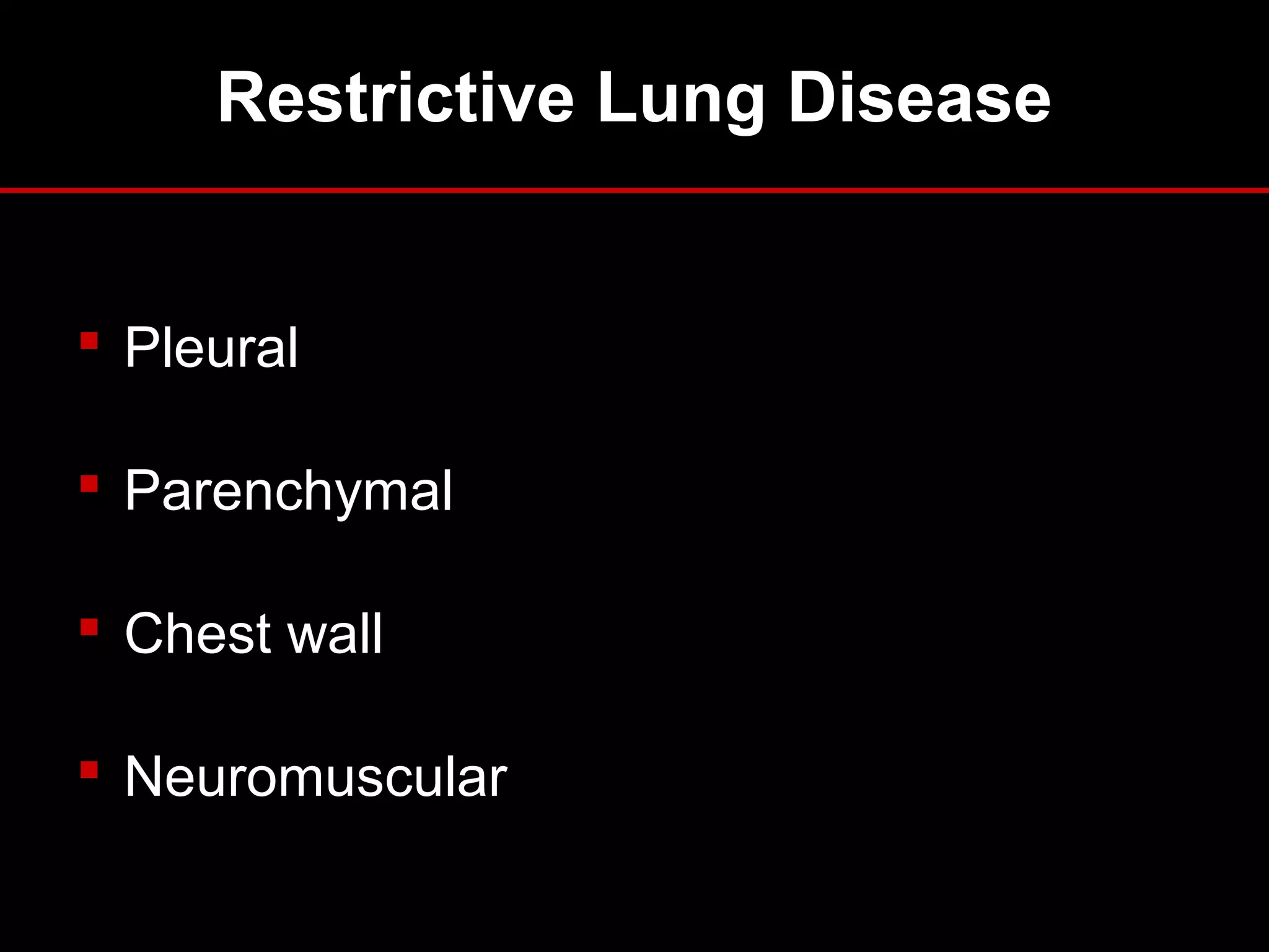
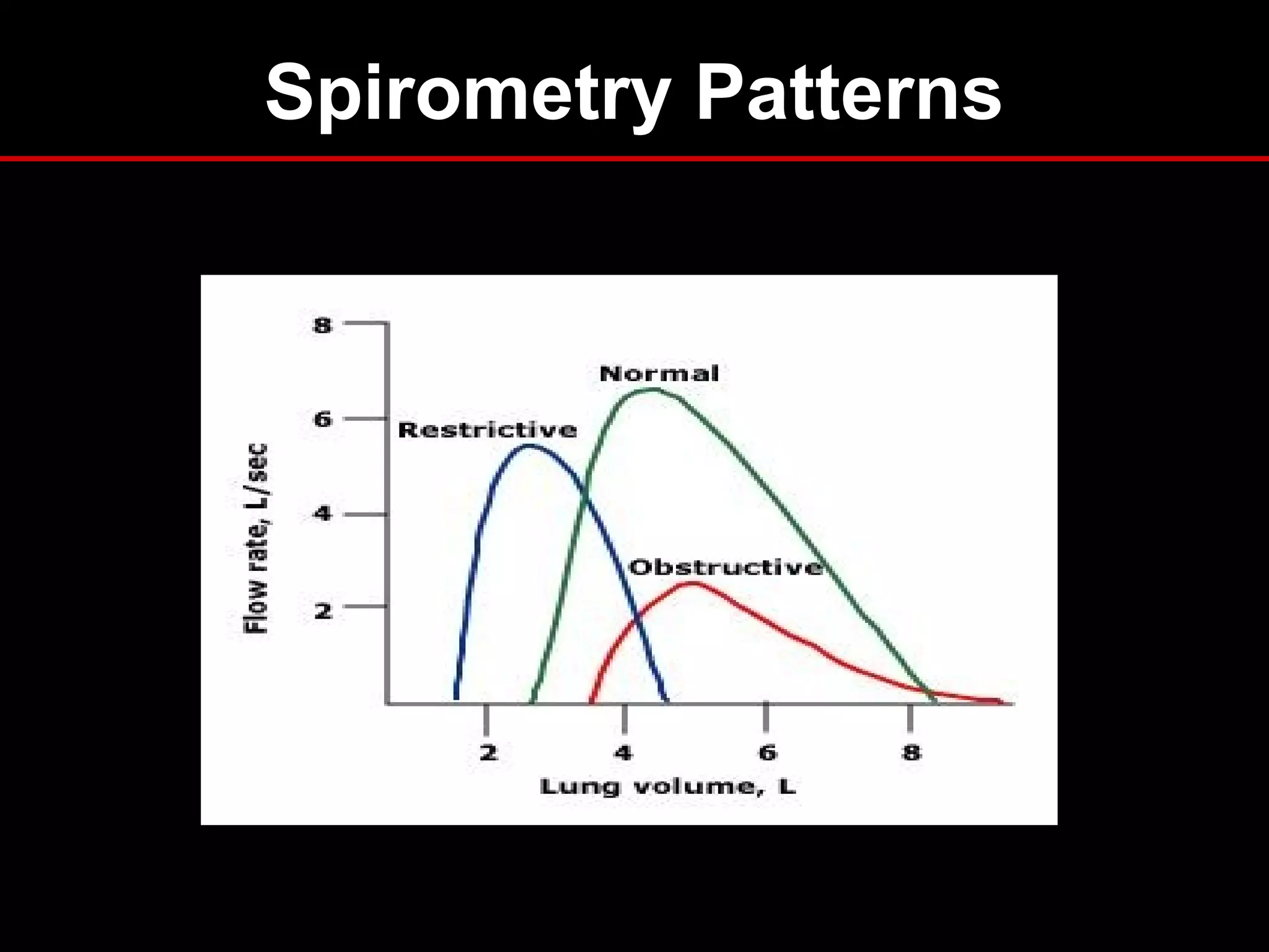
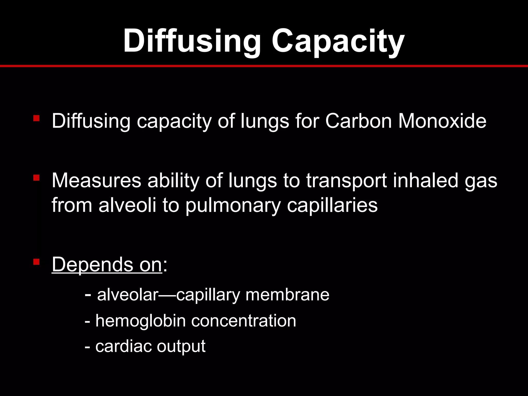
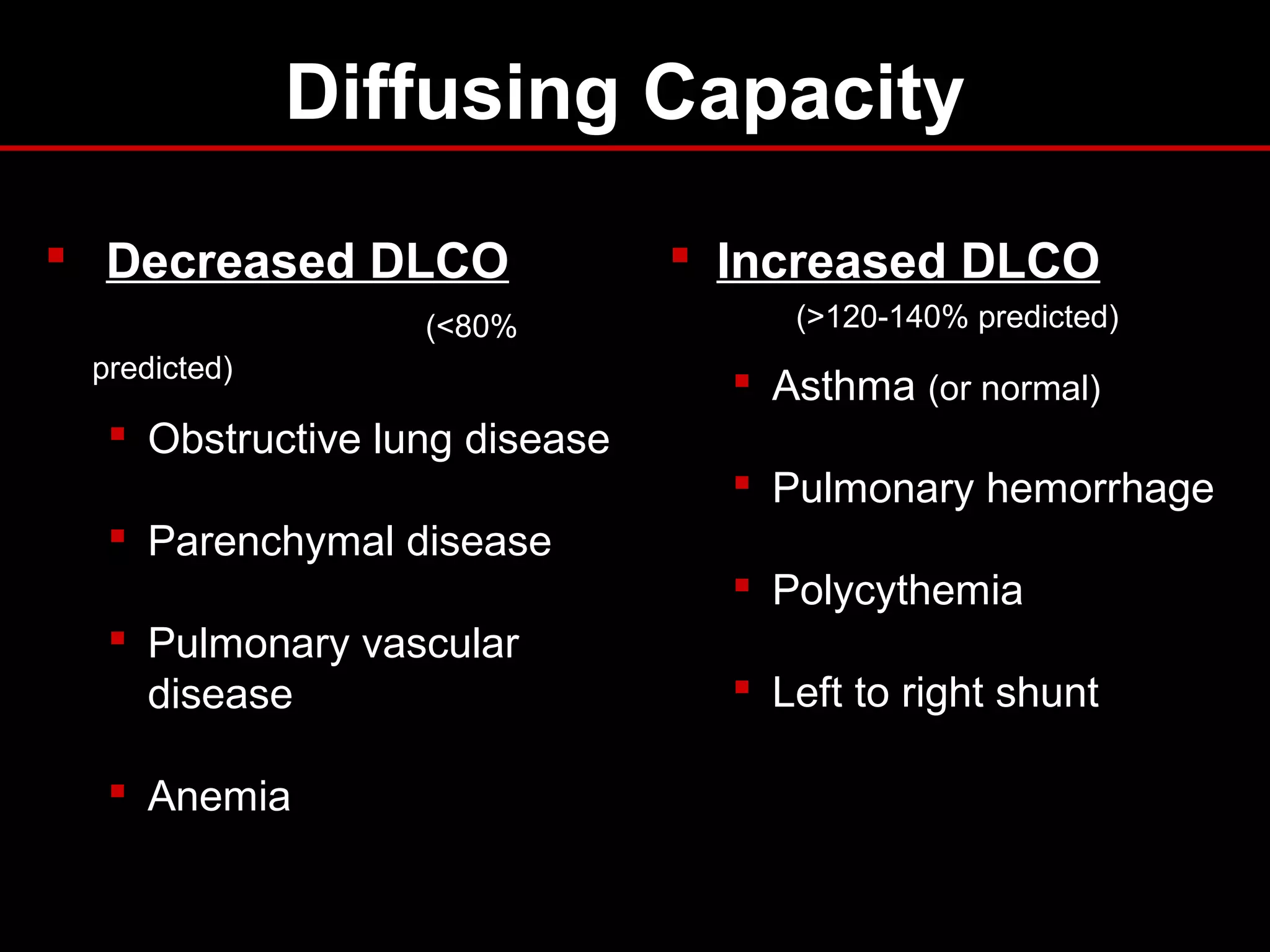
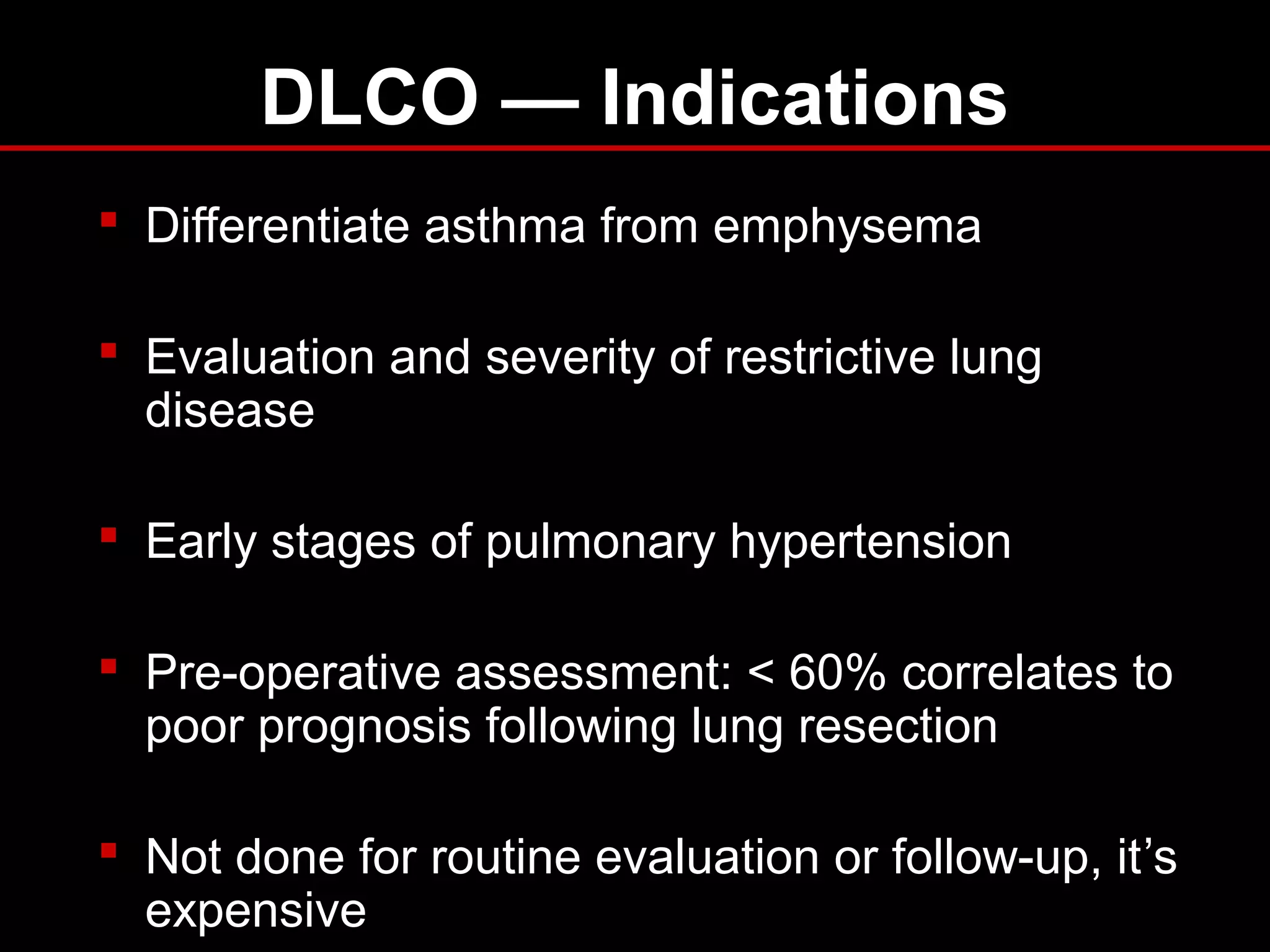
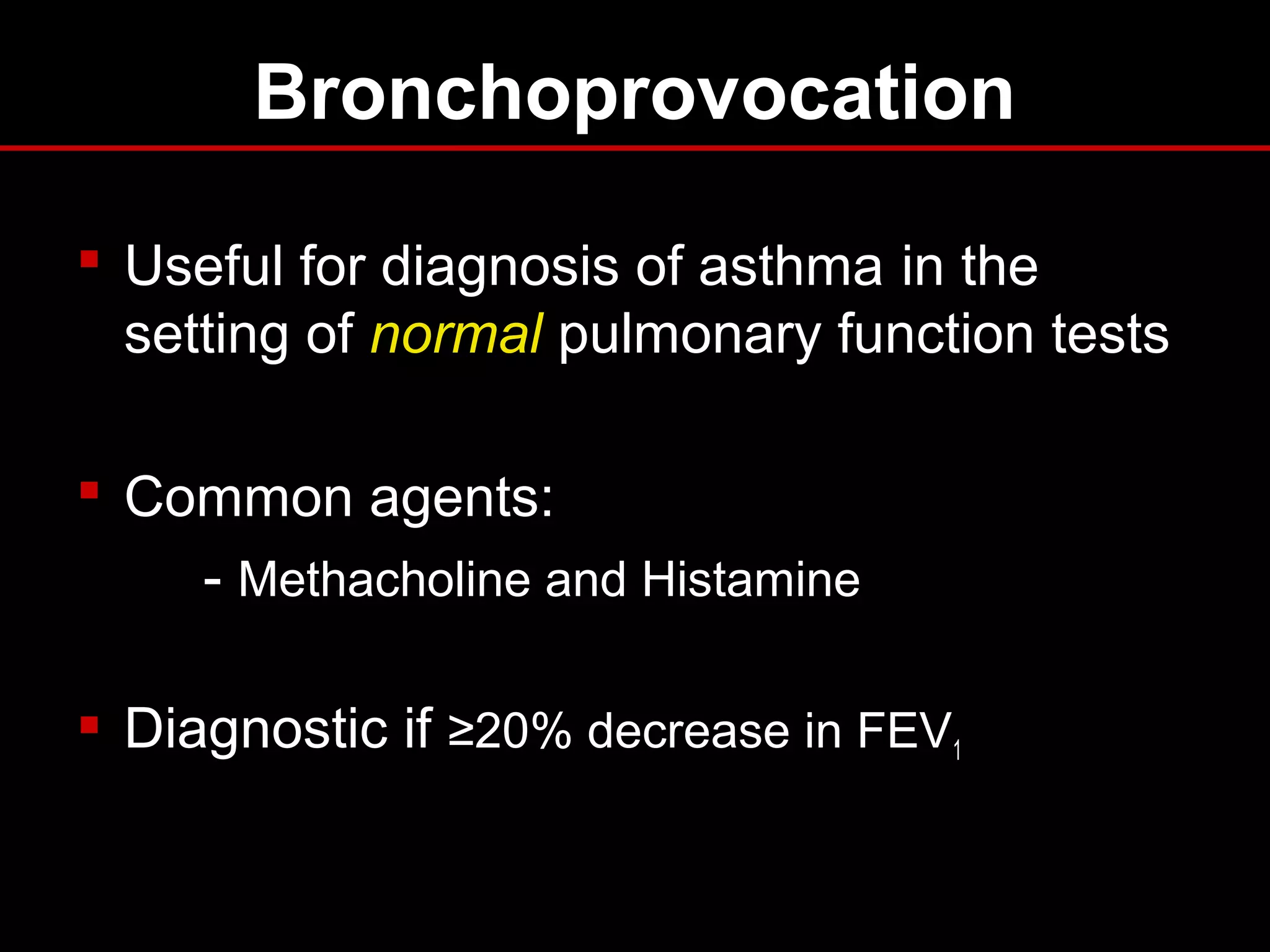
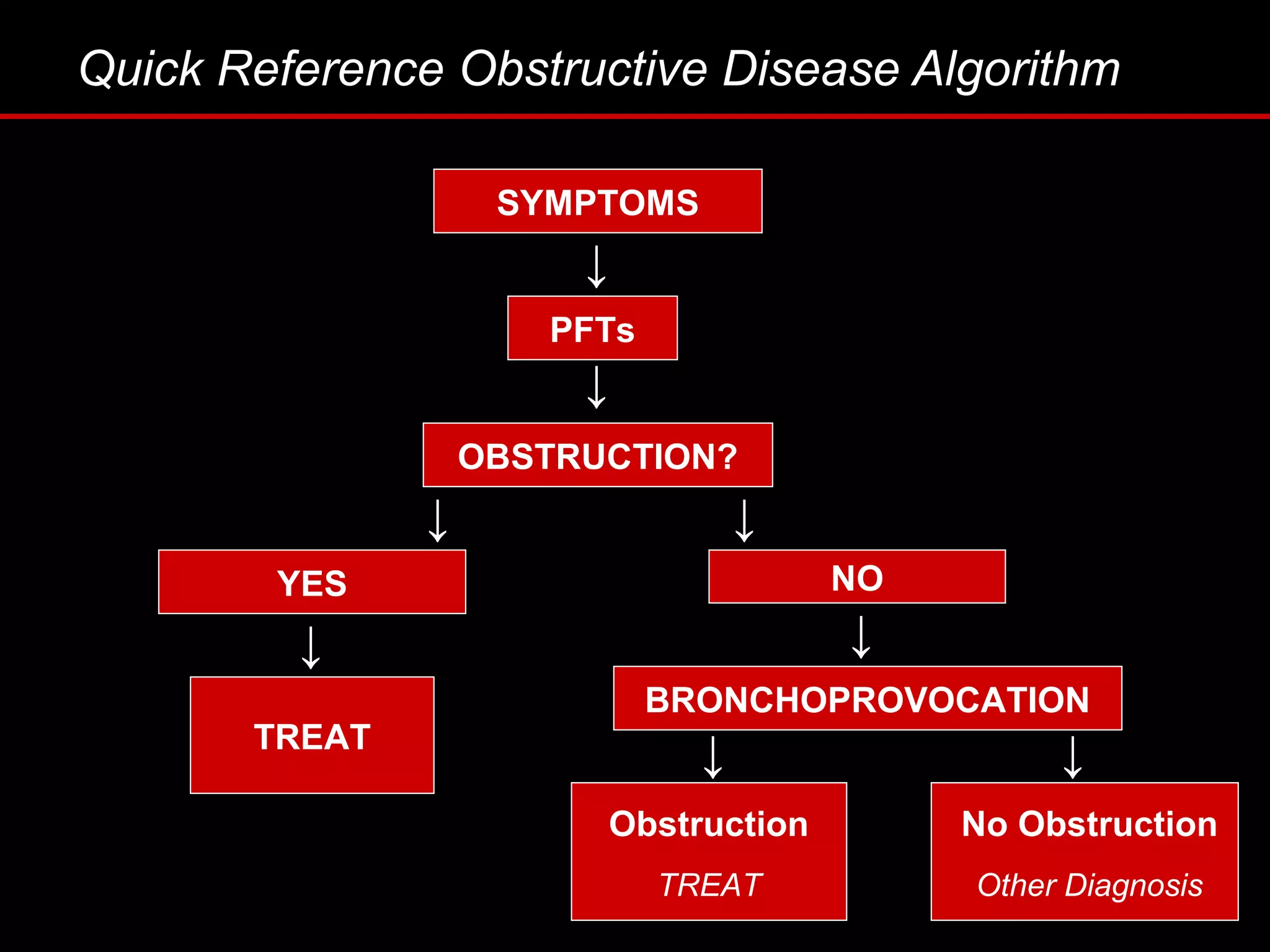
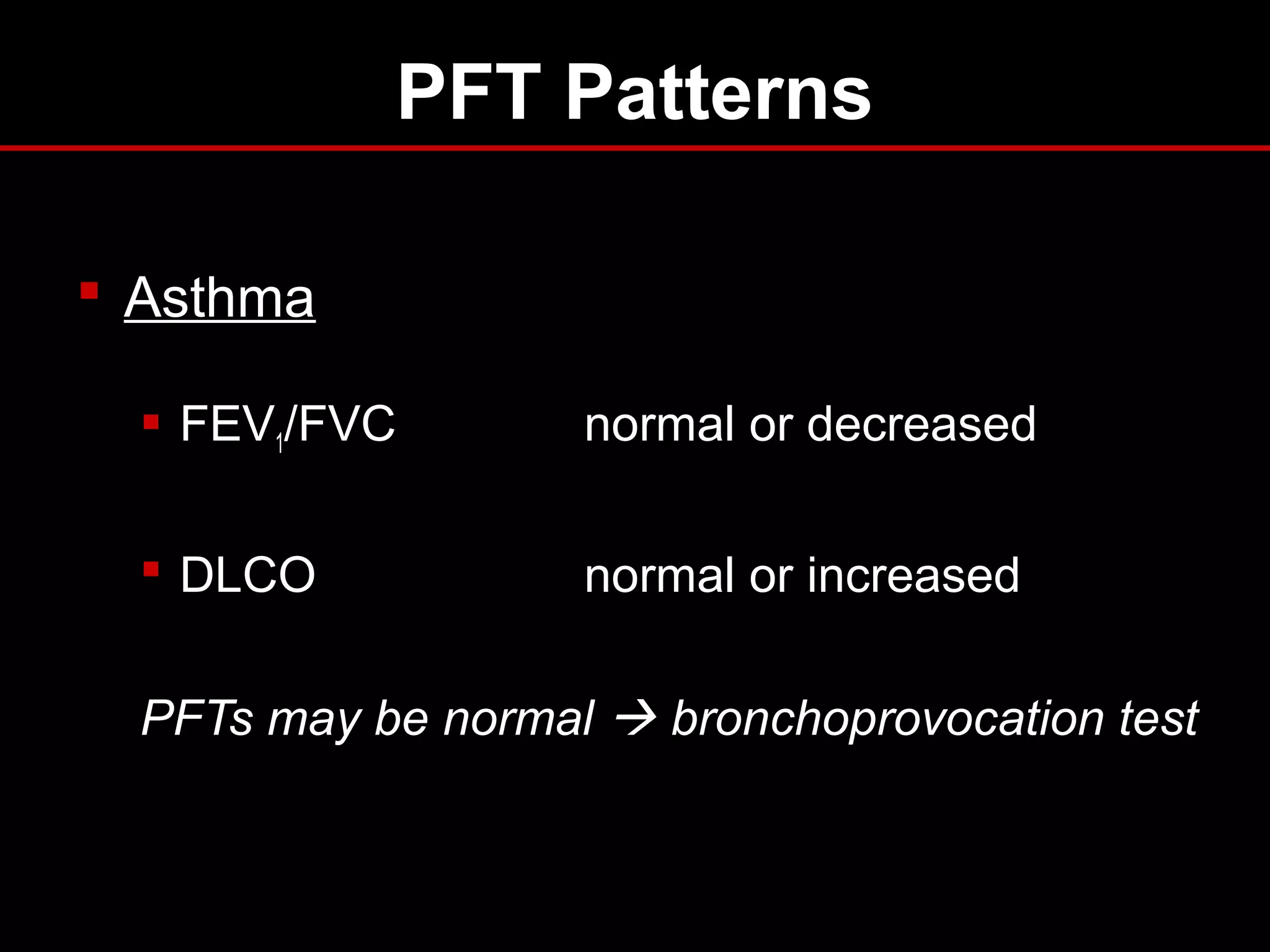
Pulmonary Function Testing:
The Basics
Dene W. Daugherty, DO
Department of Surgery
Pulmonary Function Testing
Tidal Volume (TV)
Pulmonary Function Testing
Inspiratory Reserve Volume (IRV)
Pulmonary Function Testing
Expiratory Reserve Volume (ERV)
Pulmonary Function Testing
Vital Capacity (VC)
Pulmonary Function Testing
Total Lung Capacity (TLC)
Objectives
 Identify the components
 Describe the indications
 Interpretation of results
 Recognize common patterns
 Clinical applications
The Purpose of PFT’s
To provide a quantifiable,
reproducible measurement of lung
function
Description
 Spirometry
 Flow Volume Loop
 Bronchodilator response
 Lung volumes
 Diffusion capacity (DLCO)
 Bronchoprovocation testing
 Maximum respiratory pressures
 Simple and complex cardiopulmonary exercise
testing
Indications — Diagnostic
 Evaluation of signs and symptoms
- SOB, exertional dyspnea, chronic cough
 Screening at-risk populations
 Evaluation of occupational symptoms
 Monitoring pulmonary drug toxicity
 Abnormal study
- CXR, EKG, ABG, hemoglobin
 Preoperative assessment
Indications — Prognostic
■ Assess severity
■ Follow response to therapy
■ Determine further treatment goals
■ Referral for surgery
■ Disability
Spirometry
 Simple, office-based
 Measures flow, volumes
 Volume vs. Time
 Can determine:
- Forced expiratory volume in one second (FEV1)
- Forced vital capacity (FVC)
- FEV1/FVC
- Forced expiratory flow 25%-75% (FEF25-75)
Lung Volumes
Spirometry
Obstructive Pattern
■ Decreased FEV1
■ Decreased FVC
■ Decreased FEV1/FVC
- <70% predicted
■ FEV1 used to follow severity in COPD
Obstructive Lung Disease
 Asthma
 COPD
- chronic bronchitis
- emphysema
 Bronchiectasis
 Bronchiolitis
 Upper airway obstruction
Restrictive Pattern
 Decreased FEV1
 Decreased FVC
 FEV1/FVC normal or increased
Restrictive Lung Disease
 Pleural
 Parenchymal
 Chest wall
 Neuromuscular
Spirometry Patterns
Bronchodilator Response
 Degree to which FEV1 improves with inhaled
bronchodilator
 Documents reversible airflow obstruction
 Considered a significant response if:
- FEV1 increases by 12% and >200ml
 Request if obstructive pattern on spirometry
Flow Volume Loop
 “Spirogram”
 Measures forced inspiratory and
expiratory flow rate
 Augments spirometry results
 Indications: evaluation of upper airway
obstruction (stridor, unexplained dyspnea)
Flow Volume Loop
Upper Airway Obstruction
 Variable intrathoracic obstruction
 Variable extrathoracic obstruction
 Fixed obstruction
Upper Airway Obstruction
Lung Volumes
 Measurement:
- helium
- nitrogen washout
- body plethsmography
 Indications:
- Diagnose restrictive component
- Differentiate chronic bronchitis from
emphysema
Lung Volumes – Patterns
 Obstructive
- TLC > 120% predicted
- RV > 120% predicted
 Restrictive
- TLC < 80% predicted
- RV < 80% predicted
Diffusing Capacity
 Diffusing capacity of lungs for Carbon Monoxide
 Measures ability of lungs to transport inhaled gas
from alveoli to pulmonary capillaries
 Depends on:
- alveolar—capillary membrane
- hemoglobin concentration
- cardiac output
Diffusing Capacity
 Decreased DLCO
(<80%
predicted)
 Obstructive lung disease
 Parenchymal disease
 Pulmonary vascular
disease
 Anemia
 Increased DLCO
(>120-140% predicted)
 Asthma (or normal)
 Pulmonary hemorrhage
 Polycythemia
 Left to right shunt
DLCO — Indications
 Differentiate asthma from emphysema
 Evaluation and severity of restrictive lung
disease
 Early stages of pulmonary hypertension
 Pre-operative assessment: < 60% correlates to
poor prognosis following lung resection
 Not done for routine evaluation or follow-up, it’s
expensive
Bronchoprovocation
 Useful for diagnosis of asthma in the
setting of normal pulmonary function tests
 Common agents:
- Methacholine and Histamine
 Diagnostic if ≥20% decrease in FEV1
Quick Reference Obstructive Disease Algorithm
↓
SYMPTOMS
PFTs
OBSTRUCTION?
YES NO
TREAT
BRONCHOPROVOCATION
Obstruction
TREAT
No Obstruction
Other Diagnosis
↓
↓
↓ ↓
↓
↓ ↓
Interpretation
 What is the clinical concern?
 What is “normal” or “baseline”?
 Did the test or equipment meet American
Thoracic Society (ATS) criteria?
 Don’t forget to look at the flow volume loop.
Obstructive Pattern — Evaluation
 Spirometry
 FEV1, FVC: decreased
 FEV1/FVC: decreased (<70% predicted)
 FV Loop “scooped”
 Lung Volumes
 TLC, RV: increased
 Responds to Bronchodilator
Restrictive Pattern – Evaluation
 Spirometry
 FVC, FEV1: decreased
 FEV1/FVC: normal or increased
 FV Loop “witch’s hat” pattern
 DLCO decreased
 Lung Volumes
 TLC, RV: decreased
 Muscle pressures may be helpful
PFT Patterns
 Emphysema
 FEV1/FVC <70%
 “Scooped” FV curve
 TLC increased
 Increased compliance
 DLCO decreased
 Chronic Bronchitis
 FEV1/FVC <70%
 “Scooped” FV curve
 TLC normal
 Normal compliance
 DLCO usually normal
PFT Patterns
 Asthma
 FEV1/FVC normal or decreased
 DLCO normal or increased
PFTs may be normal  bronchoprovocation test
Question
Which of the following is used to follow disease severity in
COPD patients?
a. Total lung capacity (TLC)
b. Degree of responsiveness to bronchodilators
c. Forced vital capacity (FVC)
d. Forced expiratory volume in 1 second (FEV1)
e. Diffusing capacity (DLCO)
Answer
Which of the following is used to follow disease severity in
COPD patients?
a. Total lung capacity (TLC)
b. Degree of responsiveness to bronchodilators
c. Forced vital capacity (FVC)
d. Forced expiratory volume in 1 second (FEV1)
e. Diffusing capacity (DLCO)
Question
A 36yo F, non-smoker, presents to your office for follow-up of
‘recurrent bronchitis.’ You suspect asthma and decide to
order spirometry. Which of the following would you
include in your prescription for testing?
a. Diffusing Capacity (DLCO)
b. If no obstruction present, perform trial of bronchodilator
c. If no obstruction present, perform methacholine challenge
d. Flow volume loop
e. b and c
Answer
A 36yo F, non-smoker, presents to your office for follow-up of
‘recurrent bronchitis.’ You suspect asthma and decide to
order spirometry. Which of the following would you
include in your prescription for testing?
a. Diffusing Capacity (DLCO)
b. If no obstruction present, add trial of bronchodilator
c. If no obstruction present, perform methacholine challenge
d. Flow volume loop
e. b and c
Question
A 68yo M is admitted to the ICU with acute respiratory
distress. A CXR obtained in the ED demonstrates
bilateral pulmonary infiltrates, and his DLCO is
elevated. What is the most likely diagnosis?
a. Pulmonary edema
b. Aspiration pneumonitis
c. Pulmonary emboli
d. Alveolar hemorrhage
e. Interstitial lung disease
Answer
A 68yo M is admitted to the ICU with acute respiratory
distress. A CXR obtained in the ED demonstrates
bilateral pulmonary infiltrates, and his DLCO is
elevated. What is the most likely diagnosis?
a. Pulmonary edema
b. Aspiration pneumonitis
c. Pulmonary emboli
d. Alveolar hemorrhage
e. Interstitial lung disease
References
1. Aboussouan LS, Stoller JK: Flow volume loops. UpToDate, 2006.
2. Bahhady IJ, Unterborn J: Pulmonary function tests: an update. Consultant.
2003.
3. Barreiro, TJ, Perillo I: An approach to interpreting spirometry. Am Fam
Physician. 2004 Mar 1;69(5):1107-14.
4. Chesnutt MS, Prendergast TJ. Current Medical Diagnosis and Treatment.
New York: Appleton and Lange, 2006.
5. Enright PL: Diffusing capacity for carbon monoxide. UpToDate, 2007.
6. Enright PL: Overview of pulmonary function testing in adults. UpToDate,
2007.
7. Irvin CG: Bronchoprovocation testing. UpToDate, 2006.
8. West JB. Respiratory Physiology: The Essentials. Lippincot Williams &
Wilkins, 2000.
Pulmonary Function Testing

Pulmonary Function Testing

  • 1.
    Pulmonary Function Testing: TheBasics Dene W. Daugherty, DO Department of Surgery
  • 2.
  • 3.
  • 4.
  • 5.
  • 6.
  • 7.
    Objectives  Identify thecomponents  Describe the indications  Interpretation of results  Recognize common patterns  Clinical applications
  • 8.
    The Purpose ofPFT’s To provide a quantifiable, reproducible measurement of lung function
  • 9.
    Description  Spirometry  FlowVolume Loop  Bronchodilator response  Lung volumes  Diffusion capacity (DLCO)  Bronchoprovocation testing  Maximum respiratory pressures  Simple and complex cardiopulmonary exercise testing
  • 10.
    Indications — Diagnostic Evaluation of signs and symptoms - SOB, exertional dyspnea, chronic cough  Screening at-risk populations  Evaluation of occupational symptoms  Monitoring pulmonary drug toxicity  Abnormal study - CXR, EKG, ABG, hemoglobin  Preoperative assessment
  • 11.
    Indications — Prognostic ■Assess severity ■ Follow response to therapy ■ Determine further treatment goals ■ Referral for surgery ■ Disability
  • 12.
    Spirometry  Simple, office-based Measures flow, volumes  Volume vs. Time  Can determine: - Forced expiratory volume in one second (FEV1) - Forced vital capacity (FVC) - FEV1/FVC - Forced expiratory flow 25%-75% (FEF25-75)
  • 13.
  • 14.
  • 15.
    Obstructive Pattern ■ DecreasedFEV1 ■ Decreased FVC ■ Decreased FEV1/FVC - <70% predicted ■ FEV1 used to follow severity in COPD
  • 16.
    Obstructive Lung Disease Asthma  COPD - chronic bronchitis - emphysema  Bronchiectasis  Bronchiolitis  Upper airway obstruction
  • 17.
    Restrictive Pattern  DecreasedFEV1  Decreased FVC  FEV1/FVC normal or increased
  • 18.
    Restrictive Lung Disease Pleural  Parenchymal  Chest wall  Neuromuscular
  • 19.
  • 20.
    Bronchodilator Response  Degreeto which FEV1 improves with inhaled bronchodilator  Documents reversible airflow obstruction  Considered a significant response if: - FEV1 increases by 12% and >200ml  Request if obstructive pattern on spirometry
  • 21.
    Flow Volume Loop “Spirogram”  Measures forced inspiratory and expiratory flow rate  Augments spirometry results  Indications: evaluation of upper airway obstruction (stridor, unexplained dyspnea)
  • 22.
  • 23.
    Upper Airway Obstruction Variable intrathoracic obstruction  Variable extrathoracic obstruction  Fixed obstruction
  • 24.
  • 25.
    Lung Volumes  Measurement: -helium - nitrogen washout - body plethsmography  Indications: - Diagnose restrictive component - Differentiate chronic bronchitis from emphysema
  • 26.
    Lung Volumes –Patterns  Obstructive - TLC > 120% predicted - RV > 120% predicted  Restrictive - TLC < 80% predicted - RV < 80% predicted
  • 27.
    Diffusing Capacity  Diffusingcapacity of lungs for Carbon Monoxide  Measures ability of lungs to transport inhaled gas from alveoli to pulmonary capillaries  Depends on: - alveolar—capillary membrane - hemoglobin concentration - cardiac output
  • 28.
    Diffusing Capacity  DecreasedDLCO (<80% predicted)  Obstructive lung disease  Parenchymal disease  Pulmonary vascular disease  Anemia  Increased DLCO (>120-140% predicted)  Asthma (or normal)  Pulmonary hemorrhage  Polycythemia  Left to right shunt
  • 29.
    DLCO — Indications Differentiate asthma from emphysema  Evaluation and severity of restrictive lung disease  Early stages of pulmonary hypertension  Pre-operative assessment: < 60% correlates to poor prognosis following lung resection  Not done for routine evaluation or follow-up, it’s expensive
  • 30.
    Bronchoprovocation  Useful fordiagnosis of asthma in the setting of normal pulmonary function tests  Common agents: - Methacholine and Histamine  Diagnostic if ≥20% decrease in FEV1
  • 31.
    Quick Reference ObstructiveDisease Algorithm ↓ SYMPTOMS PFTs OBSTRUCTION? YES NO TREAT BRONCHOPROVOCATION Obstruction TREAT No Obstruction Other Diagnosis ↓ ↓ ↓ ↓ ↓ ↓ ↓
  • 32.
    Interpretation  What isthe clinical concern?  What is “normal” or “baseline”?  Did the test or equipment meet American Thoracic Society (ATS) criteria?  Don’t forget to look at the flow volume loop.
  • 33.
    Obstructive Pattern —Evaluation  Spirometry  FEV1, FVC: decreased  FEV1/FVC: decreased (<70% predicted)  FV Loop “scooped”  Lung Volumes  TLC, RV: increased  Responds to Bronchodilator
  • 34.
    Restrictive Pattern –Evaluation  Spirometry  FVC, FEV1: decreased  FEV1/FVC: normal or increased  FV Loop “witch’s hat” pattern  DLCO decreased  Lung Volumes  TLC, RV: decreased  Muscle pressures may be helpful
  • 35.
    PFT Patterns  Emphysema FEV1/FVC <70%  “Scooped” FV curve  TLC increased  Increased compliance  DLCO decreased  Chronic Bronchitis  FEV1/FVC <70%  “Scooped” FV curve  TLC normal  Normal compliance  DLCO usually normal
  • 36.
    PFT Patterns  Asthma FEV1/FVC normal or decreased  DLCO normal or increased PFTs may be normal  bronchoprovocation test
  • 37.
    Question Which of thefollowing is used to follow disease severity in COPD patients? a. Total lung capacity (TLC) b. Degree of responsiveness to bronchodilators c. Forced vital capacity (FVC) d. Forced expiratory volume in 1 second (FEV1) e. Diffusing capacity (DLCO)
  • 38.
    Answer Which of thefollowing is used to follow disease severity in COPD patients? a. Total lung capacity (TLC) b. Degree of responsiveness to bronchodilators c. Forced vital capacity (FVC) d. Forced expiratory volume in 1 second (FEV1) e. Diffusing capacity (DLCO)
  • 39.
    Question A 36yo F,non-smoker, presents to your office for follow-up of ‘recurrent bronchitis.’ You suspect asthma and decide to order spirometry. Which of the following would you include in your prescription for testing? a. Diffusing Capacity (DLCO) b. If no obstruction present, perform trial of bronchodilator c. If no obstruction present, perform methacholine challenge d. Flow volume loop e. b and c
  • 40.
    Answer A 36yo F,non-smoker, presents to your office for follow-up of ‘recurrent bronchitis.’ You suspect asthma and decide to order spirometry. Which of the following would you include in your prescription for testing? a. Diffusing Capacity (DLCO) b. If no obstruction present, add trial of bronchodilator c. If no obstruction present, perform methacholine challenge d. Flow volume loop e. b and c
  • 41.
    Question A 68yo Mis admitted to the ICU with acute respiratory distress. A CXR obtained in the ED demonstrates bilateral pulmonary infiltrates, and his DLCO is elevated. What is the most likely diagnosis? a. Pulmonary edema b. Aspiration pneumonitis c. Pulmonary emboli d. Alveolar hemorrhage e. Interstitial lung disease
  • 42.
    Answer A 68yo Mis admitted to the ICU with acute respiratory distress. A CXR obtained in the ED demonstrates bilateral pulmonary infiltrates, and his DLCO is elevated. What is the most likely diagnosis? a. Pulmonary edema b. Aspiration pneumonitis c. Pulmonary emboli d. Alveolar hemorrhage e. Interstitial lung disease
  • 43.
    References 1. Aboussouan LS,Stoller JK: Flow volume loops. UpToDate, 2006. 2. Bahhady IJ, Unterborn J: Pulmonary function tests: an update. Consultant. 2003. 3. Barreiro, TJ, Perillo I: An approach to interpreting spirometry. Am Fam Physician. 2004 Mar 1;69(5):1107-14. 4. Chesnutt MS, Prendergast TJ. Current Medical Diagnosis and Treatment. New York: Appleton and Lange, 2006. 5. Enright PL: Diffusing capacity for carbon monoxide. UpToDate, 2007. 6. Enright PL: Overview of pulmonary function testing in adults. UpToDate, 2007. 7. Irvin CG: Bronchoprovocation testing. UpToDate, 2006. 8. West JB. Respiratory Physiology: The Essentials. Lippincot Williams & Wilkins, 2000.

Editor's Notes

  • #10 The 6min walk test is great to evaluate physical function and can be used to assess therapeutic response in COPD and idiopathic pulmonary fibrosis (IPF) patients. If oxygen sats fall by &amp;gt;4% (ending below 93%), this indicates significant desaturation, and need confirmatory ABGs. Maximum respiratory pressures are used to identify a neuromuscular cause of restrictive lung disease. Simple and complex cardiopulmonary exercise testing will not be addressed in this lecture.
  • #11 Preop assessment is rarely to tell surgeon not to operate, but to prepare for pulmonary complications such as pneumonia, prolonged mechanical ventilation, etc. Also for screening: this includes all current and former smokers &amp;gt;45yoa, known COPD or asthma pts, also those scheduled for thoracic or upper abdominal surgery. If mod-severe obstruction identified and surgery can be delayed, can start prophylactic program of pulmonary hygiene, stop smoking, give inhaled bronchodilators or steroids, etc.
  • #14 Image source: http://en.wikipedia.org/wiki/Main_Page
  • #15 Image source: http://www.nationalasthma.org.au/html/management/spiro_book/index.asp
  • #16 Image source: http://www.spirxpert.com/index.html FEV1 is decreased out of proportion to FVC, which causes the ratio to decrease as well.
  • #17 This is not a complete list, just some of the most common diseases that should be on your differential for obstructive lung disease.
  • #18 Image source: http://www.spirxpert.com/index.html FEV1 decreases in proportion to decrease in FVC, so ratio remains normal or even slightly increased
  • #19 Restrictive lung disease is made up of intrinsic lung disease (causes inflammation and scarring (interstitial lung diseases) or fill the airspaces w/ debris, inflammation (exudate); extrinsic causes are chest wall or pleural diseases that mechanically compress the lung and prevent expansion. Neuromuscular causes decreases ability of respiratory muscles to inflate and deflate the lungs.
  • #21 Lack of observed response to bronchodilator does not preclude use, b/c patients may have symptomatic benefit. Can give 6-8wk trial of bronchodilator and/or inhaled corticosteroids (ICS) and reassess clinically, can also obtain FEV1 at that time. HOLD MDI THE MORNING PRIOR TO TESTING.
  • #22 Have patient breath out at max effort, then breath in quickly at max effort, creates a loop w/ differing patterns. Upper airway = pharynx, larynx, trachea.
  • #23 Image source: http://www.nationalasthma.org.au/html/management/spiro_book/index.asp Vocal cord dysfunction: variable extrathoracic obstruction. Tracheal stenosis: fixed obstruction (hx frequent intubations). Rapid rise to peak flow rate, followed by fall in flow as pt exhales toward residual volume. Inspiratory curve is symmetrical.
  • #24 Example of someone grabbing trachea—causes problems w/ inspiration and expiration = fixed obstruction Vocal cord dysfunction: variable extrathoracic obstruction. Endobronchial carcinoma: variable intrathoracic obstruction. (Rare to diagnose this on flow volume loop).
  • #26 FVC is decreased in both obstructive and restrictive disease, so usually need to obtain lung volumes to see if restrictive component present (increased TLC).
  • #28 Measure of gas exchange at alveolar-capillary membrane. Changes in DLCO are one of the earliest signs of interstitial lung disease (ILD).
  • #29 Pulmonary vascular disease = pulmonary emboli, pulmonary HTN. Low DLCO is also a major predictor of desaturation during exercise.
  • #30 So you have restrictive disease by spirometry and lung volumes. You get a DLCO and see it is normal. Thinking back to your differential diagnosis of restrictive lung disease (what are the four things on your differential?), what can you probably rule out? Answer = Interstitial lung disease. This is where you would order max respiratory pressures, to evaluate for NM disease. Max inspiratory pressures are recorded as patientt is breathing through a blocked tube, also done for expiration. Should be decreased in NM disease.
  • #31 Can always send patient home and tell them to come back when having symptoms, but this delays diagnosis. Another alternative is measure peak flow variability at home. If suspected asthma but has not responded to therapy, think of obtaining flow volume loop to see if there is vocal cord dysfunction = variable extrathoracic obstruction.
  • #33 Now we’re going to put it all together…
  • #34 Don’t need a DLCO, but if were decreased would make you think emphysema, if normal then chronic bronchitis.
  • #35 IF restrictive pattern, you’re going to want to get DLCO b/c it tells you whether the restriction is due to parenchymal disease (which will change your management), or NM, pleural or CW disease
  • #36 Remember that DLCO should be normal in chronic bronchitis because it affects the more proximal airways which is not where your gas exchange takes place.
  • #39 In COPD patients, the FEV1 is used to classify severity of obstructive lung disease, and followed to assess progression.
  • #41 This is kind of tricky b/c technically the recommendation is if no obstruction, then no bronchodilator (can omit this since costs an extra $40-50). Flow volume loops will usually come w/ your testing but you need to ask if you want the full max inspiratory and expiratory curves.
  • #43 Intrinsic lung diseases that cause inflammation or scarring of the lung tissue OR that fill the airspaces w/ exudate or debris (pneumonitis or pneumonia) can cause decreased lung volumes and DLCO but NOT increased DLCO. This only comes from blood in the alveoli, polycythemia, L to R shunt or possibly asthma. So if you see bilateral pulmonary infiltrates on CXR and you get a DLCO that is elevated, then there is only one answer alveolar hemorrhage.