
More Related Content
What's hot
What's hot (20)
Similar to Niosomes
Similar to Niosomes (20)
More from Anil Pethe
More from Anil Pethe (20)
Recently uploaded
Recently uploaded (20)
Niosomes
- 1. 1 NIOSOMES Dr. Anil Pethe Shobhaben Pratapbhai Patel School of Pharmacy & Technology Management, SVKM’S NMIMS, Mumbai
- 2. Introduction Structure Niosomes Vs. Liposome Advantages & Disadvantages Properties of Niosomes Method of Manufacturing Evaluation of Niosomes Applications Marketed products Contents
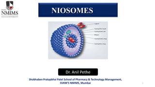
- 3. • Niosomes are non-ionic surfactant based unilamellar or multilamellar bilayer vesicles upon hydration of non ionic surfactants with or without incorporation of cholesterol . • The niosomes are very small, and microscopic in size. Their size lies in the nanometric scale. • Niosomes are a novel drug delivery system, in which the medication is encapsulated in a vesicle. • Both hydrophilic & lipophilic drugs ,entrap either in the aqueous layer or in lipid layer. Introduction
- 4. • These vesicular systems are similar to liposomes that can be used as carriers of amphiphilic and lipophilic drugs. • It is less toxic and improves the therapeutic index of drug by restricting its action to target cells. Structure of Niosomes
- 5. Niosomes Vs. Liposome Liposomes Niosomes Vesicles made up of concentric bilayer of phospholipids Vesicles made up of surfactants with or without incorporation of cholesterol. Size ranges from 10-3000nm Size ranges from 10-100nm Comparatively expensive Inexpensive Special storage condition are required No such special requirement Phospholipids used are unstable Non-ionic surfactants are stable Comparatively more toxic Less toxic
- 6. • They are osmotically active and stable. • They increase the stability of the entrapped drug. • The vesicle suspension being water based offers greater patient compliance over oil based systems • Since the structure of the niosome offers place to accommodate hydrophilic, lipophilic as well as ampiphilic drug moieties, they can be used for a variety of drugs. • The vesicles can act as a depot to release the drug slowly and of controlled release. • Biodegradable, non-immunogenic and biocompatible. Advantages
- 7. • Aggregation • Fusion • Leaking of entrapped drug • Hydrolysis of encapsulated drugs which limiting the shelf life of the dispersion. Disadvantages
- 8. Classification of Niosomes The niosomes are classified as a function of the number of bilayer (e.g. MLV, SUV) or as a function of size. (e.g. LUV, SUV) or as a function of the method of preparation The various types of niosomes are described below: i) Multi lamellar vesicles (MLV, Size=>0.05 µm) ii) Large unilamellar vesicles (LUV, Size=>0.10 µm). iii) Small unilamellar vesicles (SUV, Size=0.025-0.05 µm)
- 9. 1. Multilamellar vesicles (MLV): It consists of a number of bilayer surrounding the aqueous lipid compartment separately. The approximate size of these vesicles is 0.5-10 µm diameter. Multilamellar vesicles are the most widely used niosomes. These vesicles are highly suited as drug carrier for lipophilic compounds. 2. Large unilamellar vesicles (LUV): Niosomes of this type have a high aqueous/lipid compartment ratio, so that larger volumes of bio-active materials can be entrapped with a very economical use of membrane lipids. 3. Small unilamellar vesicles (SUV): These small unilamellar vesicles are mostly prepared from multilamellar vesicles by sonication method, French press extrusion electrostatic stabilization is the inclusion of dicetyl phosphate in 5(6)-carboxyfluorescein (CF) loaded Span 60 based niosomes Classification of Niosomes
- 10. Niosomes mainly contains following types of components: Non-ionic surfactants: • Selection of surfactant should be done on the basis of HLB value. • As Hydrophilic Lipophilic Balance (HLB) is a good indicator of the vesicle forming ability of any surfactant, HLB number in between 4 and 8 was found to be compatible with vesicle formation. • It is also reported that the hydrophilic surfactant owing to high aqueous solubility. on hydration do not reach a state of concentrated systems in order to allow free hydrated units to exist aggregates and coalesced to form lamellar structure. Components of Niosomes
- 11. a) Alkyl ethers: some surfactants for the preparation of niosomes containing drugs/chemicals as: 1) Surfactant-I (Mol.Wt.473) is C16 monoalkyl glycerol ether with average of three glycerol units. 2) Surfactant-II (Mol.Wt.972) is diglycerol ether with average of the seven glycerol units. 3) Surfactant III (Mol.Wt.393) is ester linked surfactant. a) Alkyl esters: Sorbitan esters are most preferred surfactant used for the preparation of niosomes amongst this category of surfactants. Vesicles prepared by the polyoxyethylene sorbitan monolaurate are relatively soluble than other surfactant vesicles]. For example polyoxyethylene (polysorbate 60) has been utilized for encapsulation of diclofenac sodium. b) Alkyl amides: Alkyl amide (e.g. galactosides and glucosides) have been utilized to produce niosomal vesicles c) Fatty acid and amino acid compounds: Long chain fatty acids and amino acid moieties have also been used in some niosomes preparation. Components of Niosomes (cont.)
- 12. Cholesterol: • Steroids are important components of the cell membrane and their presence in membrane affect the bilayer fluidity and permeability. Cholesterol is a steroid derivative, which is mainly used for the formulation of niosomes. • Although it may not show any role in the formation of bilayer, its importance in formation of niosomes and manipulation of layer characteristics can not be discarded. In general, incorporation of cholesterol affect properties of niosomes like membrane permeability, rigidity, encapsulation efficiency, ease of rehydration of freeze dried niosomes and their toxicity. • As a result of this, the niosome become less leaky in nature.
- 13. Charged molecule: • Some charged molecules are added to niosomes to increase stability of niosomes by electrostatic repulsion which prevents coalescence. • The negatively charged molecules used are diacetyl phosphate (DCP) and phosphotidic acid. Similarly, stearylamine (STR) and stearyl pyridinium chloride are the well known positively charged molecules used in niosomal preparations. • These charged molecules are used mainly to prevent aggregation of niosomes.
- 14. Factors affecting Niosomes formation Nature of surfactant Inclusion of a charged molecule Factors Structure of surfactant Temperature of hydration Nature of encapsulated drug
- 15. • A surfactant used for preparation of niosomes must have a hydrophilic head and hydrophobic tail. The hydrophobic tail may consist of one or two alkyl or perfluoroalkyl groups or in some cases a single steroidal group. • The ether type surfactants with single chain alkyl as hydrophobic tail is more toxic than corresponding dialkyl ether chain. • The ester type surfactants are chemically less stable than ether type surfactants and the former is less toxic than the latter due to ester-linked surfactant degraded by esterase’s to triglycerides and fatty acid in vivo. • The surfactants with alkyl chain length from C12- C18 are suitable for preparation of niosomes. • Surfactants such as C16EO5 (poly-oxyethylene cetyl ether) or C18EO5 (polyoxyethylene steryl ether) are used for preparation of polyhedral vesicles. • Span series surfactants having HLB number of between 4 and 8 can form vesicles. Nature of surfactant
- 16. • The geometry of vesicle to be formed from surfactants is affected by its structure, which is related to critical packing parameters. • On the basis of critical packing parameters of surfactants, we can predict geometry of vesicle to be formed. • Critical packing parameters can be defined using following equation, CPP (Critical Packing Parameters) = v/lc ×a0 • Where v = hydrophobic group volume, • lc = the critical hydrophobic group length, • a0= the area of hydrophilic head group. • From the critical packing parameter value type of miceller structure formed can be ascertained as given below, If CPP < ½, then formation of spherical micelles, If ½ < CPP < 1, then formation of bilayer micelles, If CPP > 1, then formation inverted micelles. Structure of surfactant
- 17. • The mean size of niosomes increases proportionally with increase in the HLB of surfactants like Span 85 (HLB 1.8) to Span 20 (HLB 8.6) because the surface free energy decreases with an increase in hydrophobicity of surfactant. • The bilayers of the vesicles are either in the so-called liquid state or in gel state, depending on the temperature, the type of lipid or surfactant and the presence of other components such as cholesterol. • In the gel state, alkyl chains are present in a well-ordered structure, and in the liquid state, the structure of the bilayers is more disordered. • The surfactants and lipids are characterized by the gel-liquid phase transition temperature (TC). • Phase transition temperature (TC) of surfactant also effects entrapment efficiency i.e. Span 60 having higher TC, provides better entrapment. Amount and type of surfactant
- 18. • The stable niosomes can be prepared with addition of different additives along with surfactants and drugs. • Niosomes formed have a number of morphologies and their permeability and stability properties can be altered by manipulating membrane characteristics by different additives. • In case of polyhedral niosomes formed from C16G2, the shape of these polyhedral niosome remains unaffected by adding low amount of solulan C24 (cholesteryl poly-24-oxyethylene ether), which prevents aggregation due to development of steric hindrance. • The mean size of niosomes is influenced by membrane composition such as Polyhedral niosomes formed by C16G2: solulan C24 in ratio (91:9) having bigger size (8.0 ±0.03mm) than spherical/tubular niosomes formed by C16G2: cholesterol: solulan C24 in ratio (49:49:2)(6.6±0.2mm). • Addition of cholesterol molecule to niosomal system provides rigidity to the membrane and reduces the leakage of drug from niosome. Membrane Composition
- 19. • The physico-chemical properties of encapsulated drug influence charge and rigidity of the niosome bilayer. • The drug interacts with surfactant head groups and develops the charge that creates mutual repulsion between surfactant bilayers and hence increases vesicle size. • The aggregation of vesicles is prevented due to the charge development on bilayer. • In polyoxyethylene glycol (PEG) coated vesicles, some drug is entrapped in the long PEG chains, thus reducing the tendency to increase the size. • The hydrophilic lipophilic balance of the drug affects degree of entrapment. Nature of Encapsulated Drug
- 20. • Hydration temperature influences the shape and size of the niosome. • For ideal condition it should be above the gel to liquid phase transition temperature of system. • Temperature change of niosomal system affects assembly of surfactants into vesicles and also induces vesicle shape transformation. • Arunothayanun et al. reported that a polyhedral vesicle formed by C16G2: solulan C24 (91:9) at 25°C which on heating transformed into spherical vesicle at 48°C, but on cooling from 55°C, the vesicle produced a cluster of smaller spherical niosomes at 49°C before changing to the polyhedral structures at 35°C. • In contrast vesicle formed by C16G2: cholesterol: solulan C24 (49:49:2) shows no shape transformation on heating or cooling. • Along with the above mentioned factors, volume of hydration medium and time of hydration of niosomes are also critical factors. Improper selection of these factors may result in formation of fragile niosomes or creation of drug leakage problems. Temperature of Hydration
- 21. Hand Shaking method Reverse phase evaporation technique Ether Injection method Multiple membrane extrusion method Bubble method Sonication From Proniosomes Method of Preparation of Niosomes
- 22. Common stages of all Method of Preparation of Niosomes
- 23. • The mixture of vesicles forming ingredients like surfactant and cholesterol are dissolved in a volatile organic solvent (diethyl ether, chloroform or methanol) in a round bottom flask. • The organic solvent is removed at room temperature (20°C)using rotary evaporator leaving a thin layer of solid mixture deposited on the wall of the flask. • The dried surfactant film can be rehydrated with aqueous phase at 0- 60°C with gentle agitation. • This process forms typical multilamellar Niosomes. Hand shaking method
- 24. • Cholesterol and surfactant (1:1) are dissolved in a mixture of ether and chloroform. • An aqueous phase containing drug is added to this and the resulting two phases are sonicated at 4-5°C. • The clear gel formed is further sonicated after the addition of a small amount of phosphate buffered saline (PBS). • The organic phase is removed at 40°C under low pressure. • The resulting viscous niosome suspension is diluted with PBS and heated on a water bath at60°C for 10 min to yield Niosomes. • It was reported that the preparation of Diclofenac Sodium Niosomes using Tween 85 by this method Reverse phase evaporation technique
- 25. • This method provides a means of making Niosomes by slowly introducing a solution of surfactant dissolved in diethyl ether into warm water maintained at 60°C. • The surfactant mixture in ether is injected through 14-gauge needle into an aqueous solution of material. • Vaporization of ether leads to formation of single layered vesicles. • Depending upon the conditions used the diameter of the vesicle range from 50 to 1000 nm Ether Injection method
- 26. • A mixture of surfactant, cholesterol, and diacetyl phosphate in chloroform is made into thin film by evaporation. • The film is hydrated with aqueous drug solution and the resultant suspension extruded through polycarbonate membranes, which are placed in a series for up to eight passages. • This is a good method for controlling niosome size. Multiple membrane extrusion method
- 27. • The oil in water (o/w) emulsion is prepared from an organic solution of surfactant, cholesterol, and an aqueous solution of the drug. • The organic solvent is then evaporated, leaving niosomes dispersed in the aqueous phase. Emulsion method
- 28. • This method does not require expensive organic phase. • Here, the mixture of lipids and surfactant is first melted and then injected into a highly agitated heated aqueous phase containing dissolved drug. • Here, the drug can be dissolved in molten lipid and the mixture will be injected into agitated, heated aqueous phase containing surfactant. Lipid injection method
- 29. • It is novel technique for the one step preparation of liposomes and niosomes without the use of organic solvents. • The bubbling unit consists of round-bottomed flask with three necks positioned in water bath to control the temperature. Water-cooled reflux and thermometer is positioned in the first and second neck and nitrogen supply through the third neck. • Cholesterol and surfactant are dispersed together in this buffer (pH 7.4) at70°C, the dispersion mixed for 15 seconds with high shear homogenizer and immediately afterwards “bubbled” at 70°C using nitrogen gas. Bubble Method
- 30. From Proniosomes Another method of producing niosomes is to coat a water soluble carrier such as sorbitol with surfactant The result of the coating process is a dry formulation. In which each water soluble particle is covered with thin film of dry surfactant This preparation is called as “proniosomes” The niosomes are recognized by the addition of aqueous phase at T˃Tm and brief agitation (where T- Temperature and TM- Phase transition temperature)
- 31. • A typical method of production of the vesicles is by Sonication of solution. • In this method an aliquot of drug solution in buffer is added to the surfactant/cholesterol mixture in a 10- ml glass vial. • The mixture is probe sonicated at 60°C for 3 minutes using a sonicator with a titanium probe to yield Niosomes. Sonication Method
- 32. • Micro fluidization is a recent technique to prepare unilamellar vesicles of defined size distribution. • This method is based on submerged jet principle in which two fluidized streams interact at ultra high velocities, in precisely defined micro channels within the interaction chamber. • The impingement of thin liquid sheet along a common front is arranged such that the energy supplied to the system remains within the area of niosomes formation. • The result is a greater uniformity, smaller size and better reproducibility of niosomes formed. Micro fluidization Method
- 33. Dialysis • The aqueous niosomal dispersion is dialyzed in dialysis tubing against phosphate buffer or normal saline or glucose solution. Gel Filtration • The unentrapped drug is removed by gel filtration of niosomal dispersion through a Sephadex-G-50 column and elution with phosphate buffered saline or normal saline. Centrifugation • The niosomal suspension is centrifuged and the supernatant is evaporated. The pellet is washed and then re-suspended to obtain a niosomal suspension free from un-entrapped drug. Separation of unentrapped drug:
- 34. a) Size, Shape and Morphology Freeze Fracture Electron Microscopy:- Visualize the vesicular structure of surfactant based vesicles. Photon Correlation spectroscopy :- Determine mean diameter of the vesicles. Electron Microscopy :- Morphological studies of vesicles. b) Entrapment efficiency After preparing niosomal dispersion, unentrapped drug is separated by dialysis and the drug remained entrapped in niosomes is determined by complete vesicle disruption using 50% n-propanol or 0.1% Triton X-100 and analysing the resultant solution by appropriate assay method for the drug. c) Vesicle Surface Charge Determined by measurement of electrophoretic mobility and expressed in expressed in terms of zeta potential d) In vitro studies Evaluation of Niosomes
- 35. Ophthalmic drug delivery From ocular dosage form like ophthalmic solution, suspension and ointment it is difficult to achieve excellent bioavailability of drug due to the tear production, impermeability of corneal epithelium, non-productive absorption and transient residence time. But niosomal and liposomal delivery systems can be used to achieve good bioavailability of drug. Bio adhesive-coated niosomal formulation of acetazolamide prepared from span 60, cholesterol stearylamine or dicetyl phosphate exhibits more tendencies for reduction of intraocular pressure as compared to marketed formulation (Dorzolamide) Application of Niosome
- 36. Localized Drug Action Drug delivery through Niosomes is one of the approaches to achieve localized drug action, since their size and low penetrability through epithelium and connective tissue keeps the drug localized at the site of administration. Localized drug action results in enhancement of efficacy of potency of the drug and at the same time reduces its systemic toxic effects e.g. Antimonials encapsulated within niosomes are taken up by mononuclear cells resulting in localization of drug, increase in potency and hence decrease both in dose and toxicity. The evolution of niosomal drug delivery technology is still at an infancy stage, but this type of drug delivery system has shown promise in cancer chemotherapy and anti-leishmanial therapy. Application of Niosome
- 37. Diagnostic imaging with niosomes Niosomal system can be used as diagnostic agents. Conjugated niosomal formulation of gadobenatedimeglcemine with [N- palmitoylglucosamine (NPG)], PEG4400, and both PEG and NPG exhibit significantly improved tumor targeting of an encapsulated paramagnetic agent assessed with MR imaging Application of Niosome
- 38. Transdermal delivery of drugs by niosomes An increase in the penetration rate has been achieved by transdermal delivery of drug incorporated in niosomes as slow penetration of drug through skin is the major drawback of transdermal route of delivery for other dosage forms. The topical delivery of erythromycin from various formulations including niosomes has studied on hair less mouse and from the studies, and confocal microscopy, it was found that nonionic vesicles could be formulated to target pilosebaceous glands Application of Niosome
- 39. Niosome as a carrier for Hemoglobin Niosomal suspension shows a visible spectrum superimposable onto that of free hemoglobin so can be used as a carrier for hemoglobin. Vesicles are also permeable to oxygen and hemoglobin dissociation curve can be modified similarly to non-encapsulated hemoglobin Application of Niosome
- 40. Application of Niosome Delivery of peptide drugs Niosomal entrapped oral delivery of 9-desglycinamide, 8- arginine vasopressin was examined in an in-vitro intestinal loop model and reported that stability of peptide increased significantly. Immunological applications of niosomes For studying the nature of the immune response provoked by antigens niosomes have been used. Niosomes have been reported as potent adjuvant in terms of immunological selectivity, low toxicity and stability
- 41. Application of Niosome Targeting of bioactive agents To reticulo-endothelial system (RES) The vesicles occupy preferentially to the cells of RES. It is due to circulating serum factors known as opsonins, which mark them for clearance. Such localized drug accumulation has, however, been exploited in treatment of animal tumours known to metastasize to the liver and spleen and in parasitic infestation of liver. To organs other than reticulo-endothelial system(RES) By use of antibodies, carrier system can be directed to specific sites in the body. Immunoglobulins seem to have affection to the lipid surface, thus providing a convenient means for targeting of drug carrier. Many cells have the intrinsic ability to recognize and bind particular carbohydrate determinants and this property can be used to direct carriers system to particular cells.
- 42. • Niosomes provide incorporating the drug into for a better targeting of the drug at appropriate tissue destination . • They presents a structure similar to liposome and hence they can represent alternative vesicular systems with respect to liposomes • Niosomes are thoughts to be better candidates drug delivery as compared to liposomes due to various factors like cost, stability etc. • Various type of drug deliveries can be possible using niosomes like targeting, ophthalmic, topical, parenteral etc. Summary of Niosomes