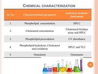
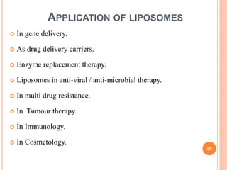
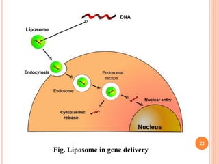
TYPES, PREPARATION AND
EVALUATIONS OF LIPOSOMES
1
Presented by,
Mr. Akash U. Thakur
M. Pharm IInd sem
Dept. of Pharmaceutics
Guide,
Dr. N. M. Mahajan
Head, Dept. of Pharmaceutics
DBCOP, Besa, Nagpur.
CONTENTS
 Introduction
 Structure of liposome
 Advantages
 Disadvantages
 Types of liposomes
 Method of liposome preparation
 Characterization of liposomes
 Application of liposomes
 References
2
LIPOSOMES
 Liposomes first described by Dr. A. D. Bingham in 1965.
 The Liposomes are the micro-particular drug carrier one or
more concentric phospholipids bi-layer separated by aqueous
buffer compartments known as “Liposomes”..
 Liposomes is Greek words means
‘Lipo’ mean ‘Fat’ and
‘Somes’ mean ‘Body’.
 Their diameter ranges from 80nm to 100µm.
3
4
ADVANTAGES
 Liposomes increased efficacy and therapeutic index of drug.
 Liposomes increased stability via. encapsulation.
 Provide selective passive targeting to tumour tissues.
 Improved pharmacokinetic effects (reduced elimination,
increased circulation life time).
 Liposomes reduce the toxicity of the encapsulation.
 Facilitation of transport across membranes. 5
DISADVANTAGES
 Low solubility in water.
 Sometimes phospholipid undergoes oxidation and hydrolysis
like reaction.
 Production cost is high.
6
1. BASED ON STRUCTURAL PARAMETER
 MLV (Multi-lamellar large vesicles),
 OLV (Oligo-lamellar vesicles),
 UV (Uni-lamellar vesicles),
 SUV (Small uni-lamellar vesicles),
 MUV (Medium sized unilamellar vesicles),
 LUV (Large unilamellar vesicles),
 MV (Multivesicular vesicles).
7
TYPES OF LIPOSOMES
2. BASED ON METHOD OF LIPOSOME PREPARATION
 REV (Single or Oligolamellar vesicles made by reverse
phase evaporation method).
 MLV-REV (Multi-lamellar vesicles made by reverse
phase evaporation method).
 SPLV (Stable Pluri-lamellar vesicles).
 DRM (Dehydration rehydration method).
8
3. BASED UPON COMPOSITION AND APPLICATIONS
 CL (Conventional liposomes),
 Fusogenic liposomes,
 pH sensitive liposomes,
 Cationic liposomes,
 Immuno-liposomes.
9
10
Method of liposome preparation
Passive loading
techniques
Mechanical
dispersion
methods.
1. Hand shaking
2. Sonication
3. Micro-
emulsification
4. French pressure
cell
Solvent
dispersion
methods.
1.Ethanol injection
2. Ether injection
3. Double
emulsion vesicles
4. Reverse phase
evaporation
vesicles
Detergent
removal
methods.
1. Detergent
2. Dialysis
3. Column
chromatography
4. Dilution
Active loading
techniques
1. MECHANICAL DISPERSION METHODS.
11
LIPID FILM HYDRATION BY HAND SHAKING
12
Fig. Multilaminar vesicles (MLVs) formed by hand shaking technique
13
2. SOLVENT DISPERSION METHOD
14
REVERSE PHASE EVAPORATION VESICLES
3. DETERGENT REMOVAL METHODS
15
 The conc. of detergent at which micelles are formed is
called as CMC.
16
 Phospholipids in the form of either sonicated vesicles or as a
dry film, at a molar ratio of 2:1 with deoxycholate form
unilamellar vesicles of 100 nm on removal of deoxycholate
by column chromatography.
 This could be achieved by passing the dispersion over a
Sephadex G-25 column pre-saturated with constitutive lipids
and pre-equilibrated with hydrating buffer.
COLUMN CHROMATOGRAPHY
CHARACTERIZATION OF
LIPOSOMES
Physical characterization.
Chemical characterization.
17
PHYSICAL CHARACTERIZATION
Sr. No. Characterization parameter Analytical methods / Instrument
1. Vesicle shape and surface
morphology
Electron Spectroscopy.
2. size and size distribution Electron microscopy.
3. Surface charge Free- flow electrophoresis.
4. Electrical surface potential
and surface pH
Zeta-potential.
5. Lamellarity X- ray scattering, 31 P-NMR, Freeze-electron
microscopy.
6. Phase behaviour of liposomes Electron microscopy.
7. % of free drug Mini-coloumn centrifugation, ion-exchange
chromatography, radio labeling.
8. Drug release Diffusion cell.
18
CHEMICAL CHARACTERIZATION
Sr. No. Characterization parameter
Analytical methods /
Instrument
1. Phospholipid concentration HPLC
2. Cholesterol concentration
Cholesterol Oxidase
assay and HPLC
3. Phospholipid peroxidation UV absorbance
4.
Phospholipid hydrolysis, Cholesterol
auto-oxidation
HPLC and TLC
5. Osmolarity Osmometer
19
 In gene delivery.
 As drug delivery carriers.
 Enzyme replacement therapy.
 Liposomes in anti-viral / anti-microbial therapy.
 In multi drug resistance.
 In Tumour therapy.
 In Immunology.
 In Cosmetology.
20
APPLICATION OF LIPOSOMES
LIPOSOME IN GENE DELIVERY
 variety of physical and biological methods have been
developed for transferring gene into cells.
 The most widely used type of vehicles for gene delivery
are viral and non-viral.
 The non-viral vector systems, liposomes and lipid
complexes especially engineered liposome such as pH
sensitive liposomes, cationic liposomes, fusogenic
liposome, genosomes, lipoplex, and lipopolyplex have
been extensively investigated for their gene delivery
potential.
 However, most of the commercially available non-viral
gene vectors used for transfection are cationic liposomes-
DNA complexes. 21
22
Fig. Liposome in gene delivery
LIPOSOME AS DRUG DELIVERY CARRIERS
Several modes of drug delivery applications have been
proposed for the liposomal system for some of the pathogen caused
diseases. The major ones are -
 Enhanced drug solubility.
E.g. Amphotericin B.
 Protection of sensitive drug molecules.
E.g. DNA, RNA, Ribosomes.
 Enhanced intracellular uptake.
E.g. Anticancer drugs, Antiviral and Antimicrobial drugs.
23
LIPOSOME IN TUMOUR THERAPY
 Most of the medical application of liposomes have reached the
preclinical and clinical stages are in cancer treatments.
 It has been demonstrated that small and stable liposomes can
passively target several different tumour because they can
circulates for prolonged times.
 Targeting liposomes can be using in different ways -
• Natural targeting of liposomes,
• Use of long circulatory,
• Use of ligand mediated targeting.
24
LIPOSOME IN COSMETOLOGY AND DERMATOLOGY
 In early studies, liposomes containing stratum corneum lipids
have been tested in order to enable better skin penetration.
 Two mechanism for enhanced topical localization or targeting of
liposome encapsulated bioactives.
 The transepidermal pathway. (skin layers)
 The transfollicular pathway. (hair follicles and glands)
 Liposome based on anti-aging formulations (e.g. creams, lotions,
gels and hydrogels) have been formulated and launched in the
cosmetic market by Loreal in 1986.
 Liposomal preparation reduce the roughness because of its
interaction with the intracellular lipid resulting in skin softening
and smoothing.
25
MARKETED PRODUCTS
26
REFERENCES
 S. P. Vyas & R. K. Khar, “Targeted & Controlled Drug
Delivery Novel Carrier Systems” First edition 2002, CBS
Publishers & Distributors Pvt. Ltd., Page no. 173-205.
 www.wikipedia.com
27
28
ANY
QUESTIONS…?
29

Liposomes

  • 1.
    TYPES, PREPARATION AND EVALUATIONSOF LIPOSOMES 1 Presented by, Mr. Akash U. Thakur M. Pharm IInd sem Dept. of Pharmaceutics Guide, Dr. N. M. Mahajan Head, Dept. of Pharmaceutics DBCOP, Besa, Nagpur.
  • 2.
    CONTENTS  Introduction  Structureof liposome  Advantages  Disadvantages  Types of liposomes  Method of liposome preparation  Characterization of liposomes  Application of liposomes  References 2
  • 3.
    LIPOSOMES  Liposomes firstdescribed by Dr. A. D. Bingham in 1965.  The Liposomes are the micro-particular drug carrier one or more concentric phospholipids bi-layer separated by aqueous buffer compartments known as “Liposomes”..  Liposomes is Greek words means ‘Lipo’ mean ‘Fat’ and ‘Somes’ mean ‘Body’.  Their diameter ranges from 80nm to 100µm. 3
  • 4.
  • 5.
    ADVANTAGES  Liposomes increasedefficacy and therapeutic index of drug.  Liposomes increased stability via. encapsulation.  Provide selective passive targeting to tumour tissues.  Improved pharmacokinetic effects (reduced elimination, increased circulation life time).  Liposomes reduce the toxicity of the encapsulation.  Facilitation of transport across membranes. 5
  • 6.
    DISADVANTAGES  Low solubilityin water.  Sometimes phospholipid undergoes oxidation and hydrolysis like reaction.  Production cost is high. 6
  • 7.
    1. BASED ONSTRUCTURAL PARAMETER  MLV (Multi-lamellar large vesicles),  OLV (Oligo-lamellar vesicles),  UV (Uni-lamellar vesicles),  SUV (Small uni-lamellar vesicles),  MUV (Medium sized unilamellar vesicles),  LUV (Large unilamellar vesicles),  MV (Multivesicular vesicles). 7 TYPES OF LIPOSOMES
  • 8.
    2. BASED ONMETHOD OF LIPOSOME PREPARATION  REV (Single or Oligolamellar vesicles made by reverse phase evaporation method).  MLV-REV (Multi-lamellar vesicles made by reverse phase evaporation method).  SPLV (Stable Pluri-lamellar vesicles).  DRM (Dehydration rehydration method). 8
  • 9.
    3. BASED UPONCOMPOSITION AND APPLICATIONS  CL (Conventional liposomes),  Fusogenic liposomes,  pH sensitive liposomes,  Cationic liposomes,  Immuno-liposomes. 9
  • 10.
    10 Method of liposomepreparation Passive loading techniques Mechanical dispersion methods. 1. Hand shaking 2. Sonication 3. Micro- emulsification 4. French pressure cell Solvent dispersion methods. 1.Ethanol injection 2. Ether injection 3. Double emulsion vesicles 4. Reverse phase evaporation vesicles Detergent removal methods. 1. Detergent 2. Dialysis 3. Column chromatography 4. Dilution Active loading techniques
  • 11.
  • 12.
    LIPID FILM HYDRATIONBY HAND SHAKING 12 Fig. Multilaminar vesicles (MLVs) formed by hand shaking technique
  • 13.
  • 14.
  • 15.
    3. DETERGENT REMOVALMETHODS 15  The conc. of detergent at which micelles are formed is called as CMC.
  • 16.
    16  Phospholipids inthe form of either sonicated vesicles or as a dry film, at a molar ratio of 2:1 with deoxycholate form unilamellar vesicles of 100 nm on removal of deoxycholate by column chromatography.  This could be achieved by passing the dispersion over a Sephadex G-25 column pre-saturated with constitutive lipids and pre-equilibrated with hydrating buffer. COLUMN CHROMATOGRAPHY
  • 17.
  • 18.
    PHYSICAL CHARACTERIZATION Sr. No.Characterization parameter Analytical methods / Instrument 1. Vesicle shape and surface morphology Electron Spectroscopy. 2. size and size distribution Electron microscopy. 3. Surface charge Free- flow electrophoresis. 4. Electrical surface potential and surface pH Zeta-potential. 5. Lamellarity X- ray scattering, 31 P-NMR, Freeze-electron microscopy. 6. Phase behaviour of liposomes Electron microscopy. 7. % of free drug Mini-coloumn centrifugation, ion-exchange chromatography, radio labeling. 8. Drug release Diffusion cell. 18
  • 19.
    CHEMICAL CHARACTERIZATION Sr. No.Characterization parameter Analytical methods / Instrument 1. Phospholipid concentration HPLC 2. Cholesterol concentration Cholesterol Oxidase assay and HPLC 3. Phospholipid peroxidation UV absorbance 4. Phospholipid hydrolysis, Cholesterol auto-oxidation HPLC and TLC 5. Osmolarity Osmometer 19
  • 20.
     In genedelivery.  As drug delivery carriers.  Enzyme replacement therapy.  Liposomes in anti-viral / anti-microbial therapy.  In multi drug resistance.  In Tumour therapy.  In Immunology.  In Cosmetology. 20 APPLICATION OF LIPOSOMES
  • 21.
    LIPOSOME IN GENEDELIVERY  variety of physical and biological methods have been developed for transferring gene into cells.  The most widely used type of vehicles for gene delivery are viral and non-viral.  The non-viral vector systems, liposomes and lipid complexes especially engineered liposome such as pH sensitive liposomes, cationic liposomes, fusogenic liposome, genosomes, lipoplex, and lipopolyplex have been extensively investigated for their gene delivery potential.  However, most of the commercially available non-viral gene vectors used for transfection are cationic liposomes- DNA complexes. 21
  • 22.
    22 Fig. Liposome ingene delivery
  • 23.
    LIPOSOME AS DRUGDELIVERY CARRIERS Several modes of drug delivery applications have been proposed for the liposomal system for some of the pathogen caused diseases. The major ones are -  Enhanced drug solubility. E.g. Amphotericin B.  Protection of sensitive drug molecules. E.g. DNA, RNA, Ribosomes.  Enhanced intracellular uptake. E.g. Anticancer drugs, Antiviral and Antimicrobial drugs. 23
  • 24.
    LIPOSOME IN TUMOURTHERAPY  Most of the medical application of liposomes have reached the preclinical and clinical stages are in cancer treatments.  It has been demonstrated that small and stable liposomes can passively target several different tumour because they can circulates for prolonged times.  Targeting liposomes can be using in different ways - • Natural targeting of liposomes, • Use of long circulatory, • Use of ligand mediated targeting. 24
  • 25.
    LIPOSOME IN COSMETOLOGYAND DERMATOLOGY  In early studies, liposomes containing stratum corneum lipids have been tested in order to enable better skin penetration.  Two mechanism for enhanced topical localization or targeting of liposome encapsulated bioactives.  The transepidermal pathway. (skin layers)  The transfollicular pathway. (hair follicles and glands)  Liposome based on anti-aging formulations (e.g. creams, lotions, gels and hydrogels) have been formulated and launched in the cosmetic market by Loreal in 1986.  Liposomal preparation reduce the roughness because of its interaction with the intracellular lipid resulting in skin softening and smoothing. 25
  • 26.
  • 27.
    REFERENCES  S. P.Vyas & R. K. Khar, “Targeted & Controlled Drug Delivery Novel Carrier Systems” First edition 2002, CBS Publishers & Distributors Pvt. Ltd., Page no. 173-205.  www.wikipedia.com 27
  • 28.
  • 29.