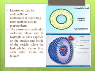
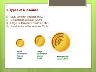
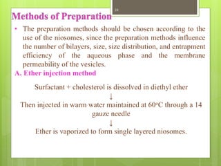
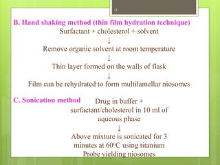
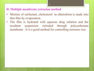
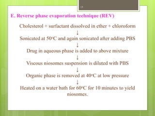
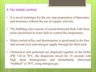
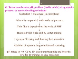
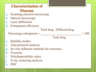
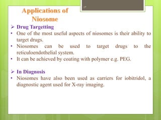
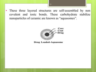
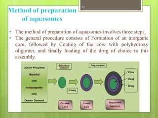
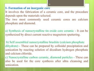
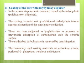
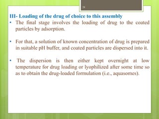
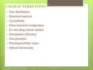
A seminar on
Niosomes
and
Aquasomes
Department of Pharmaceutics
DBCOP
Besa, Nagpur
Presented by
Syed Imran
(M Pharm sem II )
Guided by
Dr. N.M.Mahajan
1
Contents
Niosomes
 Introduction
 Structure
 Types
 Composition
 Methods of preparation
 Characterization
 Applications
Aquasomes
 Introduction
 Principle of self assembly
 Methods of preparation
 Characterization
 Applications
2
3
• Niosomes are a novel drug delivery system, in which the
medication is encapsulated in a vesicle. The vesicle is
composed of a bilayer of non-ionic surface active agents
and hence the name niosomes.
• The niosomes are very small, and microscopic in size.
Their size lies in the nanometric scale. Although
structurally similar to liposomes, they offer several
advantages over them.
• Niosomes have recently been shown to greatly increase
transdermal drug delivery and also can be used in
targeted drug delivery, and thus increased study in these
structures can provide new methods for drug delivery.
4
• Structurally, niosomes are similar to liposomes, in that
they are also made up of a bilayer.
• However, the bilayer in the case of niosomes is made up
of non-ionic surface active agents rather than
phospholipids as seen in the case of liposomes.
• Most surface active agents when immersed in water
yield micellar structures however some surfactants can
yield bilayer vesicles which are niosomes.
5
• Liposomes may be
unilamellar or
multilamellar depending
upon method used to
prepare them.
• The niosome is made of a
surfactant bilayer with its
hydrophilic ends exposed
on the outside and inside
of the vesicle, while the
hydrophobic chains face
each other within the
bilayer.
6
 Types of Niosomes
1) Multi lamellar vesicles (MLV)
2) Unilamellar vesicles (ULV)
ii) Large unilamellar vesicles (LUV)
iii) Small unilamellar vesicles (SUV)
7
The two major components used for the preparation of niosomes
are,
1. Cholesterol
2. Nonionic surfactants
1. Cholesterol
Cholesterol is used to provide rigidity and proper shape,
conformation to the niosomes preparations.
2. Nonionic surfactants
The role surfactants play a major role in the formation of
niosomes. The following non-ionic surfactants are generally used
for the preparation of niosomes.
 Composition of Niosome
8
E.g.
Spans (span 60, 40, 20, 80)
Tweens (tween 20, 40, 60, 80)
The non ionic surfactants possess a hydrophilic head and a
hydrophobic tail.
9
• The preparation methods should be chosen according to the
use of the niosomes, since the preparation methods influence
the number of bilayers, size, size distribution, and entrapment
efficiency of the aqueous phase and the membrane
permeability of the vesicles.
A. Ether injection method
Surfactant + cholesterol is dissolved in diethyl ether
↓
Then injected in warm water maintained at 60oC through a 14
gauze needle
↓
Ether is vaporized to form single layered niosomes.
10
B. Hand shaking method (thin film hydration technique)
Surfactant + cholesterol + solvent
↓
Remove organic solvent at room temperature
↓
Thin layer formed on the walls of flask
↓
Film can be rehydrated to form multilamellar niosomes
C. Sonication method Drug in buffer +
surfactant/cholesterol in 10 ml of
aqueous phase
↓
Above mixture is sonicated for 3
minutes at 60oC using titanium
Probe yielding niosomes
11
D. Multiple membrane extrusion method
• Mixture of surfactant, cholesterol in chloroform is made into
thin film by evaporation.
• The film is hydrated with aqueous drug solution and the
resultant suspension extruded through polycarbonate
membrane . It is a good method for controlling noisome size.
12
E. Reverse phase evaporation technique (REV)
Cholesterol + surfactant dissolved in ether + chloroform
↓
Sonicated at 50oC and again sonicated after adding PBS
↓
Drug in aqueous phase is added to above mixture
↓
Viscous niosomes suspension is diluted with PBS
↓
Organic phase is removed at 40oC at low pressure
↓
Heated on a water bath for 60oC for 10 minutes to yield
niosomes.
13
F. The bubble method
• It is novel technique for the one step preparation of liposomes
and niosomes without the use of organic solvents.
• The bubbling unit consists of round-bottomed flask with three
necks positioned in water bath to control the temperature.
• Water-cooled reflux and thermometer is positioned in the first
and second neck and nitrogen supply through the third neck.
• Cholesterol and surfactant are dispersed together in the buffer
(PH 7.4) at 70°C, the dispersion mixed for 15 seconds with
high shear homogenizer and immediately afterwards
“bubbled” at 70°C using nitrogen gas.
14
G. Trans membranes pH gradient (inside acidic) drug uptake
process: or remote loading technique
Surfactant + cholesterol in chloroform
↓
Solvent is evaporated under reduced pressure
↓
Thin film is deposited on the walls of RBF
↓
Hydrated with citric acid by vortex mixing
↓
3 cycles of freezing and thawing then sonication
↓
Addition of aqueous drug solution and vortexing
↓
pH raised to 7.0-7.2 by 1M disodium phosphate and heated at
60°c for 10 minutes so give niosomes
15
a. Scanning electron microscopy
b. Optical microscopy
c. Lazer diffraction
d. Entrapment efficiency
Total drug –Diffused drug
Percentage entrapment = _________________________ × 100
Total drug
e. Stability studies
f. Zeta potential analysis
g. In-vitro diffusion methods for niosomes
h. Viscosity
i. Polydispersibility index
j. X-ray scattering analysis
k. DSC
16
 Drug Targetting
• One of the most useful aspects of niosomes is their ability to
target drugs.
• Niosomes can be used to target drugs to the
reticuloendothelial system.
• It can be achieved by coating with polymer e.g. PEG.
 In Diagnosis
• Niosomes have also been used as carriers for iobitridol, a
diagnostic agent used for X-ray imaging.
17
 Anti-neoplastic Treatment
• Most antineoplastic drugs cause severe side effects.
• Niosomes can alter the metabolism; prolong circulation and
half life of the drug, thus decreasing the side effects of the
drugs.
• Niosomes, is decreased rate of proliferation of tumor and
higher plasma levels accompanied by slower elimination.
 Leishmaniasis
• Leishmaniasis is a disease in which a parasite of the genus
Leishmania invades the cells of the liver and spleen.
• Use of niosomes in tests conducted showed that it was possible
to administer higher levels of the drug without the triggering
of the side effects, and thus allowed greater efficacy in
treatment.
18
 Delivery of Peptide Drugs
• Oral peptide drug delivery has long been faced with a
challenge of bypassing the enzymes which would breakdown
the peptide.
• Use of niosomes to successfully protect the peptides from
gastrointestinal peptide breakdown is being investigated.
• In an in vitro study conducted by oral delivery of a vasopressin
derivative entrapped in niosomes showed that entrapment of
the drug significantly increased the stability of the peptide.
 Niosomes as Carriers for Haemoglobin
• Niosomes can be used as carriers for haemoglobin within the
blood.
• The niosomal vesicle is permeable to oxygen and hence can
act as a carrier for haemoglobin in anaemic patients.
19
20
 To organs other than RES
• It has been suggested that carrier system can be directed to
specific sites in the body by use of antibodies.
• Immunoglobulins seem to bind quite readily to the lipid
surface, thus offering a convenient means for targeting of drug
carrier.
• Many cells possess the intrinsic ability to recognize and bind
particular carbohydrate determinants and this can be exploited
to direct carriers system to particular cells.
21
• Aquasomes are nanoparticulate carrier system but instead of
being simple nanoparticle these are three layered self
assembled structures.
• It comprised of a solid phase nanocrystalline core coated with
oligomeric film on which biochemically active molecules are
adsorbed with or without modification.
• Aquasomes are like “bodies of water" and their water like
properties protect and preserve fragile biological molecules,
and this property of maintaining conformational integrity as
well as high degree of surface exposure is exploited in
targeting of bioactive molecules.
22
• These three layered structures are self-assembled by non
covalent and ionic bonds. These carbohydrate stabilize
nanoparticles of ceramic are known as “aquasomes”.
23
Principle of self assembly
• Self assembly implies that the constituent parts of some final
product assume spontaneously prescribed structural
orientations in two or three dimensional space.
• The self assembly of macromolecules in the aqueous
environment for the purpose of creating smart nanostructure
materials is governed basically by three physicochemical
processes:
1. The interactions of charged groups
2. Hydrogen bonding
3. structural stability
24
• The method of preparation of aquasomes involves three steps.
• The general procedure consists of Formation of an inorganic
core, followed by Coating of the core with polyhydroxy
oligomer, and finally loading of the drug of choice to this
assembly.
25
I- Formation of an inorganic core
It involves the fabrication of a ceramic core, and the procedure
depends upon the materials selected.
The two most commonly used ceramic cores are calcium
phosphate and diamond.
a) Synthesis of nanocrystalline tin oxide core ceramic - It can be
synthesized by direct current reactive magnetron sputtering.
b) Self assembled nanocrystalline brushite (calcium phosphate
dihydrate) - These can be prepared by colloidal precipitation and
sonication by reacting solution of disodium hydrogen phosphate
and calcium chloride.
c) Nanocrystalline carbon ceramic, diamond particles - These can
also be used for the core synthesis after ultra cleansing and
sonication.
26
II- Coating of the core with polyhydroxy oligomer
• In the second step, ceramic cores are coated with carbohydrate
(polyhydroxyl oligomer).
• The coating is carried out by addition of carbohydrate into an
aqueous dispersion of the cores under sonication.
• These are then subjected to lyophilization to promote an
irreversible adsorption of carbohydrate onto the ceramic
surface.
• The unadsorbed carbohydrate is removed by centrifugation.
• The commonly used coating materials are cellobiose, citrate,
pyridoxal-5- phosphate, trehalose and sucrose.
27
III- Loading of the drug of choice to this assembly
• The final stage involves the loading of drug to the coated
particles by adsorption.
• For that, a solution of known concentration of drug is prepared
in suitable pH buffer, and coated particles are dispersed into it.
• The dispersion is then either kept overnight at low
temperature for drug loading or lyophilized after some time so
as to obtain the drug-loaded formulation (i.e., aquasomes).
28
CHARACTERIZATION
 Size distribution
 Structural analysis
 Crystallinity
 Glass transition temperature
 In vitro drug release studies
 Entrapment efficiency
 Zeta potential
 Polydispersibility index
 Optical microscopy
29
Applications
 Insulin Delivery
• Aquasomes for pharmaceuticals delivery i.e. insulin,
developed because drug activity is conformationally specific.
Bio activity preserved and activity increased to 60% as
compared to i.v administration and toxicity not reported.
 Oral Delivery of Enzyme
• Aquasomes also used for delivery of enzymes like DNAase
and pigments/dyes because enzymes activity fluctuates with
molecular conformation and cosmetic properties of pigments
are sensitive to molecular conformation.
30
 Antigen Delivery
• The adjuvants generally used to enhance the immunity to
antigens have a tendency either to alter the conformation of the
antigen through surface adsorption or to shield the functional
groups.
• So Kossovsky et al demonstrated the efficacy of a new
organically modified ceramic antigen delivery vehicle.
• These particles consisted of diamond substrate coated with a
glassy carbohydrate (cellobiose) film.
• These aquasomes provided conformational stabilization as
well as a high degree of surface exposure to protein antigen.
• Diamond, being a material with high surface energy, was the
first choice for adsorption and adhesion of cellobiose. It
provided a colloidal surface capable of hydrogen bonding to
the proteinaceous antigen.
31
 As oxygen Carrier
• Aquasomes as red blood cell substitutes, haemoglobin
immobilized on oligomer surface because release of oxygen by
haemoglobin is conformationally sensitive.
• By this toxicity is reduced, haemoglobin concentration of 80%
achieved.
32
References
 N.K. Jain “Advances in Controlled and Novel Drug Delivery”
CBS Publisher and Distributors, First edition, p.no: 317-328.
 S.P. Vyas, R.K. Khar “Targeted and Controlled Drug Delivery-
Novel Carrier System” CBS Publisher and Distributers, First
edition p.no: 249-276.
 Kshitij B. Makeshwar, Suraj R. Wasankar “Niosome: a Novel
Drug Delivery System” Asian J. Pharm. Res. 2013; Vol. 3:
Issue 1, p.no:16-20
 Patel JK, Patel KN, Patel HK “Aquasomes: A Self Assembling
Nanobiopharmaceutical Carrier System for Bio-Active
Molecules: A Review” (IJPRS); V-1, I-1, 2012
33
34
 Snehal Patel, Chintan Aundhia “ Aquasomes: a novel approach
in drug carrier system” EJPMR, 2016,3(9);198-201.
 Sanjay S. Jain, Pramod S. Jagtap, Neha M. Dand “Aquasomes:
a novel drug carrier” Journal of Applied Pharmaceutical
Science 02 (01); 2012:184-192.
ANY QUESTION
35
THANK YOU
36

Niosomes and Aquasomes

  • 1.
    A seminar on Niosomes and Aquasomes Departmentof Pharmaceutics DBCOP Besa, Nagpur Presented by Syed Imran (M Pharm sem II ) Guided by Dr. N.M.Mahajan 1
  • 2.
    Contents Niosomes  Introduction  Structure Types  Composition  Methods of preparation  Characterization  Applications Aquasomes  Introduction  Principle of self assembly  Methods of preparation  Characterization  Applications 2
  • 3.
  • 4.
    • Niosomes area novel drug delivery system, in which the medication is encapsulated in a vesicle. The vesicle is composed of a bilayer of non-ionic surface active agents and hence the name niosomes. • The niosomes are very small, and microscopic in size. Their size lies in the nanometric scale. Although structurally similar to liposomes, they offer several advantages over them. • Niosomes have recently been shown to greatly increase transdermal drug delivery and also can be used in targeted drug delivery, and thus increased study in these structures can provide new methods for drug delivery. 4
  • 5.
    • Structurally, niosomesare similar to liposomes, in that they are also made up of a bilayer. • However, the bilayer in the case of niosomes is made up of non-ionic surface active agents rather than phospholipids as seen in the case of liposomes. • Most surface active agents when immersed in water yield micellar structures however some surfactants can yield bilayer vesicles which are niosomes. 5
  • 6.
    • Liposomes maybe unilamellar or multilamellar depending upon method used to prepare them. • The niosome is made of a surfactant bilayer with its hydrophilic ends exposed on the outside and inside of the vesicle, while the hydrophobic chains face each other within the bilayer. 6
  • 7.
     Types ofNiosomes 1) Multi lamellar vesicles (MLV) 2) Unilamellar vesicles (ULV) ii) Large unilamellar vesicles (LUV) iii) Small unilamellar vesicles (SUV) 7
  • 8.
    The two majorcomponents used for the preparation of niosomes are, 1. Cholesterol 2. Nonionic surfactants 1. Cholesterol Cholesterol is used to provide rigidity and proper shape, conformation to the niosomes preparations. 2. Nonionic surfactants The role surfactants play a major role in the formation of niosomes. The following non-ionic surfactants are generally used for the preparation of niosomes.  Composition of Niosome 8
  • 9.
    E.g. Spans (span 60,40, 20, 80) Tweens (tween 20, 40, 60, 80) The non ionic surfactants possess a hydrophilic head and a hydrophobic tail. 9
  • 10.
    • The preparationmethods should be chosen according to the use of the niosomes, since the preparation methods influence the number of bilayers, size, size distribution, and entrapment efficiency of the aqueous phase and the membrane permeability of the vesicles. A. Ether injection method Surfactant + cholesterol is dissolved in diethyl ether ↓ Then injected in warm water maintained at 60oC through a 14 gauze needle ↓ Ether is vaporized to form single layered niosomes. 10
  • 11.
    B. Hand shakingmethod (thin film hydration technique) Surfactant + cholesterol + solvent ↓ Remove organic solvent at room temperature ↓ Thin layer formed on the walls of flask ↓ Film can be rehydrated to form multilamellar niosomes C. Sonication method Drug in buffer + surfactant/cholesterol in 10 ml of aqueous phase ↓ Above mixture is sonicated for 3 minutes at 60oC using titanium Probe yielding niosomes 11
  • 12.
    D. Multiple membraneextrusion method • Mixture of surfactant, cholesterol in chloroform is made into thin film by evaporation. • The film is hydrated with aqueous drug solution and the resultant suspension extruded through polycarbonate membrane . It is a good method for controlling noisome size. 12
  • 13.
    E. Reverse phaseevaporation technique (REV) Cholesterol + surfactant dissolved in ether + chloroform ↓ Sonicated at 50oC and again sonicated after adding PBS ↓ Drug in aqueous phase is added to above mixture ↓ Viscous niosomes suspension is diluted with PBS ↓ Organic phase is removed at 40oC at low pressure ↓ Heated on a water bath for 60oC for 10 minutes to yield niosomes. 13
  • 14.
    F. The bubblemethod • It is novel technique for the one step preparation of liposomes and niosomes without the use of organic solvents. • The bubbling unit consists of round-bottomed flask with three necks positioned in water bath to control the temperature. • Water-cooled reflux and thermometer is positioned in the first and second neck and nitrogen supply through the third neck. • Cholesterol and surfactant are dispersed together in the buffer (PH 7.4) at 70°C, the dispersion mixed for 15 seconds with high shear homogenizer and immediately afterwards “bubbled” at 70°C using nitrogen gas. 14
  • 15.
    G. Trans membranespH gradient (inside acidic) drug uptake process: or remote loading technique Surfactant + cholesterol in chloroform ↓ Solvent is evaporated under reduced pressure ↓ Thin film is deposited on the walls of RBF ↓ Hydrated with citric acid by vortex mixing ↓ 3 cycles of freezing and thawing then sonication ↓ Addition of aqueous drug solution and vortexing ↓ pH raised to 7.0-7.2 by 1M disodium phosphate and heated at 60°c for 10 minutes so give niosomes 15
  • 16.
    a. Scanning electronmicroscopy b. Optical microscopy c. Lazer diffraction d. Entrapment efficiency Total drug –Diffused drug Percentage entrapment = _________________________ × 100 Total drug e. Stability studies f. Zeta potential analysis g. In-vitro diffusion methods for niosomes h. Viscosity i. Polydispersibility index j. X-ray scattering analysis k. DSC 16
  • 17.
     Drug Targetting •One of the most useful aspects of niosomes is their ability to target drugs. • Niosomes can be used to target drugs to the reticuloendothelial system. • It can be achieved by coating with polymer e.g. PEG.  In Diagnosis • Niosomes have also been used as carriers for iobitridol, a diagnostic agent used for X-ray imaging. 17
  • 18.
     Anti-neoplastic Treatment •Most antineoplastic drugs cause severe side effects. • Niosomes can alter the metabolism; prolong circulation and half life of the drug, thus decreasing the side effects of the drugs. • Niosomes, is decreased rate of proliferation of tumor and higher plasma levels accompanied by slower elimination.  Leishmaniasis • Leishmaniasis is a disease in which a parasite of the genus Leishmania invades the cells of the liver and spleen. • Use of niosomes in tests conducted showed that it was possible to administer higher levels of the drug without the triggering of the side effects, and thus allowed greater efficacy in treatment. 18
  • 19.
     Delivery ofPeptide Drugs • Oral peptide drug delivery has long been faced with a challenge of bypassing the enzymes which would breakdown the peptide. • Use of niosomes to successfully protect the peptides from gastrointestinal peptide breakdown is being investigated. • In an in vitro study conducted by oral delivery of a vasopressin derivative entrapped in niosomes showed that entrapment of the drug significantly increased the stability of the peptide.  Niosomes as Carriers for Haemoglobin • Niosomes can be used as carriers for haemoglobin within the blood. • The niosomal vesicle is permeable to oxygen and hence can act as a carrier for haemoglobin in anaemic patients. 19
  • 20.
    20  To organsother than RES • It has been suggested that carrier system can be directed to specific sites in the body by use of antibodies. • Immunoglobulins seem to bind quite readily to the lipid surface, thus offering a convenient means for targeting of drug carrier. • Many cells possess the intrinsic ability to recognize and bind particular carbohydrate determinants and this can be exploited to direct carriers system to particular cells.
  • 21.
  • 22.
    • Aquasomes arenanoparticulate carrier system but instead of being simple nanoparticle these are three layered self assembled structures. • It comprised of a solid phase nanocrystalline core coated with oligomeric film on which biochemically active molecules are adsorbed with or without modification. • Aquasomes are like “bodies of water" and their water like properties protect and preserve fragile biological molecules, and this property of maintaining conformational integrity as well as high degree of surface exposure is exploited in targeting of bioactive molecules. 22
  • 23.
    • These threelayered structures are self-assembled by non covalent and ionic bonds. These carbohydrate stabilize nanoparticles of ceramic are known as “aquasomes”. 23
  • 24.
    Principle of selfassembly • Self assembly implies that the constituent parts of some final product assume spontaneously prescribed structural orientations in two or three dimensional space. • The self assembly of macromolecules in the aqueous environment for the purpose of creating smart nanostructure materials is governed basically by three physicochemical processes: 1. The interactions of charged groups 2. Hydrogen bonding 3. structural stability 24
  • 25.
    • The methodof preparation of aquasomes involves three steps. • The general procedure consists of Formation of an inorganic core, followed by Coating of the core with polyhydroxy oligomer, and finally loading of the drug of choice to this assembly. 25
  • 26.
    I- Formation ofan inorganic core It involves the fabrication of a ceramic core, and the procedure depends upon the materials selected. The two most commonly used ceramic cores are calcium phosphate and diamond. a) Synthesis of nanocrystalline tin oxide core ceramic - It can be synthesized by direct current reactive magnetron sputtering. b) Self assembled nanocrystalline brushite (calcium phosphate dihydrate) - These can be prepared by colloidal precipitation and sonication by reacting solution of disodium hydrogen phosphate and calcium chloride. c) Nanocrystalline carbon ceramic, diamond particles - These can also be used for the core synthesis after ultra cleansing and sonication. 26
  • 27.
    II- Coating ofthe core with polyhydroxy oligomer • In the second step, ceramic cores are coated with carbohydrate (polyhydroxyl oligomer). • The coating is carried out by addition of carbohydrate into an aqueous dispersion of the cores under sonication. • These are then subjected to lyophilization to promote an irreversible adsorption of carbohydrate onto the ceramic surface. • The unadsorbed carbohydrate is removed by centrifugation. • The commonly used coating materials are cellobiose, citrate, pyridoxal-5- phosphate, trehalose and sucrose. 27
  • 28.
    III- Loading ofthe drug of choice to this assembly • The final stage involves the loading of drug to the coated particles by adsorption. • For that, a solution of known concentration of drug is prepared in suitable pH buffer, and coated particles are dispersed into it. • The dispersion is then either kept overnight at low temperature for drug loading or lyophilized after some time so as to obtain the drug-loaded formulation (i.e., aquasomes). 28
  • 29.
    CHARACTERIZATION  Size distribution Structural analysis  Crystallinity  Glass transition temperature  In vitro drug release studies  Entrapment efficiency  Zeta potential  Polydispersibility index  Optical microscopy 29
  • 30.
    Applications  Insulin Delivery •Aquasomes for pharmaceuticals delivery i.e. insulin, developed because drug activity is conformationally specific. Bio activity preserved and activity increased to 60% as compared to i.v administration and toxicity not reported.  Oral Delivery of Enzyme • Aquasomes also used for delivery of enzymes like DNAase and pigments/dyes because enzymes activity fluctuates with molecular conformation and cosmetic properties of pigments are sensitive to molecular conformation. 30
  • 31.
     Antigen Delivery •The adjuvants generally used to enhance the immunity to antigens have a tendency either to alter the conformation of the antigen through surface adsorption or to shield the functional groups. • So Kossovsky et al demonstrated the efficacy of a new organically modified ceramic antigen delivery vehicle. • These particles consisted of diamond substrate coated with a glassy carbohydrate (cellobiose) film. • These aquasomes provided conformational stabilization as well as a high degree of surface exposure to protein antigen. • Diamond, being a material with high surface energy, was the first choice for adsorption and adhesion of cellobiose. It provided a colloidal surface capable of hydrogen bonding to the proteinaceous antigen. 31
  • 32.
     As oxygenCarrier • Aquasomes as red blood cell substitutes, haemoglobin immobilized on oligomer surface because release of oxygen by haemoglobin is conformationally sensitive. • By this toxicity is reduced, haemoglobin concentration of 80% achieved. 32
  • 33.
    References  N.K. Jain“Advances in Controlled and Novel Drug Delivery” CBS Publisher and Distributors, First edition, p.no: 317-328.  S.P. Vyas, R.K. Khar “Targeted and Controlled Drug Delivery- Novel Carrier System” CBS Publisher and Distributers, First edition p.no: 249-276.  Kshitij B. Makeshwar, Suraj R. Wasankar “Niosome: a Novel Drug Delivery System” Asian J. Pharm. Res. 2013; Vol. 3: Issue 1, p.no:16-20  Patel JK, Patel KN, Patel HK “Aquasomes: A Self Assembling Nanobiopharmaceutical Carrier System for Bio-Active Molecules: A Review” (IJPRS); V-1, I-1, 2012 33
  • 34.
    34  Snehal Patel,Chintan Aundhia “ Aquasomes: a novel approach in drug carrier system” EJPMR, 2016,3(9);198-201.  Sanjay S. Jain, Pramod S. Jagtap, Neha M. Dand “Aquasomes: a novel drug carrier” Journal of Applied Pharmaceutical Science 02 (01); 2012:184-192.
  • 35.
  • 36.