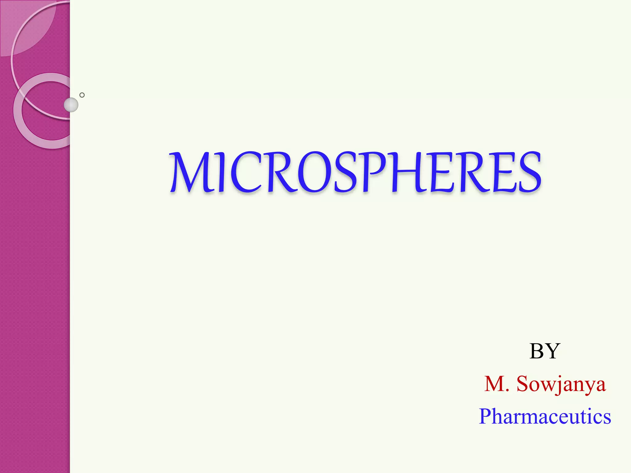
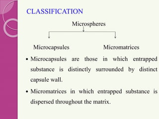
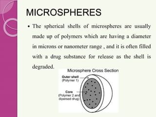
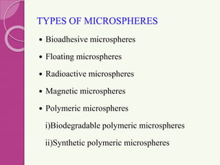
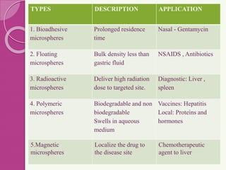
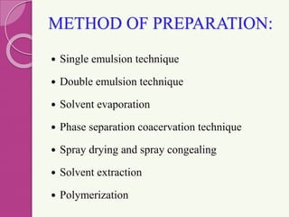
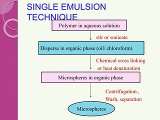
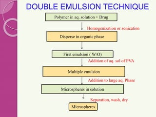
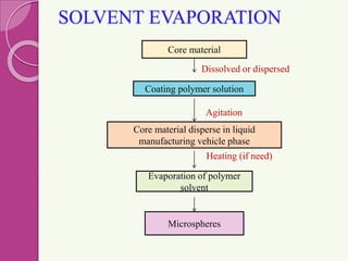
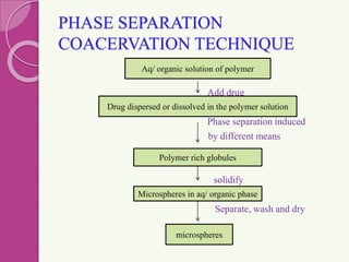
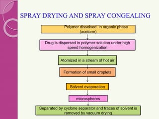
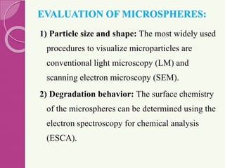
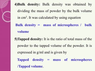
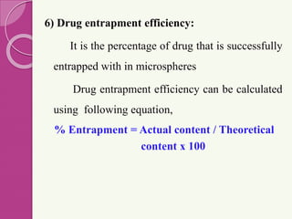
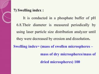
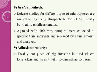
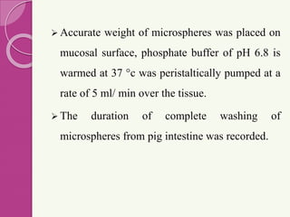
MICROSPHERES
BY
M. Sowjanya
Pharmaceutics
MICROSPHERES
 Microspheres are small spherical particles, with
diameter 1 μm to 1000 μm.
 They are spherical free flowing
particles consisting of proteins
or synthetic polymers which are
biodegradable in nature.
Fig: Microspheres
CLASSIFICATION
Microspheres
Microcapsules Micromatrices
 Microcapsules are those in which entrapped
substance is distinctly surrounded by distinct
capsule wall.
 Micromatrices in which entrapped substance is
dispersed throughout the matrix.
Fig: Microspheres and Microcapsules
ADVANTAGES
 Improve bioavailability
 Provide constant and prolonged therapeutic effect.
 Provide constant drug concentration in blood .
 Decrease dose and toxicity.
 Protect the drug from enzymatic and photolytic
cleavage so it is best for drug delivery of protein.
 Reduce the dosing frequency and thereby improve the
patient compliance
DISADVANTAGES
 The cost is more.
 Reproducibility is less.
 Process conditions like change in temperature,
pH, solvent addition, and evaporation/agitation
may influence the stability of core particles.
 Degradation of product due to heat, hydrolysis,
oxidation, solar radiation or biological agents.
MICROSPHERES
 The spherical shells of microspheres are usually
made up of polymers which are having a diameter
in microns or nanometer range , and it is often filled
with a drug substance for release as the shell is
degraded.
TYPES OF MICROSPHERES
 Bioadhesive microspheres
 Floating microspheres
 Radioactive microspheres
 Magnetic microspheres
 Polymeric microspheres
i)Biodegradable polymeric microspheres
ii)Synthetic polymeric microspheres
TYPES DESCRIPTION APPLICATION
1. Bioadhesive
microspheres
Prolonged residence
time
Nasal - Gentamycin
2. Floating
microspheres
Bulk density less than
gastric fluid
NSAIDS , Antibiotics
3. Radioactive
microspheres
Deliver high radiation
dose to targeted site.
Diagnostic: Liver ,
spleen
4. Polymeric
microspheres
Biodegradable and non
biodegradable
Swells in aqueous
medium
Vaccines: Hepatitis
Local: Proteins and
hormones
5.Magnetic
microspheres
Localize the drug to
the disease site
Chemotherapeutic
agent to liver
METHOD OF PREPARATION:
 Single emulsion technique
 Double emulsion technique
 Solvent evaporation
 Phase separation coacervation technique
 Spray drying and spray congealing
 Solvent extraction
 Polymerization
SINGLE EMULSION
TECHNIQUE
stir or sonicate
Chemical cross linking
or heat denaturation
Centrifugation ,
Wash, separation
Polymer in aqueous solution
Disperse in organic phase (oil/ chloroform)
Microspheres in organic phase
Microspheres
DOUBLE EMULSION TECHNIQUE
Homogenization or sonication
Addition of aq. sol of PVA
Addition to large aq. Phase
Separation, wash, dry
Polymer in aq. solution + Drug
Disperse in organic phase
Multiple emulsion
First emulsion ( W/O)
Microspheres in solution
Microspheres
SOLVENT EVAPORATION
Dissolved or dispersed
Agitation
Heating (if need)
Core material
Evaporation of polymer
solvent
Coating polymer solution
Core material disperse in liquid
manufacturing vehicle phase
Microspheres
PHASE SEPARATION
COACERVATION TECHNIQUE
Add drug
Phase separation induced
by different means
solidify
Separate, wash and dry
Aq/ organic solution of polymer
Drug dispersed or dissolved in the polymer solution
microspheres
Polymer rich globules
Microspheres in aq/ organic phase
SPRAY DRYING AND SPRAY CONGEALING
Polymer dissolved in organic phase
(acetone)
Solvent evaporation
Formation of small droplets
microspheres
Atomized in a stream of hot air
Drug is dispersed in polymer solution under high
speed homogenization
Separated by cyclone separator and traces of solvent is
removed by vacuum drying
SOLVENT EXTRACTION
microspheres
Organic phase is removed by extraction with water
Polymer in organic solvent
Drug is dispersed in organic solvent
(water miscible organic solvents like isopropanol)
POLYMERIZATION
polymerization
normal
Bulk
polymerization
Suspension
polymerization
Emulsion
polymerization
interfacial
BULK POLYMERIZATION
Microspheres
Heated to initiate polymerization
Polymer obtained is moulded / fragmented
Monomer / mixture of monomer + initiator
SUSPENSION
POLYMERIZATION
Microspheres
Droplets
(vigorous agitation)
Monomer or composition of monomers are
heated and dispersed in water
EMULSION POLYMERIZATION
Polymerization occurs , microspheres
are formed
Micelles solution of polymer in aq.
medium
Monomer + aq. Solution of NaOH +
initiator ( stir)
INTERFACIAL
POLYMERIZATION
High pressure
homogenization
polymerization
Monomer A + water Oil phase
Microspheres in aq. Medium
W/O emulsion
Add monomer B
Microspheres
EVALUATION OF MICROSPHERES:
1) Particle size and shape: The most widely used
procedures to visualize microparticles are
conventional light microscopy (LM) and
scanning electron microscopy (SEM).
2) Degradation behavior: The surface chemistry
of the microspheres can be determined using the
electron spectroscopy for chemical analysis
(ESCA).
3) Angle of repose:
The powder mass was allowed to flow
through the funnel orifice kept vertically to a plane
paper kept on the horizontal surface, giving a heap
angle of powder on paper. The angle of repose was
calculated by the following equation
tan θ =h/r
Where h & r are the height band radius of the
powder cone.
4)Bulk density: Bulk density was obtained by
dividing the mass of powder by the bulk volume
in cm3. It was calculated by using equation
Bulk density = mass of microspheres / bulk
volume
5)Tapped density: It is the ratio of total mass of the
powder to the tapped volume of the powder. It is
expressed in g/ml and is given by
Tapped density = mass of microspheres
/Tapped volume.
6) Drug entrapment efficiency:
It is the percentage of drug that is successfully
entrapped with in microspheres
Drug entrapment efficiency can be calculated
using following equation,
% Entrapment = Actual content / Theoretical
content x 100
7) Swelling index :
It is conducted in a phosphate buffer of pH
6.8.Their diameter is measured periodically by
using laser particle size distribution analyzer until
they were decreased by erosion and dissolution.
Swelling index= (mass of swollen microspheres –
mass of dry microspheres/mass of
dried microspheres) 100
8) In vitro methods:
 Release studies for different type of microspheres are
carried out by using phosphate buffer pH 7.4, mostly
by rotating paddle apparatus.
 Agitated with 100 rpm, samples were collected at
specific time intervals and replaced by same amount
and analyzed.
9) Adhesion property:
 Freshly cut piece of pig intestine is used (5 cm
long),clean and wash it with isotonic saline solution.
 Accurate weight of microspheres was placed on
mucosal surface, phosphate buffer of pH 6.8 is
warmed at 37 °c was peristaltically pumped at a
rate of 5 ml/ min over the tissue.
 The duration of complete washing of
microspheres from pig intestine was recorded.
APPLICATIONS
 Ophthalmic Drug Delivery
 Oral drug delivery
 Gene delivery
 Nasal drug delivery
 Buccal drug delivery
 Gastrointestinal drug delivery
 Transdermal drug delivery
 Colonic drug delivery
Recent microsphere technology
 Tretinoin microsphere gel 0.04% for acne
treatment®.
 Metronidazole mucoadhesive microspheres.
 Lupron depot® and nutropin ®genentech’s
recombinant human growth hormone (rhGH)
encapsulated with in poly(D,L-lactide -co-
glycolide) PLG microspheres using alkermes
proprietary ProLease ®( but it is withdrawn from
the market as its production costs were too
high).
Reference
 Theory and practice in novel drug delivery
system by S.P. VYAS.
 MICROSPHERES: A BRIEF REVIEW Kadam
N. R. and Suvarna V Department of Quality
Assurance, SVKM’s Dr. Bhanuben Nanavati
College of Pharmacy,Vile Parle, Maharashtra.
THANK YOU

microspheres types , preparation and evaluation

  • 1.
  • 2.
    MICROSPHERES  Microspheres aresmall spherical particles, with diameter 1 μm to 1000 μm.  They are spherical free flowing particles consisting of proteins or synthetic polymers which are biodegradable in nature. Fig: Microspheres
  • 3.
    CLASSIFICATION Microspheres Microcapsules Micromatrices  Microcapsulesare those in which entrapped substance is distinctly surrounded by distinct capsule wall.  Micromatrices in which entrapped substance is dispersed throughout the matrix.
  • 4.
    Fig: Microspheres andMicrocapsules
  • 5.
    ADVANTAGES  Improve bioavailability Provide constant and prolonged therapeutic effect.  Provide constant drug concentration in blood .  Decrease dose and toxicity.  Protect the drug from enzymatic and photolytic cleavage so it is best for drug delivery of protein.  Reduce the dosing frequency and thereby improve the patient compliance
  • 6.
    DISADVANTAGES  The costis more.  Reproducibility is less.  Process conditions like change in temperature, pH, solvent addition, and evaporation/agitation may influence the stability of core particles.  Degradation of product due to heat, hydrolysis, oxidation, solar radiation or biological agents.
  • 7.
    MICROSPHERES  The sphericalshells of microspheres are usually made up of polymers which are having a diameter in microns or nanometer range , and it is often filled with a drug substance for release as the shell is degraded.
  • 8.
    TYPES OF MICROSPHERES Bioadhesive microspheres  Floating microspheres  Radioactive microspheres  Magnetic microspheres  Polymeric microspheres i)Biodegradable polymeric microspheres ii)Synthetic polymeric microspheres
  • 9.
    TYPES DESCRIPTION APPLICATION 1.Bioadhesive microspheres Prolonged residence time Nasal - Gentamycin 2. Floating microspheres Bulk density less than gastric fluid NSAIDS , Antibiotics 3. Radioactive microspheres Deliver high radiation dose to targeted site. Diagnostic: Liver , spleen 4. Polymeric microspheres Biodegradable and non biodegradable Swells in aqueous medium Vaccines: Hepatitis Local: Proteins and hormones 5.Magnetic microspheres Localize the drug to the disease site Chemotherapeutic agent to liver
  • 10.
    METHOD OF PREPARATION: Single emulsion technique  Double emulsion technique  Solvent evaporation  Phase separation coacervation technique  Spray drying and spray congealing  Solvent extraction  Polymerization
  • 11.
    SINGLE EMULSION TECHNIQUE stir orsonicate Chemical cross linking or heat denaturation Centrifugation , Wash, separation Polymer in aqueous solution Disperse in organic phase (oil/ chloroform) Microspheres in organic phase Microspheres
  • 12.
    DOUBLE EMULSION TECHNIQUE Homogenizationor sonication Addition of aq. sol of PVA Addition to large aq. Phase Separation, wash, dry Polymer in aq. solution + Drug Disperse in organic phase Multiple emulsion First emulsion ( W/O) Microspheres in solution Microspheres
  • 13.
    SOLVENT EVAPORATION Dissolved ordispersed Agitation Heating (if need) Core material Evaporation of polymer solvent Coating polymer solution Core material disperse in liquid manufacturing vehicle phase Microspheres
  • 14.
    PHASE SEPARATION COACERVATION TECHNIQUE Adddrug Phase separation induced by different means solidify Separate, wash and dry Aq/ organic solution of polymer Drug dispersed or dissolved in the polymer solution microspheres Polymer rich globules Microspheres in aq/ organic phase
  • 15.
    SPRAY DRYING ANDSPRAY CONGEALING Polymer dissolved in organic phase (acetone) Solvent evaporation Formation of small droplets microspheres Atomized in a stream of hot air Drug is dispersed in polymer solution under high speed homogenization Separated by cyclone separator and traces of solvent is removed by vacuum drying
  • 16.
    SOLVENT EXTRACTION microspheres Organic phaseis removed by extraction with water Polymer in organic solvent Drug is dispersed in organic solvent (water miscible organic solvents like isopropanol)
  • 17.
  • 18.
    BULK POLYMERIZATION Microspheres Heated toinitiate polymerization Polymer obtained is moulded / fragmented Monomer / mixture of monomer + initiator
  • 19.
    SUSPENSION POLYMERIZATION Microspheres Droplets (vigorous agitation) Monomer orcomposition of monomers are heated and dispersed in water
  • 20.
    EMULSION POLYMERIZATION Polymerization occurs, microspheres are formed Micelles solution of polymer in aq. medium Monomer + aq. Solution of NaOH + initiator ( stir)
  • 21.
    INTERFACIAL POLYMERIZATION High pressure homogenization polymerization Monomer A+ water Oil phase Microspheres in aq. Medium W/O emulsion Add monomer B Microspheres
  • 22.
    EVALUATION OF MICROSPHERES: 1)Particle size and shape: The most widely used procedures to visualize microparticles are conventional light microscopy (LM) and scanning electron microscopy (SEM). 2) Degradation behavior: The surface chemistry of the microspheres can be determined using the electron spectroscopy for chemical analysis (ESCA).
  • 23.
    3) Angle ofrepose: The powder mass was allowed to flow through the funnel orifice kept vertically to a plane paper kept on the horizontal surface, giving a heap angle of powder on paper. The angle of repose was calculated by the following equation tan θ =h/r Where h & r are the height band radius of the powder cone.
  • 24.
    4)Bulk density: Bulkdensity was obtained by dividing the mass of powder by the bulk volume in cm3. It was calculated by using equation Bulk density = mass of microspheres / bulk volume 5)Tapped density: It is the ratio of total mass of the powder to the tapped volume of the powder. It is expressed in g/ml and is given by Tapped density = mass of microspheres /Tapped volume.
  • 25.
    6) Drug entrapmentefficiency: It is the percentage of drug that is successfully entrapped with in microspheres Drug entrapment efficiency can be calculated using following equation, % Entrapment = Actual content / Theoretical content x 100
  • 26.
    7) Swelling index: It is conducted in a phosphate buffer of pH 6.8.Their diameter is measured periodically by using laser particle size distribution analyzer until they were decreased by erosion and dissolution. Swelling index= (mass of swollen microspheres – mass of dry microspheres/mass of dried microspheres) 100
  • 27.
    8) In vitromethods:  Release studies for different type of microspheres are carried out by using phosphate buffer pH 7.4, mostly by rotating paddle apparatus.  Agitated with 100 rpm, samples were collected at specific time intervals and replaced by same amount and analyzed. 9) Adhesion property:  Freshly cut piece of pig intestine is used (5 cm long),clean and wash it with isotonic saline solution.
  • 28.
     Accurate weightof microspheres was placed on mucosal surface, phosphate buffer of pH 6.8 is warmed at 37 °c was peristaltically pumped at a rate of 5 ml/ min over the tissue.  The duration of complete washing of microspheres from pig intestine was recorded.
  • 29.
    APPLICATIONS  Ophthalmic DrugDelivery  Oral drug delivery  Gene delivery  Nasal drug delivery  Buccal drug delivery  Gastrointestinal drug delivery  Transdermal drug delivery  Colonic drug delivery
  • 30.
    Recent microsphere technology Tretinoin microsphere gel 0.04% for acne treatment®.  Metronidazole mucoadhesive microspheres.  Lupron depot® and nutropin ®genentech’s recombinant human growth hormone (rhGH) encapsulated with in poly(D,L-lactide -co- glycolide) PLG microspheres using alkermes proprietary ProLease ®( but it is withdrawn from the market as its production costs were too high).
  • 31.
    Reference  Theory andpractice in novel drug delivery system by S.P. VYAS.  MICROSPHERES: A BRIEF REVIEW Kadam N. R. and Suvarna V Department of Quality Assurance, SVKM’s Dr. Bhanuben Nanavati College of Pharmacy,Vile Parle, Maharashtra.
  • 32.