Downloaded 353 times
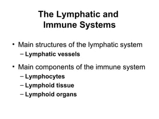
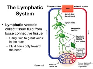
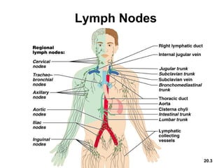
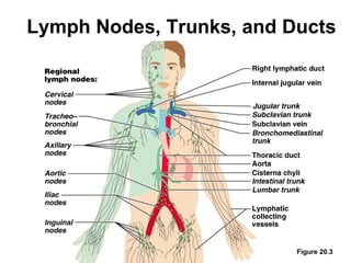

The document summarizes the lymphatic and immune systems. It describes the main structures of the lymphatic system as lymphatic vessels and lymph nodes. It then explains the main components of the immune system as lymphocytes, lymphoid tissue, and lymphoid organs. Finally, it discusses disorders that can affect these systems such as lymphangitis, mononucleosis, Hodgkin's disease, and non-Hodgkin's lymphoma.


