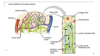
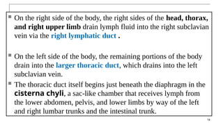
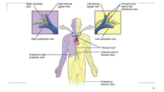
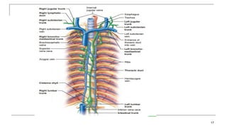
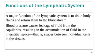
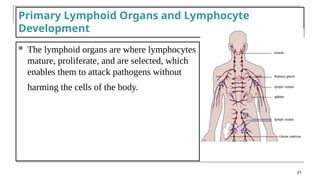
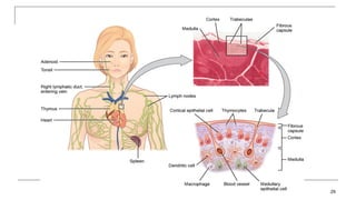
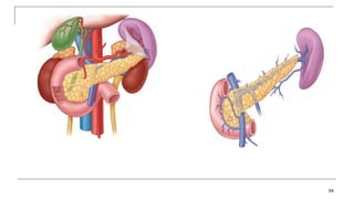
The Lymphatic System
1
Lymphatic system
 The lymphatic system is the system of vessels, cells, and
organs that carries excess fluids to the bloodstream and filters
pathogens from the blood.
 The swelling of lymph nodes during an infection and the
transport of lymphocytes via the lymphatic vessels are the two
examples of the many connections between these critical organ
systems
2
 In humans, 20 liters of plasma is released into the interstitial
space of the tissues each day due to capillary filtration.
 Once this filtrate is out of the bloodstream and in the tissue
spaces, it is referred to as interstitial fluid.
 Of this, 17 liters is reabsorbed directly by the blood vessels
 The remaining is where the lymphatic system comes into play.
 It drains the excess fluid and empties it back into the
bloodstream via a series of vessels, trunks, and ducts.
3
Lymphatic system
 Consists of:
1. Lymph
2. Lymphatic vessels
3. Structures and organs containing lymphatic tissue
4. Red bone marrow
4
 Lymph is the term used to describe interstitial fluid once it has
entered the lymphatic system.
 The transport of dietary lipids and fat-soluble vitamins
absorbed in the gut uses this system
 Lymph nodes as major staging areas for the development of
critical immune responses.
 A lymph node is one of the small, bean-shaped organs
located throughout the lymphatic system.
5
Structure of the Lymphatic System
 The lymphatic vessels begin as open-
ended capillaries, which feed into larger
and larger lymphatic vessels, and
eventually empty into the bloodstream by a
series of ducts.
 Along the way, the lymph travels through
the lymph nodes, which are commonly
found near the groin, armpits, neck, chest,
and abdomen.
6
7
 lymph is not actively pumped by the heart,
 It is moved by the movements of the body, the contraction of
skeletal muscles during body movements, and breathing.
 One-way valves (semi-lunar valves) in lymphatic vessels keep
the lymph moving toward the heart.
 Lymph flows from the lymphatic capillaries, through
lymphatic vessels, and then is dumped into the circulatory
system via the lymphatic ducts
8
Lymphatic Capillaries
9
 Lymphatic capillaries, also called the
terminal lymphatics
 are vessels where interstitial fluid enters
the lymphatic system to become lymph
fluid.
 Located in almost every tissue in the body.
 Exceptions are the central nervous system,
bone marrow, bones, teeth, and the cornea
of the eye, which do not contain lymph
vessels.
10
 Lymphatic capillaries are formed by a one cell-thick layer of
endothelial cells and represent the open end of the system,
allowing interstitial fluid to flow into them via overlapping cells
11
 In the small intestine, lymphatic
capillaries called lacteals are
critical for the transport of
dietary lipids and lipid-soluble
vitamins to the bloodstream.
 In the small intestine, dietary
triglycerides combine with other
lipids and proteins, and enter the
lacteals to form a milky fluid
called chyle.
 The chyle then travels through
the lymphatic system, eventually
entering the bloodstream
12
Larger Lymphatic Vessels, Trunks, and
Ducts
 The lymphatic capillaries empty into larger lymphatic vessels, which are
similar to veins in terms of their three-tunic structure and the presence of
valves.
 These one-way valves are located fairly close to one another, and each
one causes a bulge in the lymphatic vessel, giving the vessels a beaded
appearance .
 The superficial and deep lymphatics eventually merge to form larger
lymphatic vessels known as lymphatic trunks.
13
 On the right side of the body, the right sides of the head, thorax,
and right upper limb drain lymph fluid into the right subclavian
vein via the right lymphatic duct .
 On the left side of the body, the remaining portions of the body
drain into the larger thoracic duct, which drains into the left
subclavian vein.
 The thoracic duct itself begins just beneath the diaphragm in the
cisterna chyli, a sac-like chamber that receives lymph from
the lower abdomen, pelvis, and lower limbs by way of the left
and right lumbar trunks and the intestinal trunk.
14
 Ultimately, the lymph drains into 2 main channels:
1. Thoracic duct: a long duct that drains lymph from
the entire left half of the body and the right half
below the ribs including the right lower limb.
2. Right lymphatic duct: a short duct that drains lymph
from the right side of the body above the ribs
including the right upper limb.
15
Fig.4: Areas of lymph
drainage to the two
main lymphatic ducts.
16
17
 In general, lymphatic vessels of the subcutaneous tissues of the
skin, that is, the superficial lymphatics, follow the same routes
as veins, whereas the deep lymphatic vessels of the viscera
generally follow the paths of arteries.
18
 Each of these ducts open into the junction of
the subclavian and internal jugular veins as
they form the brachiocephalic vein.
 In this way, the lymph will return to the blood.
19
Fig.5: Opening of the lymphatic
ducts into the venous circulation.
Functions of the Lymphatic System
A major function of the lymphatic system is to drain body
fluids and return them to the bloodstream.
Blood pressure causes leakage of fluid from the
capillaries, resulting in the accumulation of fluid in the
interstitial space—that is, spaces between individual cells
in the tissues.
20
Primary Lymphoid Organs and Lymphocyte
Development
 The lymphoid organs are where lymphocytes
mature, proliferate, and are selected, which
enables them to attack pathogens without
harming the cells of the body.
21
 2 groups based on function
1. Primary lymphatic organs:
 Sites where B and T lymphocytes are formed and mature.
 Red bone marrow and thymus.
2. Secondary lymphatic organs
 Sites where most immune response occurs, meaning that they are the sites where
the mature lymphocytes perform their function.
 Lymph nodes, spleen, lymphatic nodules.
Lymphatic Organs and Tissues
22
Bone Marrow
 In the embryo, blood cells are made in the yolk sac.
 As development proceeds, this function is taken over by the
spleen, lymph nodes, and liver.
 Later, the bone marrow takes over most hematopoietic
functions.
 The B cell undergoes nearly all of its development in the red
bone marrow, whereas the immature T cell, called a
thymocyte, leaves the bone marrow and matures largely in
the thymus gland.
23
Red Bone Marrow
 Red bone marrow is the site of formation
of the blood elements: red blood cells,
white blood cells and platelets.
 Inside the red bone marrow
 B lymphocyte form and mature; T
lymphocytes are formed in the red bone
marrow but they’re immature.
 T-cells become mature in the thymus.
 Red bone marrow in adults is present in
the flat bones and the epiphyses of some
long bones.
24
1. The Thymus
 The thymus is an asymmetric bi-lobed organ where mature T-cells are
formed.
 It’s located in the superior mediastinum just behind the manubrium.
 It may descend into the anterior mediastinum to lie between the sternum
and the pericardial sac.
 Sometimes it may ascend into the neck reaching as high as the thyroid
gland.
 The fibrous capsule that surrounds the gland sends connective tissue
trabeculae into the gland dividing it into lobules.
 Each lobule is formed of a dark outer region with immature T-cells (the
cortex) and an inner lighter region with mature T-cells (the medulla).
25
 The thymus has a vascularized connective
tissue capsule that extends septa into the
parenchyma, dividing the organ into many
incompletely separated lobules.
 Each lobule has an outer darkly basophilic
cortex surrounding a more lightly stained
medulla.
26
 The staining differences reflect the much greater
density of lymphoblasts and small lymphocytes in the
cortex than the medulla.
 The thymic cortex contains an extensive population of
T lymphoblasts (or thymocytes), some newly arrived
via venules, located among numerous macrophages
and associated with the unique thymic epithelial cells
(TECs) .
27
 The thymic tissue is most
numerous in younger age.
As the person grows, this
tissue is gradually replaced
by fatty tissue.
28
Fig.6: To the left, histology of
the thymus. Below, position
of the thymus.
29
30
 Large aggregates of TECs, sometimes
concentrically
arranged, called Hassall corpuscles
 development of regulation T cells for
peripheral tolerance
31
32
33
Secondary Lymphoid Organs
In addition to circulating in the blood and lymph,
lymphocytes concentrate in secondary lymphoid organs,
which include the lymph nodes, spleen, and lymphoid
nodules.
Lymphocytes develop and mature in the primary lymphoid
organs, but they mount immune responses from the
secondary lymphoid organs.
 All of these tissues have many features in common, including the
following:
 The presence of lymphoid follicles, the sites of the formation of
lymphocytes, with specific B cell-rich and T cell-rich areas
An internal structure of reticular fibers with associated fixed
macrophages
• Germinal centers, which are the sites of rapidly dividing and
differentiating B lymphocytes
• Specialized post-capillary vessels known as high endothelial
venules
34
Lymph Nodes
 Lymph nodes function to remove debris and pathogens from the lymph, and
are thus sometimes referred to as the “filters of the lymph” .
 Any bacteria that infect the interstitial fluid are taken up by the lymphatic
capillaries and transported to a regional lymph node.
 Dendritic cells and macrophages within this organ internalize and kill many of
the pathogens that pass through, thereby removing them from the body.
 lymph nodes are surrounded by a tough capsule of connective tissue and are
separated into compartments by trabeculae, the extensions of the capsule.
 the structural support of the lymph node is provided by a series of reticular
fibers laid down by fibroblasts.
35
 Aggregations of these nodes occur near the body surface in
inguinal, axillary, and cervical regions of the body
36
Lymph Nodes
 Lymph nodes are capsulated bean shaped structures that are found along the course of the
lymphatic vessels. They’re usually present in groups and they’re scattered all over the body.
 The capsule sends trabeculae into the node dividing it into compartments.
 Beneath the capsule there’s a space called the Subcapsular Sinus.
 The node has an outer cortex with lymphatic nodules formed of B-cells and plasma cells.
 Deep to it is the inner cortex formed of T-cells with no nodules.
 Deep to the cortex is the medulla which contains B-cells, plasma cells and macrophages
embedded in reticular fibers.
 Between the lymphatic tissue in the cortex and medulla are spaces called cortical and medullary
sinuses.
37
38
Subcapsular
Sinus
Afferents
Cortical
Sinuses
Medullary
Sinuses
Efferents
 From the convex side of the lymph node several incoming lymphatic vessels enter
the node. These are called afferent lymphatic vessels.
 The concave side of the node is called the hilum.
 From it, one or two outgoing lymphatic vessels leave the node.
 These are called efferent lymphatic vessels.
 Also through the hilum arteries and nerves enter and veins exit the node.
39
The flow of lymph in a lymph node
40
Functions of lymph nodes:  Filter of lymph
1. Antigens in the lymph are trapped and the lymphocytes in the nodes react to it
and initiate the immune response.
2. Macrophages in the node may directly destroy the antigen.
 It’s important to know what lymphatic vessels drain a certain organ and
what are the lymph nodes in its course, because this represent a pathway by
which infections and cancer cells can spread.
41
42
43
 Largest single mass of lymphatic tissue in the body.
 It’s an oval, soft organ located in the left hypochondriac region.
 Its superior surface is smooth and related to the diaphragm and ribs 9,10
and 11.
 Its visceral surface is irregular and related to the stomach, pancreas, kidney
and colon.
 Stroma – capsule, trabeculae, reticular fibers, and fibroblasts.
 Parenchyma:
 White pulp – lymphatic tissue (lymphocytes and macrophages)
surrounds branches of splenic artery (the artery that supplies the spleen
and enters through the hilum).
The Spleen
44
 Red pulp – blood-filled venous sinuses surrounded by splenic cords which
contain red blood cells, macrophages, lymphocytes and plasma cells.
 Macrophages remove ruptured, worn out or defective blood cells.
 Storage of up to 1/3 of body’s platelet supply.
 Production of blood cells during fetal life.
 Function: Filter of Blood
45
46
Spleen
In addition to the lymph nodes, the spleen is a major
secondary lymphoid organ.
The spleen is a fragile organ without a strong capsule, and is
dark red due to its extensive vascularization.
The spleen also functions as the location of immune responses
to blood-borne pathogens
47
Fig.8: The spleen and its histology.
Lymphatic System
Figure34
20.6a-d
Structure of the Spleen
49
50
51
Spleen
 The spleen is connected to the :
 greater curvature of the stomach by the
gastrosplenic ligament, which contains the short
gastric and gastroomental vessels; and
■ left kidney by the splenorenal ligament ,
which contains the splenic vessels
52
53
54
 Upon entering the spleen, the splenic artery splits into several arterioles
(surrounded by white pulp) and eventually into sinusoids.
 Blood from the capillaries subsequently collects in the venous sinuses
and leaves via the splenic vein.
 The red pulp consists of reticular fibers with fixed macrophages
attached, free macrophages, and all of the other cells typical of the blood,
including some lymphocytes.
 The white pulp surrounds a central arteriole and consists of germinal
centers of dividing B cells surrounded by T cells and accessory cells,
including macrophages and dendritic cells.
 Thus, the red pulp primarily functions as a filtration system of the blood,
using cells of the relatively nonspecific immune response, and white
pulp is where adaptive T and B cell responses are mounted
55
Lymphoid Nodules
The other lymphoid tissues, the lymphoid nodules,
have a simpler architecture than the spleen and lymph
nodes in that they consist of a dense cluster of
lymphocytes without a surrounding fibrous capsule.
These nodules are located in the respiratory and
digestive tracts, areas routinely exposed to
environmental pathogens.
56
MALT (Mucosa Associated Lymphoid
Tissue
The mucosa or inner lining of the digestive,
respiratory, and genitourinary tracts is a
common site of invasion by pathogens
because their lumens open to the external
environment.
57
 Most of the immune cells in MALT are dispersed diffusely in
the connective tissue; others are found in aggregates that
form large, conspicuous structures such as the tonsils, the
Peyer patches in the ileum, and the appendix
 Consists of an aggregate of lymphoid follicles directly associated with
the mucous membrane epithelia.
 found underlying the mucosa of the gastrointestinal tract, breast tissue,
lungs, and eyes.
58
Tonsils are lymphoid nodules located along the inner surface of
the pharynx and are important in developing immunity to oral
pathogens.
Are large, irregular masses of lymphoid tissue in the
mucosa of the posterior oral cavity and nasopharynx
where their cells encounter antigens entering the
mouth and nose.
Named by their location these masses are the palatine,
lingual, and pharyngeal tonsils
59
Tonsil help children’s bodies recognize, destroy, and develop immunity to
common environmental pathogens so that they will be protected in their
later lives.
Tonsils are often removed in those children who have recurring throat
infections, especially those involving the palatine tonsils on either side of
the throat, whose swelling may interfere with their breathing and/or
swallowing
60
61
62
63
64
65
Peyer’s patches, a type of MALT in the small intestine, are especially
important for immune responses against ingested substances .
Peyer’s patches contain specialized endothelial cells called M (or microfold)
cells that sample material from the intestinal lumen and transport it to nearby
follicles so that adaptive immune responses to potential pathogens can be
mounted.
66
 Another significant collection of MALT occurs in the
mucosa of the appendix, a short, small-diameter
projection
from the cecum.
 Typically the mucosa of the appendix is almost
completely filled with lymphoid tissue
 The lumen contains the normal bacterial flora of the
large intestine
67
68
69
Bronchus-associated lymphoid tissue (BALT)
consists of lymphoid follicular structures with an overlying
epithelial layer found along the bifurcations of the bronchi,
and between bronchi and arteries.
They also have the typically less-organized structure of
other lymphoid nodules.
These tissues, in addition to the tonsils, are effective
against inhaled pathogens.
Elephantiasis

Lymphatic System.pptx Lymphatic System.pptx

  • 1.
  • 2.
    Lymphatic system  Thelymphatic system is the system of vessels, cells, and organs that carries excess fluids to the bloodstream and filters pathogens from the blood.  The swelling of lymph nodes during an infection and the transport of lymphocytes via the lymphatic vessels are the two examples of the many connections between these critical organ systems 2
  • 3.
     In humans,20 liters of plasma is released into the interstitial space of the tissues each day due to capillary filtration.  Once this filtrate is out of the bloodstream and in the tissue spaces, it is referred to as interstitial fluid.  Of this, 17 liters is reabsorbed directly by the blood vessels  The remaining is where the lymphatic system comes into play.  It drains the excess fluid and empties it back into the bloodstream via a series of vessels, trunks, and ducts. 3
  • 4.
    Lymphatic system  Consistsof: 1. Lymph 2. Lymphatic vessels 3. Structures and organs containing lymphatic tissue 4. Red bone marrow 4
  • 5.
     Lymph isthe term used to describe interstitial fluid once it has entered the lymphatic system.  The transport of dietary lipids and fat-soluble vitamins absorbed in the gut uses this system  Lymph nodes as major staging areas for the development of critical immune responses.  A lymph node is one of the small, bean-shaped organs located throughout the lymphatic system. 5
  • 6.
    Structure of theLymphatic System  The lymphatic vessels begin as open- ended capillaries, which feed into larger and larger lymphatic vessels, and eventually empty into the bloodstream by a series of ducts.  Along the way, the lymph travels through the lymph nodes, which are commonly found near the groin, armpits, neck, chest, and abdomen. 6
  • 7.
  • 8.
     lymph isnot actively pumped by the heart,  It is moved by the movements of the body, the contraction of skeletal muscles during body movements, and breathing.  One-way valves (semi-lunar valves) in lymphatic vessels keep the lymph moving toward the heart.  Lymph flows from the lymphatic capillaries, through lymphatic vessels, and then is dumped into the circulatory system via the lymphatic ducts 8
  • 9.
    Lymphatic Capillaries 9  Lymphaticcapillaries, also called the terminal lymphatics  are vessels where interstitial fluid enters the lymphatic system to become lymph fluid.  Located in almost every tissue in the body.  Exceptions are the central nervous system, bone marrow, bones, teeth, and the cornea of the eye, which do not contain lymph vessels.
  • 10.
  • 11.
     Lymphatic capillariesare formed by a one cell-thick layer of endothelial cells and represent the open end of the system, allowing interstitial fluid to flow into them via overlapping cells 11
  • 12.
     In thesmall intestine, lymphatic capillaries called lacteals are critical for the transport of dietary lipids and lipid-soluble vitamins to the bloodstream.  In the small intestine, dietary triglycerides combine with other lipids and proteins, and enter the lacteals to form a milky fluid called chyle.  The chyle then travels through the lymphatic system, eventually entering the bloodstream 12
  • 13.
    Larger Lymphatic Vessels,Trunks, and Ducts  The lymphatic capillaries empty into larger lymphatic vessels, which are similar to veins in terms of their three-tunic structure and the presence of valves.  These one-way valves are located fairly close to one another, and each one causes a bulge in the lymphatic vessel, giving the vessels a beaded appearance .  The superficial and deep lymphatics eventually merge to form larger lymphatic vessels known as lymphatic trunks. 13
  • 14.
     On theright side of the body, the right sides of the head, thorax, and right upper limb drain lymph fluid into the right subclavian vein via the right lymphatic duct .  On the left side of the body, the remaining portions of the body drain into the larger thoracic duct, which drains into the left subclavian vein.  The thoracic duct itself begins just beneath the diaphragm in the cisterna chyli, a sac-like chamber that receives lymph from the lower abdomen, pelvis, and lower limbs by way of the left and right lumbar trunks and the intestinal trunk. 14
  • 15.
     Ultimately, thelymph drains into 2 main channels: 1. Thoracic duct: a long duct that drains lymph from the entire left half of the body and the right half below the ribs including the right lower limb. 2. Right lymphatic duct: a short duct that drains lymph from the right side of the body above the ribs including the right upper limb. 15 Fig.4: Areas of lymph drainage to the two main lymphatic ducts.
  • 16.
  • 17.
  • 18.
     In general,lymphatic vessels of the subcutaneous tissues of the skin, that is, the superficial lymphatics, follow the same routes as veins, whereas the deep lymphatic vessels of the viscera generally follow the paths of arteries. 18
  • 19.
     Each ofthese ducts open into the junction of the subclavian and internal jugular veins as they form the brachiocephalic vein.  In this way, the lymph will return to the blood. 19 Fig.5: Opening of the lymphatic ducts into the venous circulation.
  • 20.
    Functions of theLymphatic System A major function of the lymphatic system is to drain body fluids and return them to the bloodstream. Blood pressure causes leakage of fluid from the capillaries, resulting in the accumulation of fluid in the interstitial space—that is, spaces between individual cells in the tissues. 20
  • 21.
    Primary Lymphoid Organsand Lymphocyte Development  The lymphoid organs are where lymphocytes mature, proliferate, and are selected, which enables them to attack pathogens without harming the cells of the body. 21
  • 22.
     2 groupsbased on function 1. Primary lymphatic organs:  Sites where B and T lymphocytes are formed and mature.  Red bone marrow and thymus. 2. Secondary lymphatic organs  Sites where most immune response occurs, meaning that they are the sites where the mature lymphocytes perform their function.  Lymph nodes, spleen, lymphatic nodules. Lymphatic Organs and Tissues 22
  • 23.
    Bone Marrow  Inthe embryo, blood cells are made in the yolk sac.  As development proceeds, this function is taken over by the spleen, lymph nodes, and liver.  Later, the bone marrow takes over most hematopoietic functions.  The B cell undergoes nearly all of its development in the red bone marrow, whereas the immature T cell, called a thymocyte, leaves the bone marrow and matures largely in the thymus gland. 23
  • 24.
    Red Bone Marrow Red bone marrow is the site of formation of the blood elements: red blood cells, white blood cells and platelets.  Inside the red bone marrow  B lymphocyte form and mature; T lymphocytes are formed in the red bone marrow but they’re immature.  T-cells become mature in the thymus.  Red bone marrow in adults is present in the flat bones and the epiphyses of some long bones. 24
  • 25.
    1. The Thymus The thymus is an asymmetric bi-lobed organ where mature T-cells are formed.  It’s located in the superior mediastinum just behind the manubrium.  It may descend into the anterior mediastinum to lie between the sternum and the pericardial sac.  Sometimes it may ascend into the neck reaching as high as the thyroid gland.  The fibrous capsule that surrounds the gland sends connective tissue trabeculae into the gland dividing it into lobules.  Each lobule is formed of a dark outer region with immature T-cells (the cortex) and an inner lighter region with mature T-cells (the medulla). 25
  • 26.
     The thymushas a vascularized connective tissue capsule that extends septa into the parenchyma, dividing the organ into many incompletely separated lobules.  Each lobule has an outer darkly basophilic cortex surrounding a more lightly stained medulla. 26
  • 27.
     The stainingdifferences reflect the much greater density of lymphoblasts and small lymphocytes in the cortex than the medulla.  The thymic cortex contains an extensive population of T lymphoblasts (or thymocytes), some newly arrived via venules, located among numerous macrophages and associated with the unique thymic epithelial cells (TECs) . 27
  • 28.
     The thymictissue is most numerous in younger age. As the person grows, this tissue is gradually replaced by fatty tissue. 28 Fig.6: To the left, histology of the thymus. Below, position of the thymus.
  • 29.
  • 30.
  • 31.
     Large aggregatesof TECs, sometimes concentrically arranged, called Hassall corpuscles  development of regulation T cells for peripheral tolerance 31
  • 32.
  • 33.
    33 Secondary Lymphoid Organs Inaddition to circulating in the blood and lymph, lymphocytes concentrate in secondary lymphoid organs, which include the lymph nodes, spleen, and lymphoid nodules. Lymphocytes develop and mature in the primary lymphoid organs, but they mount immune responses from the secondary lymphoid organs.
  • 34.
     All ofthese tissues have many features in common, including the following:  The presence of lymphoid follicles, the sites of the formation of lymphocytes, with specific B cell-rich and T cell-rich areas An internal structure of reticular fibers with associated fixed macrophages • Germinal centers, which are the sites of rapidly dividing and differentiating B lymphocytes • Specialized post-capillary vessels known as high endothelial venules 34
  • 35.
    Lymph Nodes  Lymphnodes function to remove debris and pathogens from the lymph, and are thus sometimes referred to as the “filters of the lymph” .  Any bacteria that infect the interstitial fluid are taken up by the lymphatic capillaries and transported to a regional lymph node.  Dendritic cells and macrophages within this organ internalize and kill many of the pathogens that pass through, thereby removing them from the body.  lymph nodes are surrounded by a tough capsule of connective tissue and are separated into compartments by trabeculae, the extensions of the capsule.  the structural support of the lymph node is provided by a series of reticular fibers laid down by fibroblasts. 35
  • 36.
     Aggregations ofthese nodes occur near the body surface in inguinal, axillary, and cervical regions of the body 36
  • 37.
    Lymph Nodes  Lymphnodes are capsulated bean shaped structures that are found along the course of the lymphatic vessels. They’re usually present in groups and they’re scattered all over the body.  The capsule sends trabeculae into the node dividing it into compartments.  Beneath the capsule there’s a space called the Subcapsular Sinus.  The node has an outer cortex with lymphatic nodules formed of B-cells and plasma cells.  Deep to it is the inner cortex formed of T-cells with no nodules.  Deep to the cortex is the medulla which contains B-cells, plasma cells and macrophages embedded in reticular fibers.  Between the lymphatic tissue in the cortex and medulla are spaces called cortical and medullary sinuses. 37
  • 38.
  • 39.
    Subcapsular Sinus Afferents Cortical Sinuses Medullary Sinuses Efferents  From theconvex side of the lymph node several incoming lymphatic vessels enter the node. These are called afferent lymphatic vessels.  The concave side of the node is called the hilum.  From it, one or two outgoing lymphatic vessels leave the node.  These are called efferent lymphatic vessels.  Also through the hilum arteries and nerves enter and veins exit the node. 39 The flow of lymph in a lymph node
  • 40.
  • 41.
    Functions of lymphnodes:  Filter of lymph 1. Antigens in the lymph are trapped and the lymphocytes in the nodes react to it and initiate the immune response. 2. Macrophages in the node may directly destroy the antigen.  It’s important to know what lymphatic vessels drain a certain organ and what are the lymph nodes in its course, because this represent a pathway by which infections and cancer cells can spread. 41
  • 42.
  • 43.
  • 44.
     Largest singlemass of lymphatic tissue in the body.  It’s an oval, soft organ located in the left hypochondriac region.  Its superior surface is smooth and related to the diaphragm and ribs 9,10 and 11.  Its visceral surface is irregular and related to the stomach, pancreas, kidney and colon.  Stroma – capsule, trabeculae, reticular fibers, and fibroblasts.  Parenchyma:  White pulp – lymphatic tissue (lymphocytes and macrophages) surrounds branches of splenic artery (the artery that supplies the spleen and enters through the hilum). The Spleen 44
  • 45.
     Red pulp– blood-filled venous sinuses surrounded by splenic cords which contain red blood cells, macrophages, lymphocytes and plasma cells.  Macrophages remove ruptured, worn out or defective blood cells.  Storage of up to 1/3 of body’s platelet supply.  Production of blood cells during fetal life.  Function: Filter of Blood 45
  • 46.
    46 Spleen In addition tothe lymph nodes, the spleen is a major secondary lymphoid organ. The spleen is a fragile organ without a strong capsule, and is dark red due to its extensive vascularization. The spleen also functions as the location of immune responses to blood-borne pathogens
  • 47.
    47 Fig.8: The spleenand its histology.
  • 48.
  • 49.
  • 50.
  • 51.
  • 52.
    Spleen  The spleenis connected to the :  greater curvature of the stomach by the gastrosplenic ligament, which contains the short gastric and gastroomental vessels; and ■ left kidney by the splenorenal ligament , which contains the splenic vessels 52
  • 53.
  • 54.
  • 55.
     Upon enteringthe spleen, the splenic artery splits into several arterioles (surrounded by white pulp) and eventually into sinusoids.  Blood from the capillaries subsequently collects in the venous sinuses and leaves via the splenic vein.  The red pulp consists of reticular fibers with fixed macrophages attached, free macrophages, and all of the other cells typical of the blood, including some lymphocytes.  The white pulp surrounds a central arteriole and consists of germinal centers of dividing B cells surrounded by T cells and accessory cells, including macrophages and dendritic cells.  Thus, the red pulp primarily functions as a filtration system of the blood, using cells of the relatively nonspecific immune response, and white pulp is where adaptive T and B cell responses are mounted 55
  • 56.
    Lymphoid Nodules The otherlymphoid tissues, the lymphoid nodules, have a simpler architecture than the spleen and lymph nodes in that they consist of a dense cluster of lymphocytes without a surrounding fibrous capsule. These nodules are located in the respiratory and digestive tracts, areas routinely exposed to environmental pathogens. 56
  • 57.
    MALT (Mucosa AssociatedLymphoid Tissue The mucosa or inner lining of the digestive, respiratory, and genitourinary tracts is a common site of invasion by pathogens because their lumens open to the external environment. 57
  • 58.
     Most ofthe immune cells in MALT are dispersed diffusely in the connective tissue; others are found in aggregates that form large, conspicuous structures such as the tonsils, the Peyer patches in the ileum, and the appendix  Consists of an aggregate of lymphoid follicles directly associated with the mucous membrane epithelia.  found underlying the mucosa of the gastrointestinal tract, breast tissue, lungs, and eyes. 58
  • 59.
    Tonsils are lymphoidnodules located along the inner surface of the pharynx and are important in developing immunity to oral pathogens. Are large, irregular masses of lymphoid tissue in the mucosa of the posterior oral cavity and nasopharynx where their cells encounter antigens entering the mouth and nose. Named by their location these masses are the palatine, lingual, and pharyngeal tonsils 59
  • 60.
    Tonsil help children’sbodies recognize, destroy, and develop immunity to common environmental pathogens so that they will be protected in their later lives. Tonsils are often removed in those children who have recurring throat infections, especially those involving the palatine tonsils on either side of the throat, whose swelling may interfere with their breathing and/or swallowing 60
  • 61.
  • 62.
  • 63.
  • 64.
  • 65.
    65 Peyer’s patches, atype of MALT in the small intestine, are especially important for immune responses against ingested substances . Peyer’s patches contain specialized endothelial cells called M (or microfold) cells that sample material from the intestinal lumen and transport it to nearby follicles so that adaptive immune responses to potential pathogens can be mounted.
  • 66.
  • 67.
     Another significantcollection of MALT occurs in the mucosa of the appendix, a short, small-diameter projection from the cecum.  Typically the mucosa of the appendix is almost completely filled with lymphoid tissue  The lumen contains the normal bacterial flora of the large intestine 67
  • 68.
  • 69.
    69 Bronchus-associated lymphoid tissue(BALT) consists of lymphoid follicular structures with an overlying epithelial layer found along the bifurcations of the bronchi, and between bronchi and arteries. They also have the typically less-organized structure of other lymphoid nodules. These tissues, in addition to the tonsils, are effective against inhaled pathogens.
  • 70.