Downloaded 176 times
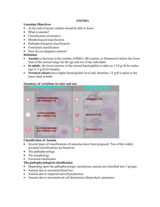
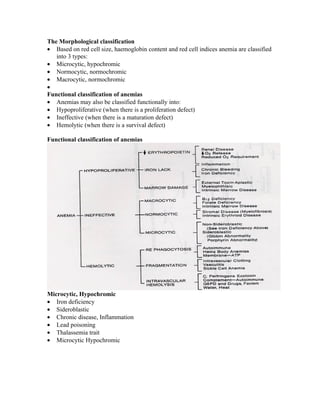

This document provides an overview of anemia, including its definition, classifications, causes, and methods of diagnosis. Anemia is classified based on pathophysiology (increased blood loss, impaired red blood cell production, increased destruction), morphology (microcytic/hypochromic, normocytic/normochromic, macrocytic/normochromic), and function (hypoproliferative, ineffective, hemolytic). Common causes of different types of anemia are outlined. Laboratory tests including red blood cell indices help diagnose the type of anemia and indicate the underlying cause.


