Learning Outcomes
• Listthe functions of the
lymphatic system
• Explain how lymph forms and
returns to the bloodstream
• Name the major cells of the
lymphatic system and state their
functions
• Name and describe the types
of lymphatic tissue
• Describe the structure and
function of the red bone
marrow, thymus, lymph
nodes, tonsils, and spleen
3.
What exactly isthe Lymphatic System?
• What is the function of the Lymphatic System?
• Which organs are involved?
• How does it work?
4.
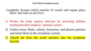
FUNCTION of LymphaticSystem:
Lymphatic System which consists of vessels and organs plays
three vital roles in our lives:
1) Protect the body against infection by activating defense
mechanism that comprise immune system.
2) Collect tissue fluids, solutes, hormones, and plasma proteins
and return them to the circulatory system.
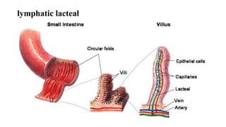
3) Absorb fat from the small intestine into the lymphatic
lacteals.
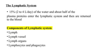
The Lymphatic System
•15% (2 to 4 L/day) of the water and about half of the
plasma proteins enter the lymphatic system and then are returned
to the blood
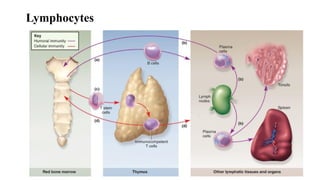
Components of Lymphatic system
• Lymph
• Lymph vessel
• Lymph organs
• Lymphocytes and phagocytes
7.
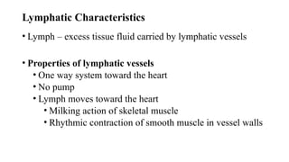
Lymphatic Characteristics
• Lymph– excess tissue fluid carried by lymphatic vessels
• Properties of lymphatic vessels
• One way system toward the heart
• No pump
• Lymph moves toward the heart
• Milking action of skeletal muscle
• Rhythmic contraction of smooth muscle in vessel walls
8.
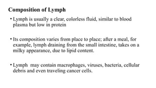
Composition of Lymph
•Lymph is usually a clear, colorless fluid, similar to blood
plasma but low in protein
• Its composition varies from place to place; after a meal, for
example, lymph draining from the small intestine, takes on a
milky appearance, due to lipid content.
• Lymph may contain macrophages, viruses, bacteria, cellular
debris and even traveling cancer cells.
9.
Flow of Lymph
•Lymph flows under forces similar to those that govern venous
return, except no pump (heart)
•Lymph flows at low pressure and slower speed than venous
blood
•Moved along by rhythmic contractions of lymphatic vessels
• Stretching of vessels stimulates contraction
10.
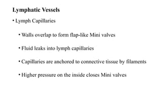
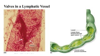
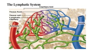
Lymphatic Vessels
• LymphCapillaries
• Walls overlap to form flap-like Mini valves
• Fluid leaks into lymph capillaries
• Capillaries are anchored to connective tissue by filaments
• Higher pressure on the inside closes Mini valves
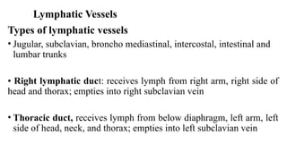
Lymphatic Vessels
Types oflymphatic vessels
• Jugular, subclavian, broncho mediastinal, intercostal, intestinal and
lumbar trunks
• Right lymphatic duct: receives lymph from right arm, right side of
head and thorax; empties into right subclavian vein
• Thoracic duct, receives lymph from below diaphragm, left arm, left
side of head, neck, and thorax; empties into left subclavian vein
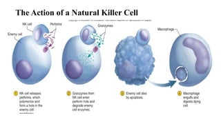
Lymphatic Cells
• Naturalkiller (NK) cells
Attack and destroy bacteria, host cells infected with viruses or that
have turned cancerous
• T-lymphocytes (T cells)
Mature in thymus
• B-lymphocytes (B cells)
Activation causes production and differentiation into plasma cells that
produce antibodies
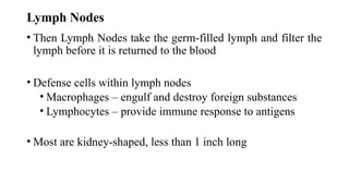
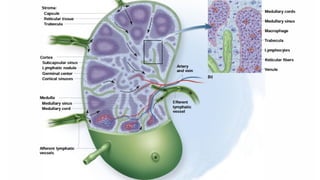
Lymph Nodes
• ThenLymph Nodes take the germ-filled lymph and filter the
lymph before it is returned to the blood
• Defense cells within lymph nodes
• Macrophages – engulf and destroy foreign substances
• Lymphocytes – provide immune response to antigens
• Most are kidney-shaped, less than 1 inch long
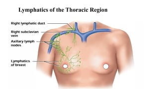
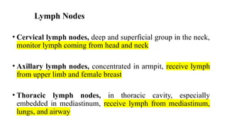
Lymph Nodes
• Cervicallymph nodes, deep and superficial group in the neck,
monitor lymph coming from head and neck
• Axillary lymph nodes, concentrated in armpit, receive lymph
from upper limb and female breast
• Thoracic lymph nodes, in thoracic cavity, especially
embedded in mediastinum, receive lymph from mediastinum,
lungs, and airway
22.
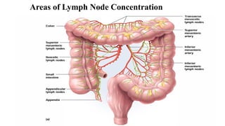
Lymph Nodes
• Abdominallymph nodes, occur in posterior abdominopelvic wall,
monitor lymph from the urinary and reproductive systems
•Intestinal and mesenteric lymph nodes, found in the mesenteries,
adjacent to the appendix and intestines, monitor lymph from the
digestive tract
Inguinal lymph nodes, in the groin and receive lymph from the
entire lower limb
23.
Lymph Nodes
• Popliteallymph nodes– Occur on the back of the knee–
• Receive lymph from the leg proper
Functions
• Phagocytic actions
• Production of antibodies
• Enlargement of lymph nodes when area of drainage is infected
• Activate T and B lymphocytes
• Filtration of lymph
Lymphatic Organs:
• LymphNode- Important lymphocytes of the immune response are
matured here.
• Spleen: destroys RBCs and Reservoir of Blood; it filter blood of
bacteria and antigen-filled cells.
• Thymus Gland-produces hormone, thymosin, functions in
programing lymphocytes T and B cells; T-cells matured here
( become immunocompetent)
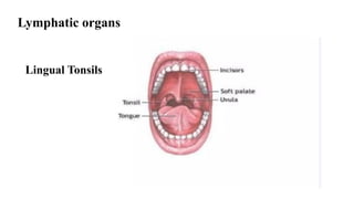
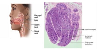
• Tonsils-Traps bacteria and other microbes in throat.
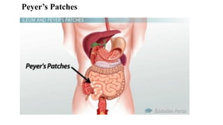
• Peyer’s Patch-capture and destroy bacteria in intestine, thereby
preventing them from penetrating the intestinal wall.
27.
Lymphatic organs
The lymphaticorgans are found in a number of situations in
the body
• Tonsils, spleen
• Bone marrow
• Thymus gland
• Peyer’s Patch-capture
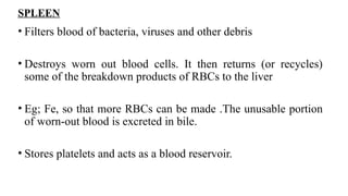
SPLEEN
• Filters bloodof bacteria, viruses and other debris
• Destroys worn out blood cells. It then returns (or recycles)
some of the breakdown products of RBCs to the liver
• Eg; Fe, so that more RBCs can be made .The unusable portion
of worn-out blood is excreted in bile.
• Stores platelets and acts as a blood reservoir.
32.
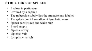
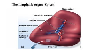
STRUCTURE OF SPLEEN
•Enclose in peritoneum
• Covered by a capsule
• The trabeculae subdivides the structure into lobules
• The spleen don’t have afferent lymphatic vessel
• Spleen consists red and white pulp
• Blood supply
• Splenic artery
• Splenic vein
• Lymphatic vessels
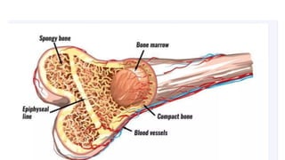
The bone marrow
•The red bone marrow is a key element of the lymphatic system
Generate lymphocytes from immature hematopoietic progenitor
cells
Constitute primary lymphoid tissue involved in the production and
early selection of lymphocytes
Performs a valve like function to prevent the back flow of
lymphatic fluid in the lymphatic system
36.
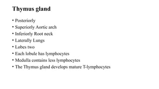
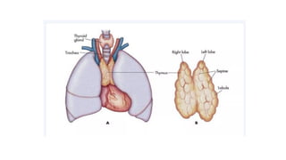
Thymus gland
• Posteriorly
•Superiorly Aortic arch
• Inferiorly Root neck
• Laterally Lungs
• Lobes two
• Each lobule has lymphocytes
• Medulla contains less lymphocytes
• The Thymus gland develops mature T-lymphocytes
38.
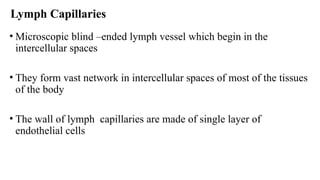
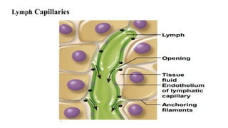
Lymph Capillaries
• Microscopicblind –ended lymph vessel which begin in the
intercellular spaces
• They form vast network in intercellular spaces of most of the tissues
of the body
• The wall of lymph capillaries are made of single layer of
endothelial cells
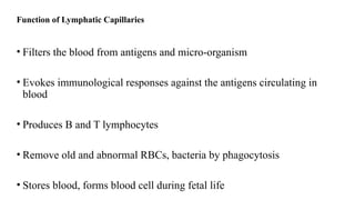
Function of LymphaticCapillaries
• Filters the blood from antigens and micro-organism
• Evokes immunological responses against the antigens circulating in
blood
• Produces B and T lymphocytes
• Remove old and abnormal RBCs, bacteria by phagocytosis
• Stores blood, forms blood cell during fetal life
43.
Function of Thymus
•The site of lymphocytes production
• It receives immunologically incompetent stem cells from bone
marrow
• Thymus secrete hormone like thymosin which support the activity
of T-lymphocytes through out the body
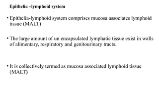
Epithelia –lymphoid system
•Epithelia-lymphoid system comprises mucosa associates lymphoid
tissue (MALT)
• The large amount of un encapsulated lymphatic tissue exist in walls
of alimentary, respiratory and genitourinary tracts.
• It is collectively termed as mucosa associated lymphoid tissue
(MALT)
46.
• The MALTtissue is generally sub divided into the following
two types
• Bronchus associated lymphoid tissue (BALT) in respiratory
system
• Gut associated lymphoid tissue (GALT)
47.
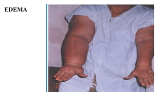
Odema
• Edema isthe excess accumulation of fluids in tissue spaces.
• This can retard normal exchange of nutrients and metabolites.
• Anything that causes increased capillary pressure, such as
decreased plasma protein, increased capillary permeability or
lymphatic blockage,
• can result in swelling and congestion of the extravascular
compartment.