The Lymphatic System: Anatomy, Function, and Clinical Terms
•
2 likes•291 views

Dr Shashikant and Team IM WELL Pvt Ltd Akshara Family Clinic and Integrated Medical Center www.Imwellyoga.com

Recommended
More Related Content
What's hot
What's hot (20)
Similar to The Lymphatic System: Anatomy, Function, and Clinical Terms
Similar to The Lymphatic System: Anatomy, Function, and Clinical Terms (20)
More from Dr Shashikant S Kumbar
More from Dr Shashikant S Kumbar (20)
Recently uploaded
Recently uploaded (20)
The Lymphatic System: Anatomy, Function, and Clinical Terms
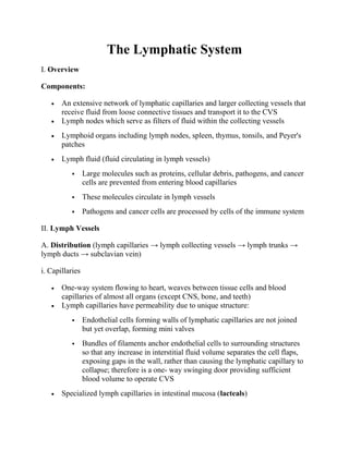
- 1. The Lymphatic System I. Overview Components: • An extensive network of lymphatic capillaries and larger collecting vessels that receive fluid from loose connective tissues and transport it to the CVS • Lymph nodes which serve as filters of fluid within the collecting vessels • Lymphoid organs including lymph nodes, spleen, thymus, tonsils, and Peyer's patches • Lymph fluid (fluid circulating in lymph vessels) Large molecules such as proteins, cellular debris, pathogens, and cancer cells are prevented from entering blood capillaries These molecules circulate in lymph vessels Pathogens and cancer cells are processed by cells of the immune system II. Lymph Vessels A. Distribution (lymph capillaries → lymph collecting vessels → lymph trunks → lymph ducts → subclavian vein) i. Capillaries • One-way system flowing to heart, weaves between tissue cells and blood capillaries of almost all organs (except CNS, bone, and teeth) • Lymph capillaries have permeability due to unique structure: Endothelial cells forming walls of lymphatic capillaries are not joined but yet overlap, forming mini valves Bundles of filaments anchor endothelial cells to surrounding structures so that any increase in interstitial fluid volume separates the cell flaps, exposing gaps in the wall, rather than causing the lymphatic capillary to collapse; therefore is a one- way swinging door providing sufficient blood volume to operate CVS • Specialized lymph capillaries in intestinal mucosa (lacteals)
- 2. ii. Lymphatic collecting vessels • Lymph flowing from capillaries enter lymph collecting vessels • Have the same three tunics as veins, but are thinner walled, have more internal valves, and anastomose more • Receive nutrient blood supply from a branching vasa vasorum iii. Lymphatic trunk Formed by the union of the largest collecting vessels • Named mostly for the regions from which they collect lymph • Lumbar, bronchomediastinal, subclavian, jugular and intestinal iv. Lymphatic ducts Lymph is delivered to one of two large ducts • Right lymphatic duct drains lymph from the right upper arm and the right side of the head and thorax • Thoracic duct receives lymph from the rest of the body • Intestinal trunk and lumbar trunks merge to form the cisterna chyli of the thoracic trunk • Lymph empties into venous circulation at or near the junction of internal jugular vein and subclavian vein B. Transport • Transport influenced by smooth muscle contraction, pressure changes, presence of valves • Over a 24 hr period, the volume lost from the blood stream equals the volume entering and circulating the lypmh system III. Lymph Nodes Small organs clustered along lymph vessels and embedded in connective tissue • Occur near the body surface • Located in inguinal, axillary, and cervical regions of the body • Functions
- 3. Filters lymph via phagocytic macrophages Activate immune system; lymphocytes in nodes monitor the lymph for antigens and mount an attack against them • Structure Capsule surrounds node and its connective tissue fibers extend into node Trabeculae are formed from the extended capsule fivers and compartmentalize the node Afferent vessels bring lymph into the node, passing through the cortex Sub scapular sinus forms betwen the capsule and the cortex Lymphatic Follicles found in the cortex contain reticular cells and fibers as well as lymphocytes in the germinal center of the follcile Medullary cords extend inward and contain both types of lymphocytes Medularry sinus is formed by lymph capillaries Efferent vessels in the hilus transport filtered lymph toward venous circulation IV. Lymphoid Organs A. Spleen • Blood-rich, size of fist, left side of abdominal cavity beneath diaphragm • Site of lymphocyte proliferation and immune response • Three functions: Stores some products of RBC degradation Site of erythrocyte production in fetus Stores thrombocytes • Sturcture: Fibrous capsule Trabeculae Red pulp (RBCs and macrophage)
- 4. White pulp (lymphocytes) B. Thymus • Located in the lower neck extending into mediastinum of thorax • Secretes hormones (thymosins) that stimulate the development and cell differentiation of T lymphocytes • T cells become immunocompetent in infants (increases immune responses) • Atrophies after adolescence C. Tonsils • Ring of lymph tissue around entrance of pharynx • Gather and remove many pathogens entering the pharynx • Types: palatine, lingual, pharyngeal, and tubal tonsils D. Peyer's patches • Located in the wall of the distal portion (ileum) of small intestine • Contains macrophages that capture and destroy bacteria thereby preventing microbes from breaching the intestinal wall V. Clinical Terms • Lymphadenitis/lymphangitis - nfection of the lymph nodes (also called lymph glands) and lymph channels • Lymphedema - an accumulation of lymphatic fluid in the interstitial tissue that causes swelling • Hodgkin's lymphoma - (also called Hodgkin's Disease) is a malignant growth of cells in the lymph system; it is differentiated from Nonhodgkin's lymphoma be the presence of special cells called Reed-Sternberg cells • Nonhodgkin's lymphoma - is a malignant growth of B or T cells in the lymph system • Splenomegaly - enlargement of the spleen due to accumulation of infectious microorganisms • Tonsilitis - inflammation of tonsils due to bacterial infection
- 5. • Elephantasis - lympatics (of lower limbs) become clogged due to filariasis (parasitic roundworms infection) Dr Shashikant and Team IM WELL Pvt Ltd Medical Yoga TTC Akshara Family Clinic and Integrated Medical Center www.Imwellyoga.com
- 6. • Elephantasis - lympatics (of lower limbs) become clogged due to filariasis (parasitic roundworms infection) Dr Shashikant and Team IM WELL Pvt Ltd Medical Yoga TTC Akshara Family Clinic and Integrated Medical Center www.Imwellyoga.com