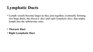
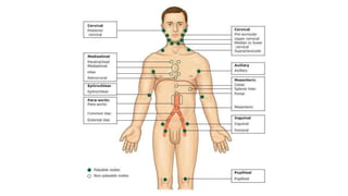
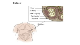
The Lymphatic
System
Mehmood ul Hassan
Nursing Faculty
Bahria Town School Of Nursing
Lahore, Pakistan
Objectives
At the completion of this unit, learners will be able to:
1. Define lymph & the lymphatic system
2. Identify the organs of lymphatic system
3. Describe the general functions of the lymphatic system
4. Describe how lymph is formed
5. Describe the lymph vessels & how lymph is returned to the blood
vessels
6. Describe the structure and functions of the lymph nodes, nodules,
spleen and the thymus glands.
Lymphatic System
• The lymphatic system contributes to homeostasis by draining
interstitial fluid as well as providing the mechanisms for defense
against disease.
Or
• The lymphatic system is the system of vessels, cells, and organs that
carries excess fluids to the bloodstream and filters pathogens from
the blood.
Functions of The Lymphatic System
• TISSUE DRAINAGE
• ABSORPTION IN THE SMALL INTESTINE
• IMMUNITY
TISSUE DRAINAGE
• Every day, around 21 liters of plasma fluid, carrying dissolved
substances and some plasma protein, escape from the arterial end of
the capillaries and into the tissues.
• Most of this fluid is returned directly to the bloodstream via the
capillary at its venous end, but 3-4 liters of fluid are drained away by
the lymphatic vessels.
• Without this, the tissues would rapidly become waterlogged, and the
cardiovascular system would begin to fail as the blood volume falls.
Interstitial fluid
CapillariesArterial end of capillaries Venous end of capillaries
Blood
Hydrostatic pressure
32 26
32 – 26 = 6
Blood
Hydrostatic pressure
15 2615 – 26 = -11
Close ended capillaries
Cells
Interstitial
fluid
FAT ABSORPTION IN THE SMALL
INTESTINE
• Fat and fat-soluble materials, e.g. the fat-soluble vitamins, are
absorbed into the central lacteals (lymphatic vessels) of the villi.
FAT ABSORPTION IN THE SMALL
INTESTINE
• The free fatty acids and monoacylglycerides that enter the epithelial
cells are reincorporated into triglycerides. The triglycerides are mixed
with phospholipids and cholesterol, and surrounded with a protein
coat. This new complex, called a chylomicron, is a water-soluble
lipoprotein. After being processed by the Golgi apparatus,
chylomicrons are released from the cell. Too big to pass through the
basement membranes of blood capillaries, chylomicrons instead enter
the large pores of lacteals. The lacteals come together to form the
lymphatic vessels. The chylomicrons are transported in the lymphatic
vessels and empty through the thoracic duct into the subclavian vein of
the circulatory system.
FAT ABSORPTION
IN THE SMALL
INTESTINE
IMMUNITY
• The lymphatic organs are concerned with the production and
maturation of lymphocytes, the white blood cells that are primarily
responsible for provision of immunity.
• Bone marrow is therefore considered to be lymphatic tissue, since
lymphocytes are produced there.
IMMUNITY
• lymphocytes are the primary cells of adaptive immune responses.
• The two basic types of lymphocytes, B cells and T cells. While B cells
mature in red bone marrow and T cells mature in the thymus, they
both initially develop from bone marrow.
• T-cells migrate from bone marrow to the thymus gland where they
further mature.
• B cells and T cells are found in many parts of the body, circulating in
the bloodstream and lymph, and residing in secondary lymphoid
organs, including the spleen and lymph nodes,
Organs of Lymphatic System
1. Lymph
2. Lymph vessels
3. Lymph nodes
4. Lymph organs, e.g. Spleen and thymus
5. Diffuse lymphoid tissue, e.g. Tonsils
6. Bone marrow.
Lymph
• Lymph is the term used to describe interstitial fluid once it has entered
the lymphatic system.
• Lymph is a clear watery fluid, similar in composition to plasma, with
the important exception of plasma proteins, and identical in
composition to interstitial fluid.
Functions of Lymph
• Lymph transports the plasma proteins that seep out of the capillary
beds back to the bloodstream.
• It also carries away larger particles, e.g. bacteria and cell debris from
damaged tissues, which can then be filtered out and destroyed by the
lymph nodes.
• Lymph contains lymphocytes, which circulate in the lymphatic system
allowing them to patrol the different regions of the body.
• In the lacteals of the small intestine, fats absorbed into the lymphatics
give the lymph (now called chyle), a milky appearance.
Factors Effecting on Lymph Flow
• Valves:
• Lymph vessels have numerous cup-shaped valves which ensure that
lymph flows in one way only, i.e. towards the thorax.
• Muscle Tissue In Tunica Media
• There is no 'pump', like the heart, involved in the onward movement of
lymph but the muscle tissue in the walls of the large lymph vessels has
an intrinsic ability to contract rhythmically (the lymphatic pump).
Factors Effecting on Lymph Flow
• Skeletal muscle pump
• The “milking action” of skeletal muscle contractions compresses lymphatic
vessels (as well as veins) and forces lymph toward the junction of the internal
jugular and subclavian veins.
• Respiratory pump.
• Lymph flow is also maintained by pressure changes that occur during
inhalation (breathing in). Lymph flows from the abdominal region, where the
pressure is higher, toward the thoracic region, where it is lower. When the
pressures reverse during exhalation (breathing out), the valves prevent
backflow of lymph. In addition, when a lymphatic vessel
Lymph vessels
• Lymph capillaries
• Larger Lymphatic Vessels (Trunks)
• Lymphatic Ducts
Lymph capillaries
• Lymphatic capillaries, are vessels where interstitial fluid enters the
lymphatic system to become lymph fluid.
• Location:
• almost every tissue in the body, these vessels are interlaced among the
arterioles and venules of the circulatory system in the soft connective tissues
of the body
• Exceptions are the central nervous system, bone marrow, bones, teeth,
and the cornea of the eye, which do not contain lymph vessels.
Lymph capillaries
• Structure:
• Made up of Unicellular endothelial cell
• Lage pores and more permeable to interstitial fluid
• Allow passage for protein and large molecules
• Function:
• Provide passage for the flow of interstitial fluid into lymphatic system
Lymph capillaries
Larger Lymphatic Vessels (Trunks)
• The tiny capillaries join up to form larger lymph vessels.
• Structure:
• The walls of lymph vessels are about the same thickness as those of
small veins and have the same layers of tissue:
• Tunica Adventitia
• Consist on Fibrous Layer
• Tunica Media
• Consist on elastin and smooth muscular
• Tunica Intima
• Endothelium
Lymphatic Ducts
• Lymph vessels become larger as they join together, eventually forming
two large ducts, the thoracic duct and right lymphatic duct, that empty
lymph into the subclavian veins.
• Thoracic Duct
• Right Lymphatic Duct
Lymphatic Ducts
Thoracic Duct
• This duct begins at the cisterna chyli, which is a dilated lymph channel
situated in front of the bodies of the first two lumbar vertebrae. The
duct is about 40 cm long and opens into the left subclavian vein in the
root of the neck. It drains lymph from both legs, the pelvic and
abdominal cavities, the left half of the thorax, head and neck and the
left arm.
(Waugh
, Grant,
& Ross,
2001,
pp. 1–
3)
(Waugh, Grant, & Ross, 2001, pp. 130, Figure 6.1)
Right Lymphatic Duct
• This is a dilated lymph vessel about 1 cm long. It lies in the root of the
neck and opens into the right subclavian vein. It drains lymph from the
right half of the thorax, head and neck and the right arm
Lymph nodes
• Lymph nodes are oval or bean-shaped organs that lie, often in groups,
along the length of lymph vessels. The lymph drains through a number
of nodes, usually 8 to 10, before returning to the venous circulation.
These nodes vary considerably in size: some are as small as a pin head
and the largest are about the size of an almond
Structure of Lymph Nodes
• Trabeculae
• Lymph nodes have an outer capsule of fibrous tissue which dips down into the
node substance forming partitions
• Reticular and lymphatic tissue
• Containing many lymphocytes and macrophages
• Afferent Lymph Vessels
• Efferent Lymph Vessel
• Hilum
• concave surface of Node
Location Of Nodes
• Situated throughout the body
• Arranged in deep and superficial groups. Some examples are,
• Cervical Nodes
• Axillary Nodes
• Pelvic Nodes
• Abdominal Nodes
• Inguinal Nodes
Functions of Lymph Nodes
• Filtering and Phagocytosis
• Proliferation of Lymphocytes
Filtering and Phagocytosis
• Filtering
• Lymph is filtered by the reticular and lymphoid tissue as it passes through
lymph nodes.
• Particulate matter may microbes, dead and live phagocytes containing ingested
microbes, cells from malignant tumours, wornout and damaged tissue cells and
inhaled particles
Filtering and Phagocytosis
• Phagocytosis:
• Organic material is destroyed in lymph nodes by macrophages and antibodies.
• Some inorganic inhaled particles cannot be destroyed by phagocytosis. These
remain inside the macrophages, either causing no damage or killing the cell.
• Material not filtered off and dealt with in one lymph node passes on to
successive nodes and by the time lymph enters the blood it has usually been
cleared of foreign matter and cell debris.
• In some cases where phagocytosis of microbes is incomplete they may
stimulate inflammation and enlargement of the node (lymphadenopathy).
Proliferation of Lymphocytes
• Activated T- and B-lymphocytes multiply in lymph nodes.
• Antibodies produced by sensitized B-lymphocytes enter lymph and
blood draining the node.
Spleen
• Formed by reticular and lymphatic tissue
• Largest lymph organ
• Lies in the left hypochondriac region
• It is purplish in color
• varies in size usually about 12 cm long, 7 cm wide and 2.5 cm thick
• Weighs about 200 g.
Spleen Structure
• Oval in shape with the hilum on the lower medial border
• The anterior surface is covered with peritoneum
• Trabeculae
• Fibroelastic capsule that dips into the organ
• splenic pulp
• The cellular material, consisting of lymphocytes and macrophages
• Red pulp
• The part suffused with blood
• white pulp
• Consists of areas of lymphatic tissue where there are sleeves of lymphocytes and
macrophages around blood vessels.
Spleen Structure
The structures entering and leaving the spleen at the hilum are
• Splenic artery, a branch of the coeliac artery
• Splenic vein, a branch of the portal vein
• Lymph vessels (efferent only)
• Nerves
Spleen Functions
• Phagocytosis
• Storage of Blood
• Immune Response
• Erythropoiesis
Phagocytosis
• Old and abnormal erythrocytes are destroyed in the spleen and the
breakdown products, bilirubin and iron, are passed to the liver via the
splenic and portal veins. Other cellular material, e.g. leukocytes,
platelets and microbes, are phagocytosed in the spleen. Unlike lymph
nodes, the spleen has no afferent lymphatics entering it, so it is not
exposed to diseases spread by lymph.
Storage of Blood
• The spleen contains up to 350 ml of blood, and in response to
sympathetic stimulation can rapidly return a large part of this volume
to the circulation, e.g. in haemorrhage.
Immune Response
• The spleen contains T- and B-lymphocytes, which are activated by the
presence of antigens, e.g. in infection. Lymphocyte proliferation
during serious infection can cause enlargement of the spleen
(splenomegaly}.
Erythropoiesis
• The spleen and liver are important sites of fetal blood cell production,
and the spleen can also fulfil this function in adults in times of great
need.
Thymus gland
• Lies in the upper part of the mediastinum behind the sternum and
extends upwards into the root of the neck
• It weighs about 10 to 15 g at birth
• Maximum weight, at puberty, is between 30 and 40 g
Structure of Thymus gland
• The thymus consists of two lobes joined by areolar tissue.
• The lobes are enclosed by a fibrous capsule which dips into their
substance, dividing them into lobules that consist of an irregular
branching framework of epithelial cells and lymphocytes.
Function of Thymus Gland
• Mature T-lymphocytes
References
• Waugh, A., Grant, A. W., & Ross, J. S. (2001). Ross and Wilson
Anatomy and Physiology in Health and Illness. Spain: Churchill
Livingstone.

Lymphatic system

  • 1.
    The Lymphatic System Mehmood ulHassan Nursing Faculty Bahria Town School Of Nursing Lahore, Pakistan
  • 2.
    Objectives At the completionof this unit, learners will be able to: 1. Define lymph & the lymphatic system 2. Identify the organs of lymphatic system 3. Describe the general functions of the lymphatic system 4. Describe how lymph is formed 5. Describe the lymph vessels & how lymph is returned to the blood vessels 6. Describe the structure and functions of the lymph nodes, nodules, spleen and the thymus glands.
  • 3.
    Lymphatic System • Thelymphatic system contributes to homeostasis by draining interstitial fluid as well as providing the mechanisms for defense against disease. Or • The lymphatic system is the system of vessels, cells, and organs that carries excess fluids to the bloodstream and filters pathogens from the blood.
  • 4.
    Functions of TheLymphatic System • TISSUE DRAINAGE • ABSORPTION IN THE SMALL INTESTINE • IMMUNITY
  • 5.
    TISSUE DRAINAGE • Everyday, around 21 liters of plasma fluid, carrying dissolved substances and some plasma protein, escape from the arterial end of the capillaries and into the tissues. • Most of this fluid is returned directly to the bloodstream via the capillary at its venous end, but 3-4 liters of fluid are drained away by the lymphatic vessels. • Without this, the tissues would rapidly become waterlogged, and the cardiovascular system would begin to fail as the blood volume falls.
  • 6.
    Interstitial fluid CapillariesArterial endof capillaries Venous end of capillaries Blood Hydrostatic pressure 32 26 32 – 26 = 6 Blood Hydrostatic pressure 15 2615 – 26 = -11 Close ended capillaries Cells Interstitial fluid
  • 7.
    FAT ABSORPTION INTHE SMALL INTESTINE • Fat and fat-soluble materials, e.g. the fat-soluble vitamins, are absorbed into the central lacteals (lymphatic vessels) of the villi.
  • 8.
    FAT ABSORPTION INTHE SMALL INTESTINE • The free fatty acids and monoacylglycerides that enter the epithelial cells are reincorporated into triglycerides. The triglycerides are mixed with phospholipids and cholesterol, and surrounded with a protein coat. This new complex, called a chylomicron, is a water-soluble lipoprotein. After being processed by the Golgi apparatus, chylomicrons are released from the cell. Too big to pass through the basement membranes of blood capillaries, chylomicrons instead enter the large pores of lacteals. The lacteals come together to form the lymphatic vessels. The chylomicrons are transported in the lymphatic vessels and empty through the thoracic duct into the subclavian vein of the circulatory system.
  • 9.
    FAT ABSORPTION IN THESMALL INTESTINE
  • 10.
    IMMUNITY • The lymphaticorgans are concerned with the production and maturation of lymphocytes, the white blood cells that are primarily responsible for provision of immunity. • Bone marrow is therefore considered to be lymphatic tissue, since lymphocytes are produced there.
  • 11.
    IMMUNITY • lymphocytes arethe primary cells of adaptive immune responses. • The two basic types of lymphocytes, B cells and T cells. While B cells mature in red bone marrow and T cells mature in the thymus, they both initially develop from bone marrow. • T-cells migrate from bone marrow to the thymus gland where they further mature. • B cells and T cells are found in many parts of the body, circulating in the bloodstream and lymph, and residing in secondary lymphoid organs, including the spleen and lymph nodes,
  • 12.
    Organs of LymphaticSystem 1. Lymph 2. Lymph vessels 3. Lymph nodes 4. Lymph organs, e.g. Spleen and thymus 5. Diffuse lymphoid tissue, e.g. Tonsils 6. Bone marrow.
  • 13.
    Lymph • Lymph isthe term used to describe interstitial fluid once it has entered the lymphatic system. • Lymph is a clear watery fluid, similar in composition to plasma, with the important exception of plasma proteins, and identical in composition to interstitial fluid.
  • 14.
    Functions of Lymph •Lymph transports the plasma proteins that seep out of the capillary beds back to the bloodstream. • It also carries away larger particles, e.g. bacteria and cell debris from damaged tissues, which can then be filtered out and destroyed by the lymph nodes. • Lymph contains lymphocytes, which circulate in the lymphatic system allowing them to patrol the different regions of the body. • In the lacteals of the small intestine, fats absorbed into the lymphatics give the lymph (now called chyle), a milky appearance.
  • 15.
    Factors Effecting onLymph Flow • Valves: • Lymph vessels have numerous cup-shaped valves which ensure that lymph flows in one way only, i.e. towards the thorax. • Muscle Tissue In Tunica Media • There is no 'pump', like the heart, involved in the onward movement of lymph but the muscle tissue in the walls of the large lymph vessels has an intrinsic ability to contract rhythmically (the lymphatic pump).
  • 16.
    Factors Effecting onLymph Flow • Skeletal muscle pump • The “milking action” of skeletal muscle contractions compresses lymphatic vessels (as well as veins) and forces lymph toward the junction of the internal jugular and subclavian veins. • Respiratory pump. • Lymph flow is also maintained by pressure changes that occur during inhalation (breathing in). Lymph flows from the abdominal region, where the pressure is higher, toward the thoracic region, where it is lower. When the pressures reverse during exhalation (breathing out), the valves prevent backflow of lymph. In addition, when a lymphatic vessel
  • 17.
    Lymph vessels • Lymphcapillaries • Larger Lymphatic Vessels (Trunks) • Lymphatic Ducts
  • 18.
    Lymph capillaries • Lymphaticcapillaries, are vessels where interstitial fluid enters the lymphatic system to become lymph fluid. • Location: • almost every tissue in the body, these vessels are interlaced among the arterioles and venules of the circulatory system in the soft connective tissues of the body • Exceptions are the central nervous system, bone marrow, bones, teeth, and the cornea of the eye, which do not contain lymph vessels.
  • 19.
    Lymph capillaries • Structure: •Made up of Unicellular endothelial cell • Lage pores and more permeable to interstitial fluid • Allow passage for protein and large molecules • Function: • Provide passage for the flow of interstitial fluid into lymphatic system
  • 20.
  • 21.
    Larger Lymphatic Vessels(Trunks) • The tiny capillaries join up to form larger lymph vessels. • Structure: • The walls of lymph vessels are about the same thickness as those of small veins and have the same layers of tissue: • Tunica Adventitia • Consist on Fibrous Layer • Tunica Media • Consist on elastin and smooth muscular • Tunica Intima • Endothelium
  • 22.
    Lymphatic Ducts • Lymphvessels become larger as they join together, eventually forming two large ducts, the thoracic duct and right lymphatic duct, that empty lymph into the subclavian veins. • Thoracic Duct • Right Lymphatic Duct
  • 23.
  • 24.
    Thoracic Duct • Thisduct begins at the cisterna chyli, which is a dilated lymph channel situated in front of the bodies of the first two lumbar vertebrae. The duct is about 40 cm long and opens into the left subclavian vein in the root of the neck. It drains lymph from both legs, the pelvic and abdominal cavities, the left half of the thorax, head and neck and the left arm.
  • 25.
    (Waugh , Grant, & Ross, 2001, pp.1– 3) (Waugh, Grant, & Ross, 2001, pp. 130, Figure 6.1)
  • 26.
    Right Lymphatic Duct •This is a dilated lymph vessel about 1 cm long. It lies in the root of the neck and opens into the right subclavian vein. It drains lymph from the right half of the thorax, head and neck and the right arm
  • 28.
    Lymph nodes • Lymphnodes are oval or bean-shaped organs that lie, often in groups, along the length of lymph vessels. The lymph drains through a number of nodes, usually 8 to 10, before returning to the venous circulation. These nodes vary considerably in size: some are as small as a pin head and the largest are about the size of an almond
  • 29.
    Structure of LymphNodes • Trabeculae • Lymph nodes have an outer capsule of fibrous tissue which dips down into the node substance forming partitions • Reticular and lymphatic tissue • Containing many lymphocytes and macrophages • Afferent Lymph Vessels • Efferent Lymph Vessel • Hilum • concave surface of Node
  • 31.
    Location Of Nodes •Situated throughout the body • Arranged in deep and superficial groups. Some examples are, • Cervical Nodes • Axillary Nodes • Pelvic Nodes • Abdominal Nodes • Inguinal Nodes
  • 33.
    Functions of LymphNodes • Filtering and Phagocytosis • Proliferation of Lymphocytes
  • 34.
    Filtering and Phagocytosis •Filtering • Lymph is filtered by the reticular and lymphoid tissue as it passes through lymph nodes. • Particulate matter may microbes, dead and live phagocytes containing ingested microbes, cells from malignant tumours, wornout and damaged tissue cells and inhaled particles
  • 35.
    Filtering and Phagocytosis •Phagocytosis: • Organic material is destroyed in lymph nodes by macrophages and antibodies. • Some inorganic inhaled particles cannot be destroyed by phagocytosis. These remain inside the macrophages, either causing no damage or killing the cell. • Material not filtered off and dealt with in one lymph node passes on to successive nodes and by the time lymph enters the blood it has usually been cleared of foreign matter and cell debris. • In some cases where phagocytosis of microbes is incomplete they may stimulate inflammation and enlargement of the node (lymphadenopathy).
  • 37.
    Proliferation of Lymphocytes •Activated T- and B-lymphocytes multiply in lymph nodes. • Antibodies produced by sensitized B-lymphocytes enter lymph and blood draining the node.
  • 38.
    Spleen • Formed byreticular and lymphatic tissue • Largest lymph organ • Lies in the left hypochondriac region • It is purplish in color • varies in size usually about 12 cm long, 7 cm wide and 2.5 cm thick • Weighs about 200 g.
  • 40.
    Spleen Structure • Ovalin shape with the hilum on the lower medial border • The anterior surface is covered with peritoneum • Trabeculae • Fibroelastic capsule that dips into the organ • splenic pulp • The cellular material, consisting of lymphocytes and macrophages • Red pulp • The part suffused with blood • white pulp • Consists of areas of lymphatic tissue where there are sleeves of lymphocytes and macrophages around blood vessels.
  • 44.
    Spleen Structure The structuresentering and leaving the spleen at the hilum are • Splenic artery, a branch of the coeliac artery • Splenic vein, a branch of the portal vein • Lymph vessels (efferent only) • Nerves
  • 46.
    Spleen Functions • Phagocytosis •Storage of Blood • Immune Response • Erythropoiesis
  • 47.
    Phagocytosis • Old andabnormal erythrocytes are destroyed in the spleen and the breakdown products, bilirubin and iron, are passed to the liver via the splenic and portal veins. Other cellular material, e.g. leukocytes, platelets and microbes, are phagocytosed in the spleen. Unlike lymph nodes, the spleen has no afferent lymphatics entering it, so it is not exposed to diseases spread by lymph.
  • 48.
    Storage of Blood •The spleen contains up to 350 ml of blood, and in response to sympathetic stimulation can rapidly return a large part of this volume to the circulation, e.g. in haemorrhage.
  • 49.
    Immune Response • Thespleen contains T- and B-lymphocytes, which are activated by the presence of antigens, e.g. in infection. Lymphocyte proliferation during serious infection can cause enlargement of the spleen (splenomegaly}.
  • 50.
    Erythropoiesis • The spleenand liver are important sites of fetal blood cell production, and the spleen can also fulfil this function in adults in times of great need.
  • 51.
    Thymus gland • Liesin the upper part of the mediastinum behind the sternum and extends upwards into the root of the neck • It weighs about 10 to 15 g at birth • Maximum weight, at puberty, is between 30 and 40 g
  • 53.
    Structure of Thymusgland • The thymus consists of two lobes joined by areolar tissue. • The lobes are enclosed by a fibrous capsule which dips into their substance, dividing them into lobules that consist of an irregular branching framework of epithelial cells and lymphocytes.
  • 54.
    Function of ThymusGland • Mature T-lymphocytes
  • 55.
    References • Waugh, A.,Grant, A. W., & Ross, J. S. (2001). Ross and Wilson Anatomy and Physiology in Health and Illness. Spain: Churchill Livingstone.