Downloaded 32 times


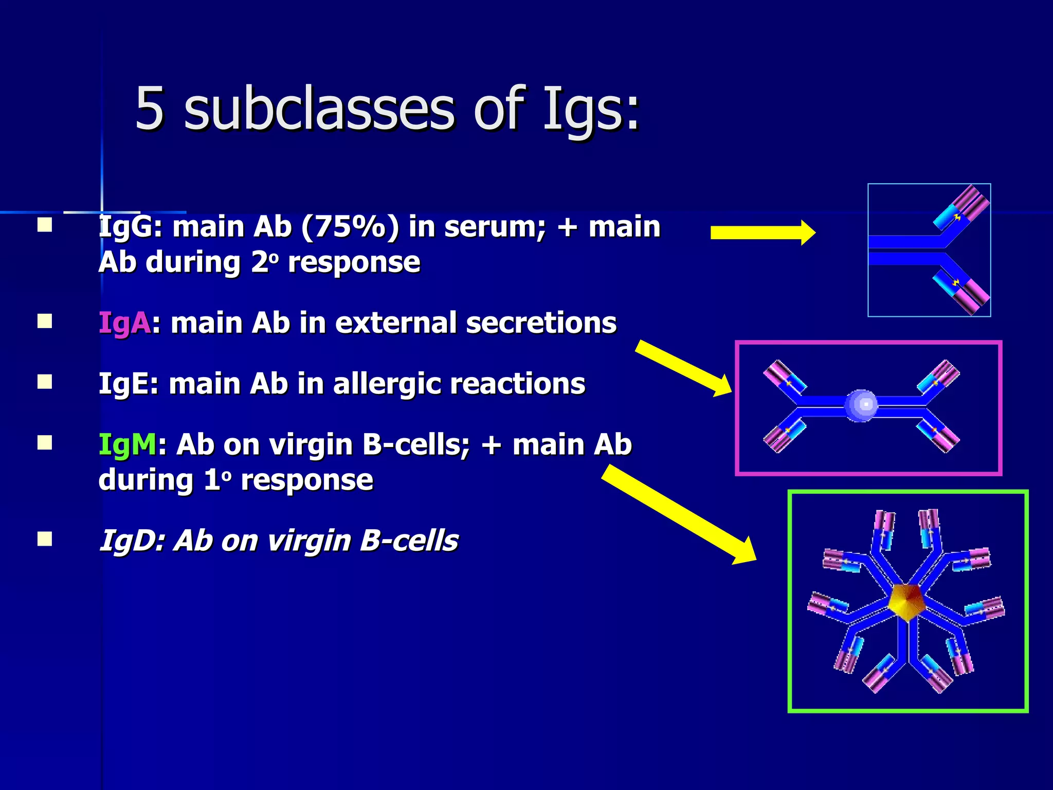
![Antibodies (Ab) AKA immunoglobulins (Ig) Proteins produced by plasma cells in response to a specific antigen (Ag) Plasma cells are derived from B-lymphocytes Antibodies [Ab] frequently measured as a diagnostic tool Serology IgG](https://image.slidesharecdn.com/chapter20lymphimmunemarieb-101020231429-phpapp02/75/Chapter20-lymphimmunemarieb-22-2048.jpg)


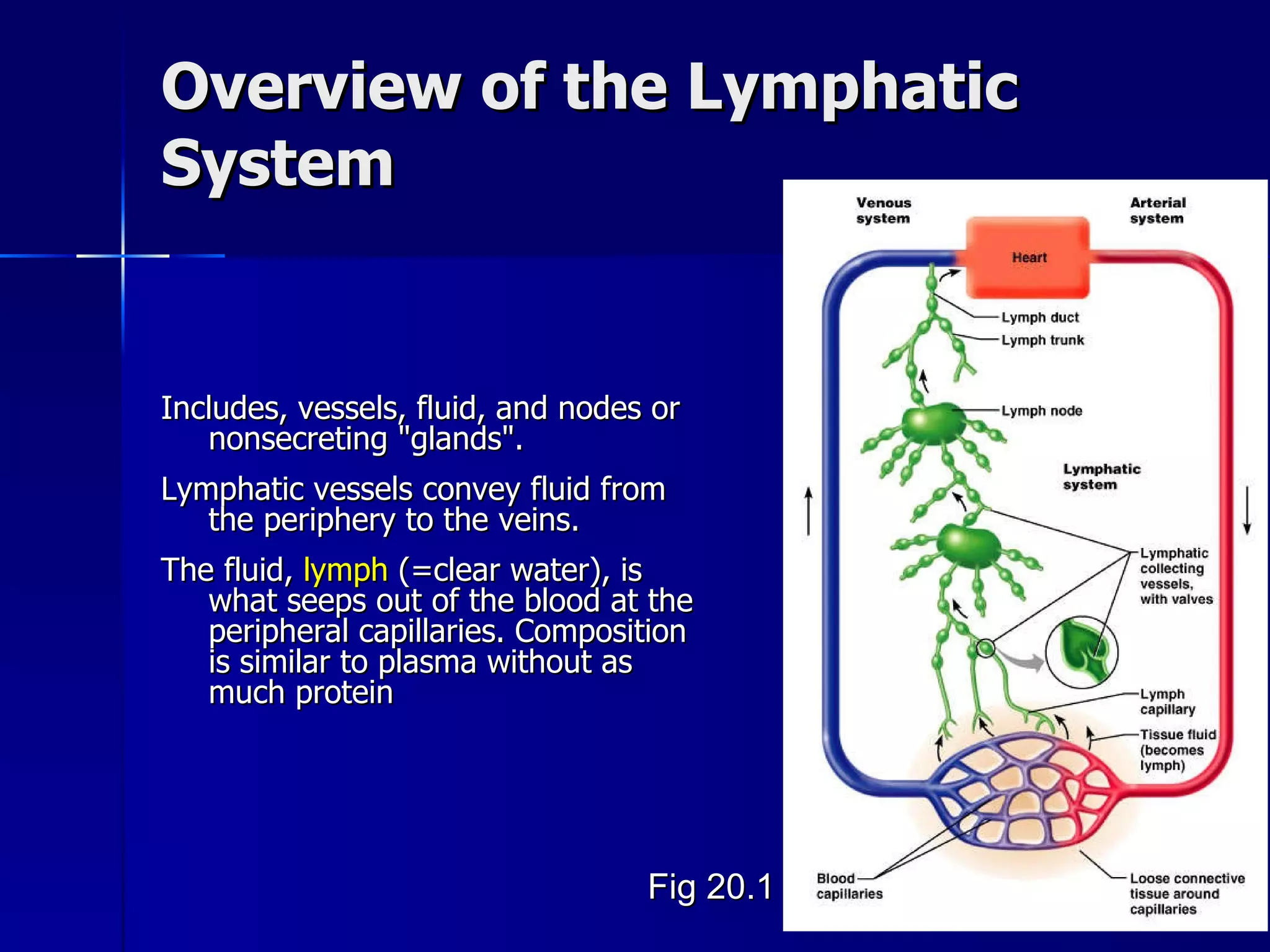
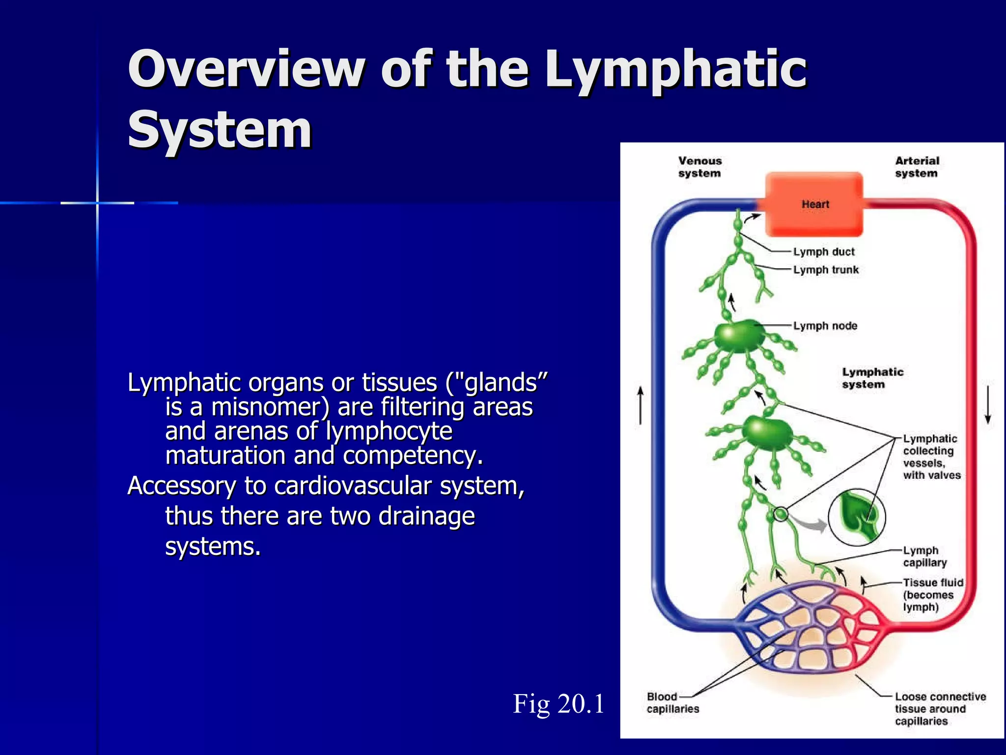
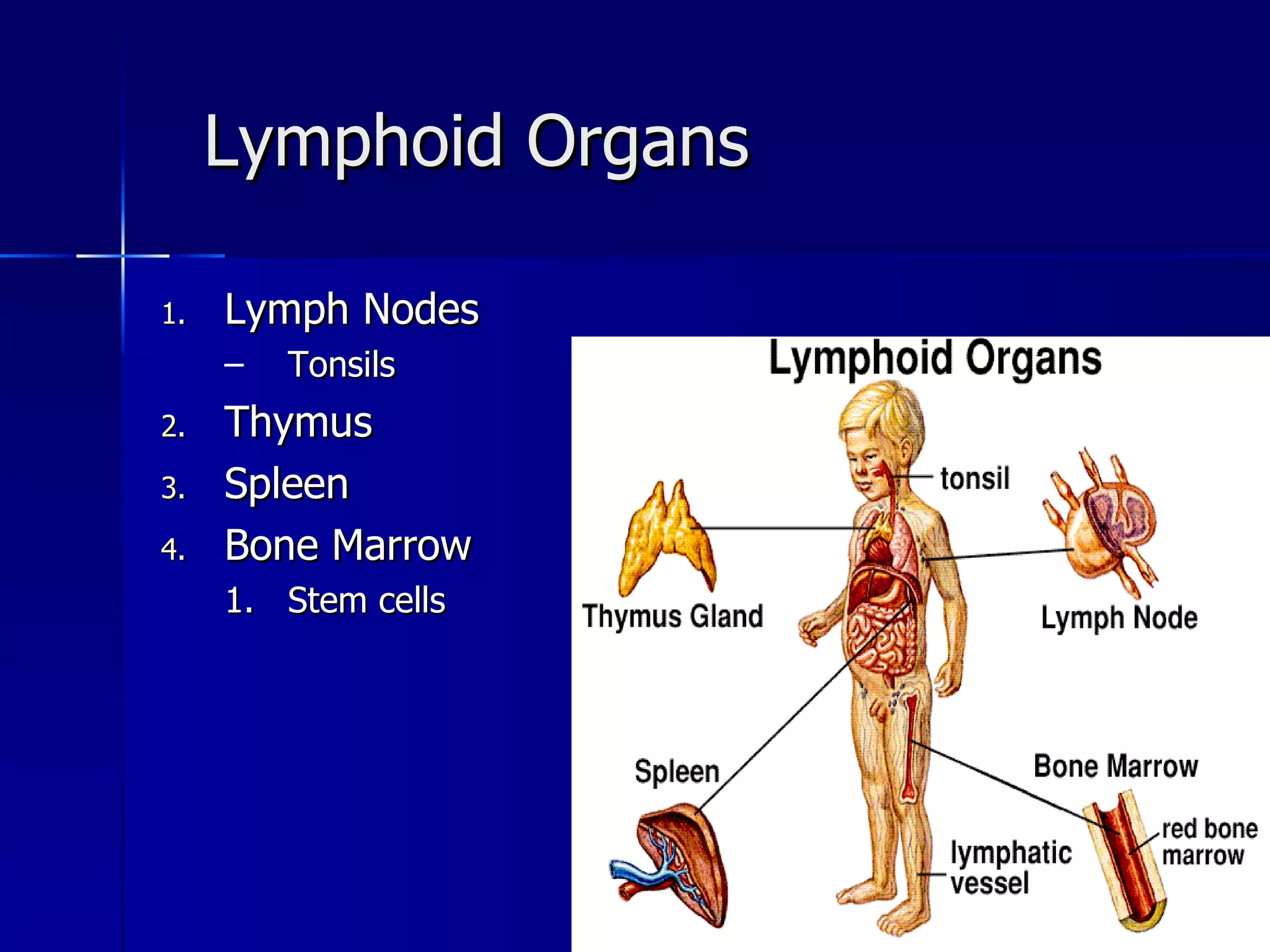
The document discusses the lymphatic and immune systems. It describes the organization of the lymphatic system including vessels, lymph nodes, thymus, and spleen. It explains the relationship between the lymphatic and circulatory systems and the role of lymphoid tissues and lymphocytes in defending the body. It provides an overview of the lymphatic system, its major functions, and lymphatic organs.


