Downloaded 1,003 times

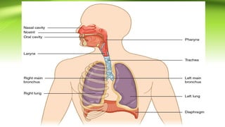
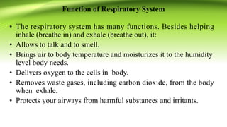


The document provides a comprehensive overview of the respiratory system, detailing its organs, functions, and mechanisms. It describes the anatomical structures involved, including the nasal cavity, pharynx, larynx, trachea, bronchi, bronchioles, and alveoli, and outlines processes such as inspiration and expiration. Additionally, it discusses the regulation of respiration, types of respiratory volumes, and abnormalities in breathing.