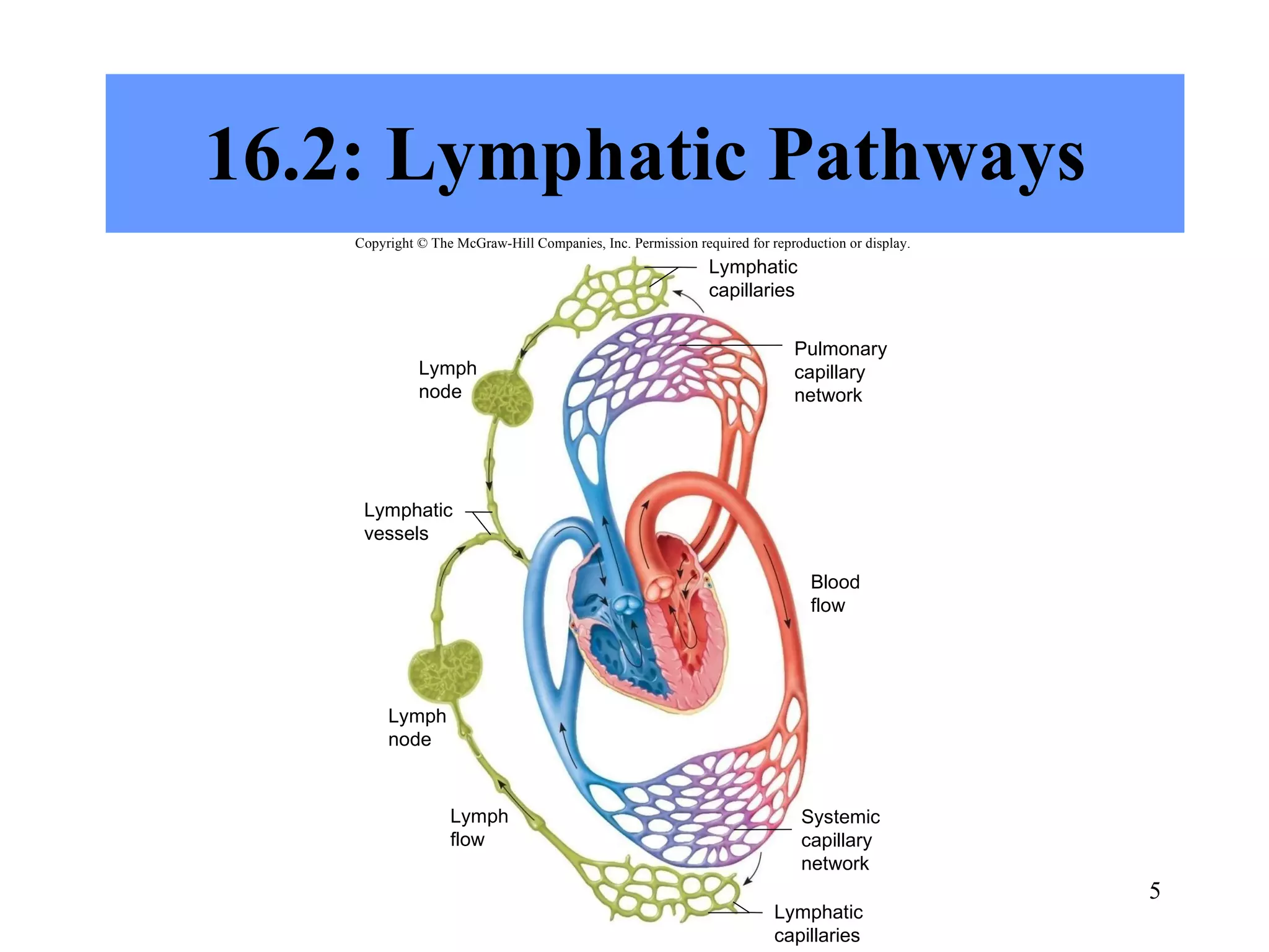
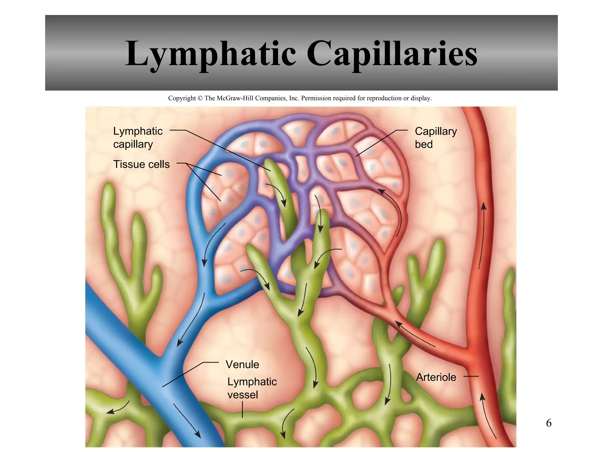
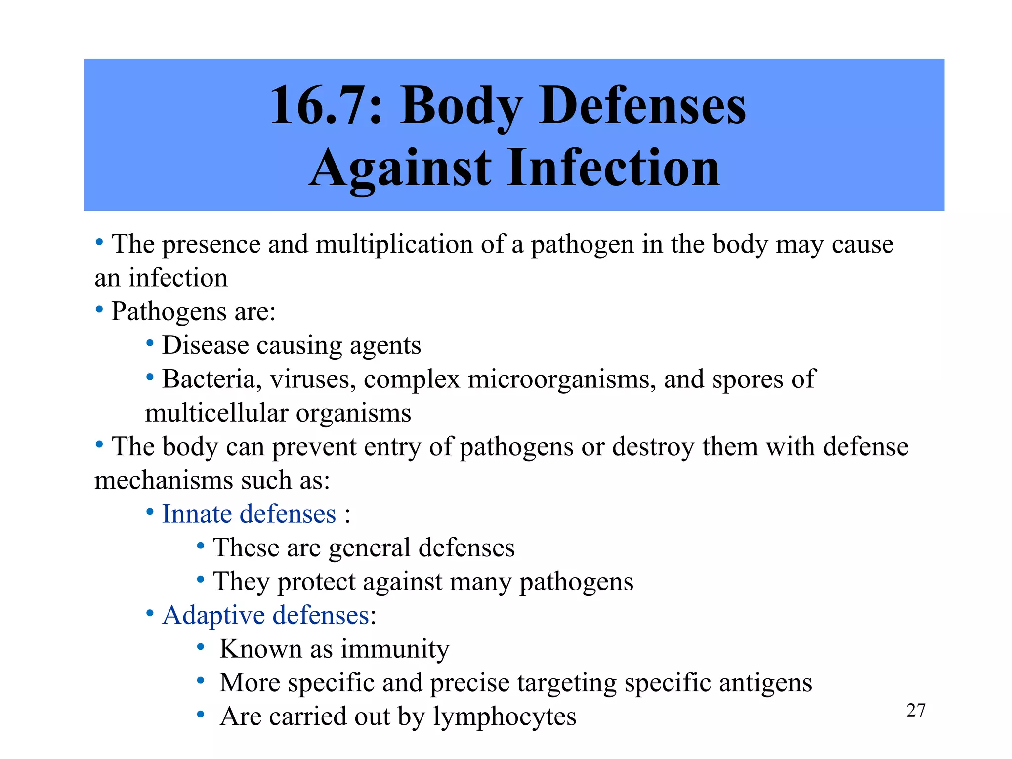
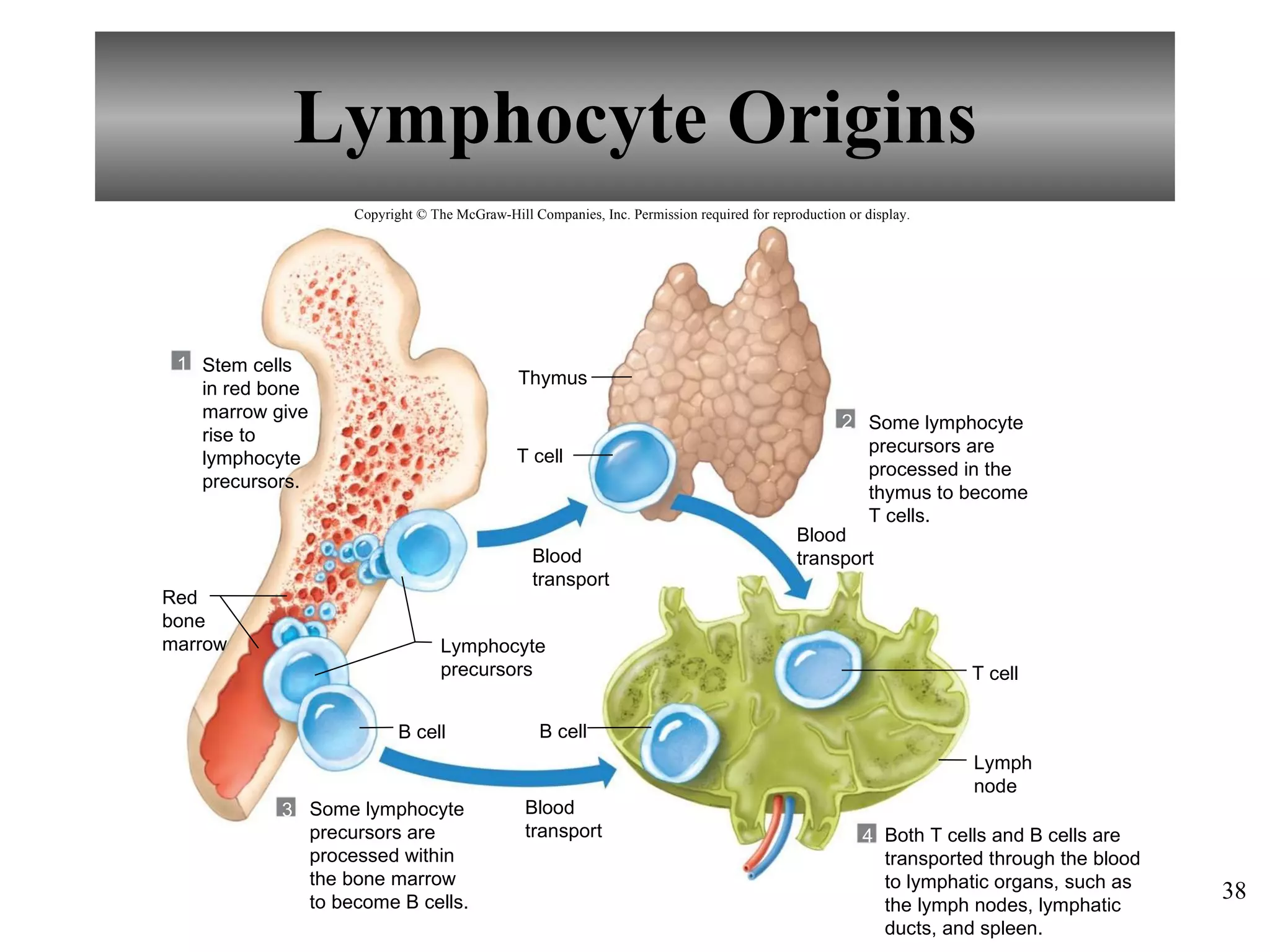
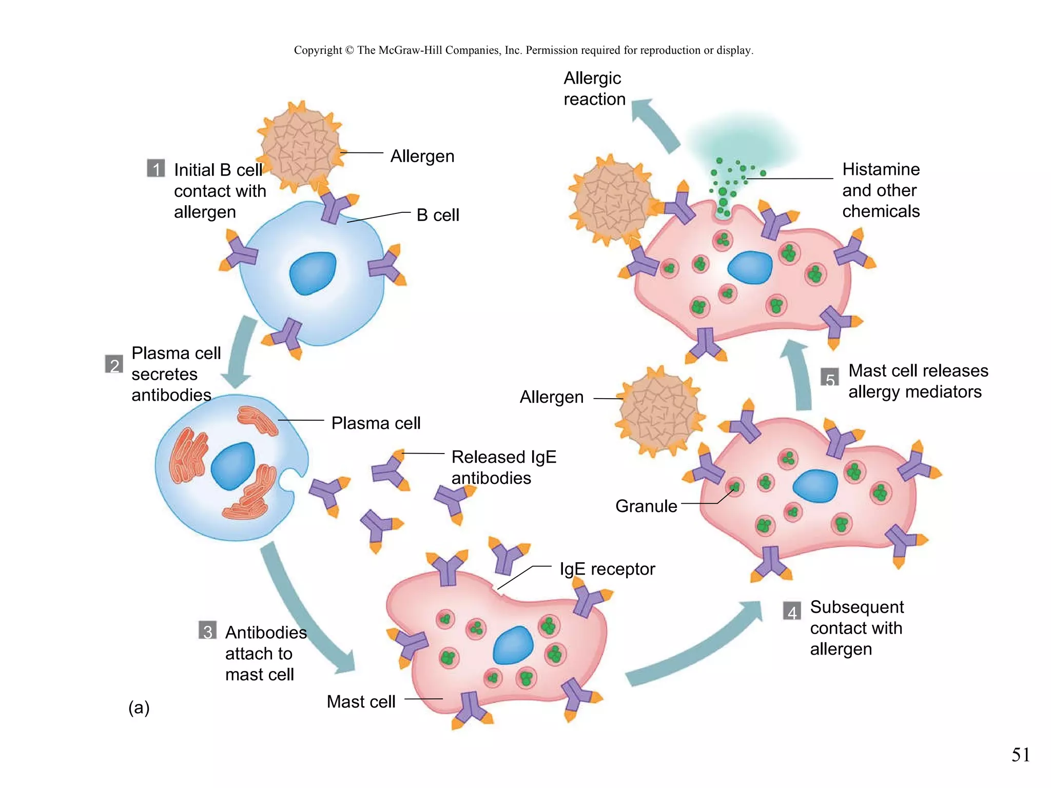
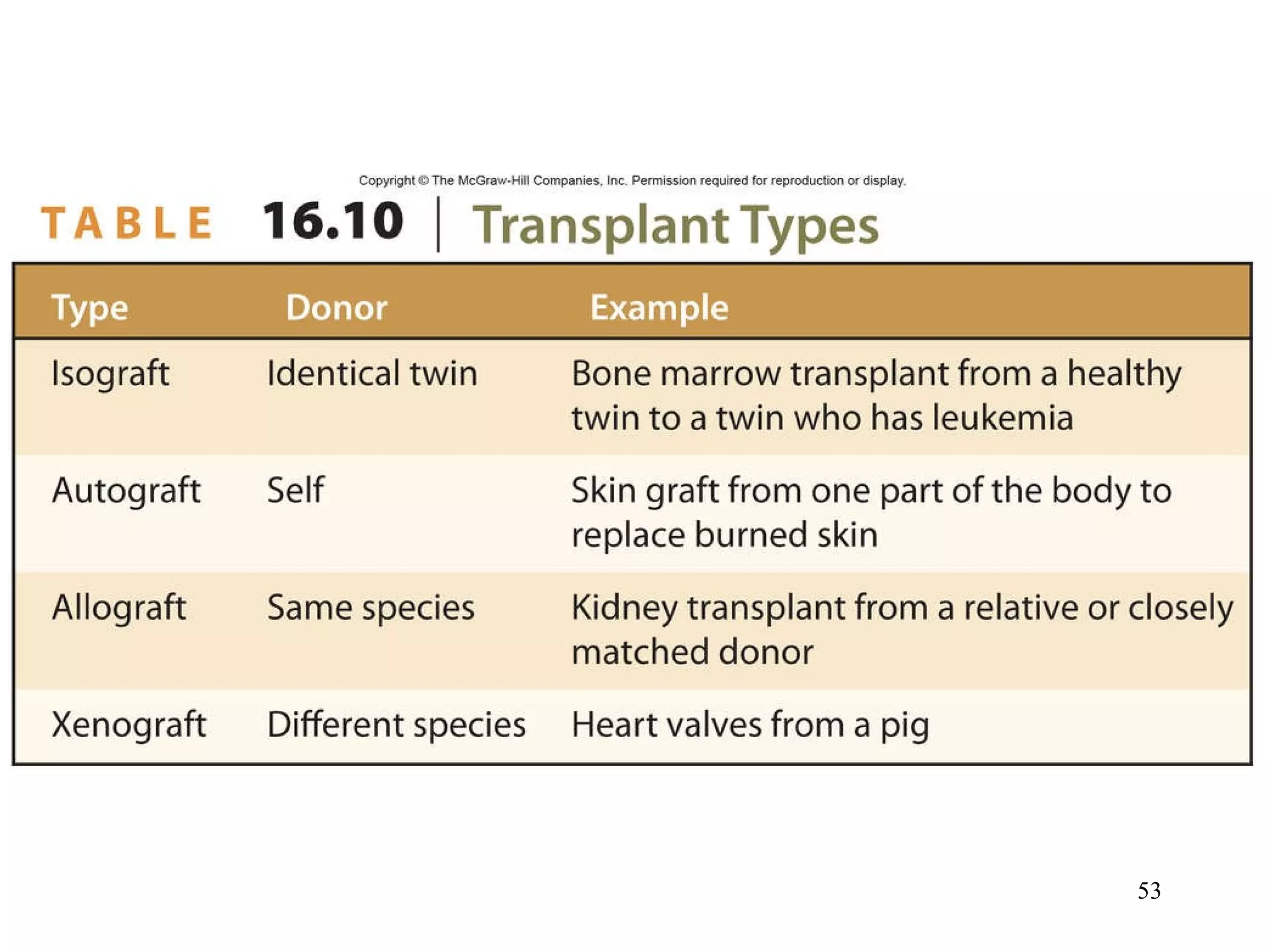
Copyright © The McGraw-Hill Companies, Inc. Permission required for reproduction or display. Chapter 16 Lecture PowerPoint
2402 Anatomy and Physiology II Chapter 16 Susan Gossett [email_address] Department of Biology Paris Junior College
Hole’s Human Anatomy and Physiology Twelfth Edition Shier    Butler    Lewis  Chapter  16 Lymphatic System and Immunity Copyright © The McGraw-Hill Companies, Inc. Permission required for reproduction or display.
16.1: Introduction The lymphatic system is a vast collection of cells and biochemicals that travel in lymphatic vessels It is a network of vessels that assist in circulating fluids It is closely associated with the cardiovascular system It transports excess fluid away from the interstitial spaces It transports fluid to the bloodstream It transports fats to the bloodstream It helps defend the body against diseases
16.2: Lymphatic Pathways Copyright © The McGraw-Hill Companies, Inc. Permission required for reproduction or display. Lymphatic capillaries Pulmonary capillary network Lymph node Lymphatic vessels Blood flow Lymph node Lymph flow Systemic capillary network Lymphatic capillaries
Lymphatic Capillaries Tissue cells Arteriole Venule Lymphatic capillary Capillary bed Lymphatic vessel Copyright © The McGraw-Hill Companies, Inc. Permission required for reproduction or display.
Lymphatic Vessels The walls are similar but thinner than those of veins Lymphatic vessels are composed of three layers: An endothelial lining (inner) Smooth muscle (middle) Connective tissue (outer) Larger vessels lead to lymph    nodes and then to larger   lymphatic trunks Copyright © The McGraw-Hill Companies, Inc. Permission required for reproduction or display. © The McGraw-Hill Companies, Inc./Dennis Strete, photographer
Lymphatic Trunks  and Collecting Ducts The trunks drain lymph from the lymphatic vessels They are named for the regions they serve such as lumbar, intestinal, intercostal, bronchomediastinal, subclavian, and jugular  Jugular trunk Right lymphatic duct Brachiocephalic vein Bronchomediastinal trunk Intercostal trunk Internal jugular vein Thoracic duct Subclavian trunk Thoracic duct Intestinal trunk Lumbar trunk Lymphatic vessels Copyright © The McGraw-Hill Companies, Inc. Permission required for reproduction or display.
Copyright © The McGraw-Hill Companies, Inc. Permission required for reproduction or display. Lymph nodes Thoracic duct Thoracic duct Right lymphatic duct Right internal jugular vein (a) (b) Area drained by right lymphatic duct Right lymphatic duct Lymphatic trunks Lymphatic vessels Left internal jugular vein Left subclavian vein Cisterna chyli Right subclavian vein Axillary lymph nodes Lymphatics of mammary gland
Copyright © The McGraw-Hill Companies, Inc. Permission required for reproduction or display. Afferent lymphatic vessel Collecting duct Subclavian vein Efferent lymphatic vessel Lymphatic capillary Lymph node Lymphatic trunk
16.3: Tissue and Fluid Lymph Lymph is essentially tissue fluid that has entered a lymphatic capillary Lymph formation depends on tissue fluid formation
Tissue Fluid Formation Capillary blood pressure filters water and small molecules from the plasma The resulting fluid has: Much the same consistency as plasma Contains water and dissolved substances Contains smaller proteins which create plasma colloid osmotic pressure
Lymph Formation Filtration from the plasma normally exceeds reabsorption, leading to the net formation of tissue fluid This increases the tissue fluid hydrostatic pressure within interstitial spaces forcing fluid into lymphatic capillaries forming lymph This process prevents accumulation of excess tissue fluid or edema
Lymph Function Lymphatic vessels play a role in: Absorption of dietary fats Delivering fats to the   bloodstream Collecting of excess   interstitial fluids Delivering excess fluids   to the bloodstream Delivering foreign   particles to the    lymph nodes Copyright © The McGraw-Hill Companies, Inc. Permission required for reproduction or display. Flow of lymph Epithelial cell Filaments anchored to connective tissue Movement of tissue fluid
16.4: Lymph Movement Hydrostatic pressure of tissue fluid drives the lymph into the lymphatic capillaries Muscle activity largely influences the movement of lymph through the lymphatic vessels via: Action of skeletal muscles Respiratory movements Smooth muscle in the larger lymphatic vessels Valves in the lymphatic vessels
Lymph Flow Copyright © The McGraw-Hill Companies, Inc. Permission required for reproduction or display. Sinus Capsule (a) Nodule Hilum Artery Medulla (macrophages, T cells) Capsule Subcapsule (macrophages, B cells) Efferent lymphatic vessel Lymph flow Germinal center (B cells) Vein Lymph flow Afferent lymphatic vessel Germinal center b: © The McGraw-Hill Companies, Inc./Al Telser, photographer (b)
Obstruction of Lymph Movement Copyright © The McGraw-Hill Companies, Inc. Permission required for reproduction or display. Blood vessels Lymph node Muscle Lymphatic vessels © Dr. Kent M. Van De Graaff
16.5: Lymph Nodes Lymph nodes or lymph glands are located along the lymphatic pathways They contain lymphocytes and macrophages to fight invading pathogens
Structure of a Lymph Node Copyright © The McGraw-Hill Companies, Inc. Permission required for reproduction or display. Lymph flow Sinus Capsule (a) Nodule Hilum Lymph flow Artery Vein Afferent lymphatic vessel Germinal center (B cells) Subcapsule (macrophages, B cells) Medulla (macrophages, T cells) Efferent lymphatic vessel
Locations of Lymph Nodes Lymph nodes are found in groups or chains along the paths of the larger lymphatic vessels throughout the body, including the: Cervical region Axillary region Supratrochlear region Inguinal region Pelvic cavity Abdominal cavity Thoracic cavity Copyright © The McGraw-Hill Companies, Inc. Permission required for reproduction or display. Thoracic lymph node Axillary lymph node Cervical lymph node Supratrochlear lymph node Abdominal lymph node Pelvic lymph node Inguinal lymph node
Functions of Lymph Nodes Lymph nodes have two primary functions: Filter potentially harmful particles from the lymph Act with immune surveillance provided by macrophages and lymphocytes Along with the red bone marrow, the lymph nodes are centers for lymphocyte production
16.6: Thymus and Spleen These are two other lymphatic organs with functions similar to those of the lymph nodes
Thymus Thymus in fetus Thymus in adult The thymus is: Larger in infancy and   during puberty Small in an adult Replaced by fat and   connective tissue in   the elderly Site of T lymphocyte   (or T cell) production Secretes protein hormones   called thymosins Copyright © The McGraw-Hill Companies, Inc. Permission required for reproduction or display.
Larynx Thymus Lung Liver (a) (b) Diaphragm Thyroid gland Heart Stomach Spleen Lobule Trachea Connective tissue b: © The McGraw-Hill Companies, Inc./Dennis Strete, photographer Copyright © The McGraw-Hill Companies, Inc. Permission required for reproduction or display.
Spleen The spleen is: The largest lymphatic organ Located in the upper left abdominal quadrant Has sinuses filled with blood Contains two tissue types:  White pulp ( lymphocytes) Red pulp ( red blood cells, lymphocytes and macrophages) Spleen Artery of pulp White pulp Red pulp (a) (b) Capillary Capsule Capsule White pulp Red pulp Splenic artery Splenic vein Connective tissue Venous sinus b: © The McGraw-Hill Companies, Inc./Al Telser, photographer Copyright © The McGraw-Hill Companies, Inc. Permission required for reproduction or display.
16.7: Body Defenses  Against Infection The presence and multiplication of a pathogen in the body may cause an infection Pathogens are: Disease causing agents Bacteria, viruses, complex microorganisms, and spores of multicellular organisms The body can prevent entry of pathogens or destroy them with defense mechanisms such as: Innate defenses  : These are general defenses They protect against many pathogens Adaptive defenses : Known as immunity More specific and precise targeting specific antigens  Are carried out by lymphocytes
16.8: Innate  (Nonspecific) Defenses
Species Resistance Refers to a given type of organism, or species, that develops diseases unique to it
Mechanical Barriers The skin and mucous membranes create mechanical barriers Mechanical barriers are considered the first line of defense (all other non-specific defenses are part of the second line of defense)
Chemical Barriers Enzymes in body fluids provide a chemical barrier to pathogens, and they may include: Interferons are horomone-like peptides and stimulate phagocytosis Defensins are peptides produced by neutrophils and other granulocytes. They cripple microbes. Collectins are proteins with broad protection against bacteria, yeast and some viruses Complement is a group of proteins in plasma and other body fluids that stimulate inflammation, attract phagocytes and enhance phagocytosis
Natural Killer (NK) Cells NK cells are a small population of lymphocytes defending against viruses and cancer by secreting cytolytic substances called perforins that destroy the infected cell NK may also enhance inflammation
Inflammation Inflammation produces local redness, swelling, heat and pain Table 16.2 summarizes the process
Phagocytosis Phagocytosis removes foreign particles from the lymph Phagocytes are also in the blood vessels and in the tissues of the spleen, liver or bone marrow The most active phagocytic cells are neutrophils and monocytes Chemicals attract these phagocytic cells to the injury and this is called chemotaxis
Fever A fever begins when a viral or bacterial infection stimulates lymphocytes to proliferate, producing cells that secrete a substance called interleukin-1 (IL-1)
16.9: Adaptive (Specific)  Defenses or Immunity This is the third line of defense and known as immunity It is resistance to particular pathogens or to their toxins or metabolic by-products It is based on the ability to distinguish molecules that are part of the body (“self” from “non-self”) Antigens are molecules that can elicit an immune response
Antigens Antigens may be: Proteins  Polysaccharides Glycoproteins Glycolipids The most effective antigens are large and complex Haptens are small molecules that are not antigenic by themselves, but when they combine with a large molecule can stimulate an immune response
Lymphocyte Origins B cell Thymus T cell T cell 1 2 3 4 B cell Stem cells in red bone marrow give rise to lymphocyte precursors. Red bone marrow Some lymphocyte precursors are processed within the bone marrow to become B cells. Both T cells and B cells are transported through the blood to lymphatic organs, such as the lymph nodes, lymphatic ducts, and spleen. Some lymphocyte precursors are processed in the thymus to become T cells. Blood transport Blood transport Lymphocyte precursors Blood transport Lymph node Copyright © The McGraw-Hill Companies, Inc. Permission required for reproduction or display.
T Cells and the  Cellular Immune Response A lymphocyte must be activated before it can respond to an antigen T cell activation requires antigen-presenting cell (accessory cell) and may include macrophages, B cells and several other types of cells Requires major histocompatibility complex (MHC) or human leukocyte antigens (HLA) to recognize “non-self” T cells can synthesize and secrete polypeptides called cytokines Types of specialized T cells include: Helper T cells Cytotoxic T cells Memory T cells
B Cells and the  Humoral Immune Response B cells can be activated when an antigen fits the shape of its receptor Most of the time B cell activation requires T cells T cells release cytokines that stimulate B cells Some B cells may become memory B cells while others differentiate into plasma cells and produce and secrete large globular proteins called antibodies or immunoglobulins
Copyright © The McGraw-Hill Companies, Inc. Permission required for reproduction or display. Cytokines Antigen B cells Activated B cell Interleukin-2 1 3 2 4 5 (a) Macrophage Helper T cell (b) Cytotoxic T cell Cytotoxic T cell contacts Displayed antigen Helper T cell contacts displayed antigen and proliferates Cytotoxic T cell Helper T cells Activated helper  T cell interacts with cytotoxic T cell (which has combined with an identical antigen) and releases interleukin-2, which activates the cytotoxic T cell Proliferation and Differentiation Memory T cell Cytotoxic T cell Antigen receptor B cell combines with antigen Macrophage displays ingested antigen on its surface Displayed antigen b: © Manfred Kage/Peter Arnold
Copyright © The McGraw-Hill Companies, Inc. Permission required for reproduction or display. Activated B cell Proliferation B cells Antigen Cytokines Activation 1 2 3 Antigen receptor Stimulation by activated helper T cell Clone of B cells
Antigen Proliferation Antigen Receptor  (antibody) Receptor-antigen combination Activated B cell Cytokines from helper T cell Clone of B cells Proliferation and Differentiation Proliferation and Differentiation Endoplasmic reticulum Released antibodies Plasma cell (antibody-secreting cell) Memory cell (dormant cell) Plasma cell (antibody-secreting cell) Memory cell (dormant cell) Clone of B cells Copyright © The McGraw-Hill Companies, Inc. Permission required for reproduction or display.
16.1 From Science to Technology Immunotherapy
Immune Responses Plasma antibody concentration 10 0 20 30 Primary response Secondary response 40 Days after exposure to antigen Copyright © The McGraw-Hill Companies, Inc. Permission required for reproduction or display.
Practical Classification  of Immunity
Allergic Reactions Type I Immediate-reaction allergy Occurs minutes after contact with allergen Symptoms include hives, hay fever, asthma, eczema, gastric disturbances, and anaphylactic shock Type II Antibody-dependent cytotoxic reaction Takes 1-3 hours to develop Transfusion reaction Type III Immune-complex reaction Takes 1-3 hours to develop Antibody complexes cannot be cleared from the body Damage of body tissues Type IV Delayed-reaction allergy Results from repeated exposure to allergen Eruptions and inflammation of the skin Takes about 48 hours to occur
Copyright © The McGraw-Hill Companies, Inc. Permission required for reproduction or display. B cell Plasma cell Mast cell IgE receptor Granule (a) 1 2 3 5 4 Allergen Allergen Allergic reaction Histamine and other chemicals Mast cell releases allergy mediators Subsequent contact with allergen Antibodies attach to mast cell Released IgE antibodies Initial B cell contact with allergen Plasma cell secretes antibodies
Transplantation  and Tissue Rejection Successfully transplanted tissues and organs: Cornea Kidney Liver Pancreas When the donor’s tissues are recognized as foreign there is a tissue rejection reaction Resembles the cellular immune response against antigens Important to match MHC antigens Immunosuppressive drugs used to prevent rejection Heart Bone marrow Skin
Autoimmunity The immune system fails to distinguish “self” from “non-self” and the body produces antibodies called autoantibodies, and cytotoxic T cells to attack the body’s tissues and organs
16.10: Lifespan Changes The immune system declines early in life as the thymus gland shrinks (only 25% as powerful as it once was) There is a higher risk of infection Antibody response to antigens becomes slower IgA and IgG antibodies increase IgM and IgE antibodies decrease Elderly may not be candidates for certain medical treatments that suppresses immunity
16.1 Clinical Application Immunity Breakdown: AIDS
Important Points in Chapter 16: Outcomes to be Assessed 16.1: Introduction  Describe the general functions of the lymphatic system. 16.2: Lymphatic Pathways Identify and describe the parts of the major lymphatic pathways. 16.3: Tissue Fluid and Lymph Describe how tissue fluid and lymph form, and explain the function of lymph. 16.4: Lymph Movement Explain how lymphatic circulation is maintained, and describe the consequence of lymphatic obstruction.
Important Points in Chapter 16: Outcomes to be Assessed 16.5: Lymph Nodes Describe a lymph node and its major functions. Describe the location of the major chains of lymph nodes. 16.6: Thymus and Spleen Discuss the locations and functions of the thymus and spleen. 16.7: Body Defenses Against Infection Distinguish between innate (nonspecific) and adaptive (specific) defenses. 16.8: Innate (Nonspecific) Defenses List seven innate body defense mechanisms, and describe the action of each mechanism.
Important Points in Chapter 16: Outcomes to be Assessed 16.9: Adaptive (Specific) Defenses, or Immunity Explain how two major types of lymphocytes are formed, activated, and how they function in immune mechanisms. Discuss the origins and actions of the five different types of antibodies. Distinguish between primary and secondary immune responses. Distinguish between active and passive immunity. Explain how allergic reactions, tissue rejection reactions, and autoimmunity arise from immune mechanisms. 16.10: Lifespan Changes Describe lifespan changes in immunity.
Quiz 16 Complete Quiz 16 now! Read Chapter 17.

Chapter 16 Lymphatic System and Immunity

  • 1.
    Copyright © TheMcGraw-Hill Companies, Inc. Permission required for reproduction or display. Chapter 16 Lecture PowerPoint
  • 2.
    2402 Anatomy andPhysiology II Chapter 16 Susan Gossett [email_address] Department of Biology Paris Junior College
  • 3.
    Hole’s Human Anatomyand Physiology Twelfth Edition Shier  Butler  Lewis Chapter 16 Lymphatic System and Immunity Copyright © The McGraw-Hill Companies, Inc. Permission required for reproduction or display.
  • 4.
    16.1: Introduction Thelymphatic system is a vast collection of cells and biochemicals that travel in lymphatic vessels It is a network of vessels that assist in circulating fluids It is closely associated with the cardiovascular system It transports excess fluid away from the interstitial spaces It transports fluid to the bloodstream It transports fats to the bloodstream It helps defend the body against diseases
  • 5.
    16.2: Lymphatic PathwaysCopyright © The McGraw-Hill Companies, Inc. Permission required for reproduction or display. Lymphatic capillaries Pulmonary capillary network Lymph node Lymphatic vessels Blood flow Lymph node Lymph flow Systemic capillary network Lymphatic capillaries
  • 6.
    Lymphatic Capillaries Tissuecells Arteriole Venule Lymphatic capillary Capillary bed Lymphatic vessel Copyright © The McGraw-Hill Companies, Inc. Permission required for reproduction or display.
  • 7.
    Lymphatic Vessels Thewalls are similar but thinner than those of veins Lymphatic vessels are composed of three layers: An endothelial lining (inner) Smooth muscle (middle) Connective tissue (outer) Larger vessels lead to lymph nodes and then to larger lymphatic trunks Copyright © The McGraw-Hill Companies, Inc. Permission required for reproduction or display. © The McGraw-Hill Companies, Inc./Dennis Strete, photographer
  • 8.
    Lymphatic Trunks and Collecting Ducts The trunks drain lymph from the lymphatic vessels They are named for the regions they serve such as lumbar, intestinal, intercostal, bronchomediastinal, subclavian, and jugular Jugular trunk Right lymphatic duct Brachiocephalic vein Bronchomediastinal trunk Intercostal trunk Internal jugular vein Thoracic duct Subclavian trunk Thoracic duct Intestinal trunk Lumbar trunk Lymphatic vessels Copyright © The McGraw-Hill Companies, Inc. Permission required for reproduction or display.
  • 9.
    Copyright © TheMcGraw-Hill Companies, Inc. Permission required for reproduction or display. Lymph nodes Thoracic duct Thoracic duct Right lymphatic duct Right internal jugular vein (a) (b) Area drained by right lymphatic duct Right lymphatic duct Lymphatic trunks Lymphatic vessels Left internal jugular vein Left subclavian vein Cisterna chyli Right subclavian vein Axillary lymph nodes Lymphatics of mammary gland
  • 10.
    Copyright © TheMcGraw-Hill Companies, Inc. Permission required for reproduction or display. Afferent lymphatic vessel Collecting duct Subclavian vein Efferent lymphatic vessel Lymphatic capillary Lymph node Lymphatic trunk
  • 11.
    16.3: Tissue andFluid Lymph Lymph is essentially tissue fluid that has entered a lymphatic capillary Lymph formation depends on tissue fluid formation
  • 12.
    Tissue Fluid FormationCapillary blood pressure filters water and small molecules from the plasma The resulting fluid has: Much the same consistency as plasma Contains water and dissolved substances Contains smaller proteins which create plasma colloid osmotic pressure
  • 13.
    Lymph Formation Filtrationfrom the plasma normally exceeds reabsorption, leading to the net formation of tissue fluid This increases the tissue fluid hydrostatic pressure within interstitial spaces forcing fluid into lymphatic capillaries forming lymph This process prevents accumulation of excess tissue fluid or edema
  • 14.
    Lymph Function Lymphaticvessels play a role in: Absorption of dietary fats Delivering fats to the bloodstream Collecting of excess interstitial fluids Delivering excess fluids to the bloodstream Delivering foreign particles to the lymph nodes Copyright © The McGraw-Hill Companies, Inc. Permission required for reproduction or display. Flow of lymph Epithelial cell Filaments anchored to connective tissue Movement of tissue fluid
  • 15.
    16.4: Lymph MovementHydrostatic pressure of tissue fluid drives the lymph into the lymphatic capillaries Muscle activity largely influences the movement of lymph through the lymphatic vessels via: Action of skeletal muscles Respiratory movements Smooth muscle in the larger lymphatic vessels Valves in the lymphatic vessels
  • 16.
    Lymph Flow Copyright© The McGraw-Hill Companies, Inc. Permission required for reproduction or display. Sinus Capsule (a) Nodule Hilum Artery Medulla (macrophages, T cells) Capsule Subcapsule (macrophages, B cells) Efferent lymphatic vessel Lymph flow Germinal center (B cells) Vein Lymph flow Afferent lymphatic vessel Germinal center b: © The McGraw-Hill Companies, Inc./Al Telser, photographer (b)
  • 17.
    Obstruction of LymphMovement Copyright © The McGraw-Hill Companies, Inc. Permission required for reproduction or display. Blood vessels Lymph node Muscle Lymphatic vessels © Dr. Kent M. Van De Graaff
  • 18.
    16.5: Lymph NodesLymph nodes or lymph glands are located along the lymphatic pathways They contain lymphocytes and macrophages to fight invading pathogens
  • 19.
    Structure of aLymph Node Copyright © The McGraw-Hill Companies, Inc. Permission required for reproduction or display. Lymph flow Sinus Capsule (a) Nodule Hilum Lymph flow Artery Vein Afferent lymphatic vessel Germinal center (B cells) Subcapsule (macrophages, B cells) Medulla (macrophages, T cells) Efferent lymphatic vessel
  • 20.
    Locations of LymphNodes Lymph nodes are found in groups or chains along the paths of the larger lymphatic vessels throughout the body, including the: Cervical region Axillary region Supratrochlear region Inguinal region Pelvic cavity Abdominal cavity Thoracic cavity Copyright © The McGraw-Hill Companies, Inc. Permission required for reproduction or display. Thoracic lymph node Axillary lymph node Cervical lymph node Supratrochlear lymph node Abdominal lymph node Pelvic lymph node Inguinal lymph node
  • 21.
    Functions of LymphNodes Lymph nodes have two primary functions: Filter potentially harmful particles from the lymph Act with immune surveillance provided by macrophages and lymphocytes Along with the red bone marrow, the lymph nodes are centers for lymphocyte production
  • 22.
    16.6: Thymus andSpleen These are two other lymphatic organs with functions similar to those of the lymph nodes
  • 23.
    Thymus Thymus infetus Thymus in adult The thymus is: Larger in infancy and during puberty Small in an adult Replaced by fat and connective tissue in the elderly Site of T lymphocyte (or T cell) production Secretes protein hormones called thymosins Copyright © The McGraw-Hill Companies, Inc. Permission required for reproduction or display.
  • 24.
    Larynx Thymus LungLiver (a) (b) Diaphragm Thyroid gland Heart Stomach Spleen Lobule Trachea Connective tissue b: © The McGraw-Hill Companies, Inc./Dennis Strete, photographer Copyright © The McGraw-Hill Companies, Inc. Permission required for reproduction or display.
  • 25.
    Spleen The spleenis: The largest lymphatic organ Located in the upper left abdominal quadrant Has sinuses filled with blood Contains two tissue types: White pulp ( lymphocytes) Red pulp ( red blood cells, lymphocytes and macrophages) Spleen Artery of pulp White pulp Red pulp (a) (b) Capillary Capsule Capsule White pulp Red pulp Splenic artery Splenic vein Connective tissue Venous sinus b: © The McGraw-Hill Companies, Inc./Al Telser, photographer Copyright © The McGraw-Hill Companies, Inc. Permission required for reproduction or display.
  • 27.
    16.7: Body Defenses Against Infection The presence and multiplication of a pathogen in the body may cause an infection Pathogens are: Disease causing agents Bacteria, viruses, complex microorganisms, and spores of multicellular organisms The body can prevent entry of pathogens or destroy them with defense mechanisms such as: Innate defenses : These are general defenses They protect against many pathogens Adaptive defenses : Known as immunity More specific and precise targeting specific antigens Are carried out by lymphocytes
  • 28.
    16.8: Innate (Nonspecific) Defenses
  • 29.
    Species Resistance Refersto a given type of organism, or species, that develops diseases unique to it
  • 30.
    Mechanical Barriers Theskin and mucous membranes create mechanical barriers Mechanical barriers are considered the first line of defense (all other non-specific defenses are part of the second line of defense)
  • 31.
    Chemical Barriers Enzymesin body fluids provide a chemical barrier to pathogens, and they may include: Interferons are horomone-like peptides and stimulate phagocytosis Defensins are peptides produced by neutrophils and other granulocytes. They cripple microbes. Collectins are proteins with broad protection against bacteria, yeast and some viruses Complement is a group of proteins in plasma and other body fluids that stimulate inflammation, attract phagocytes and enhance phagocytosis
  • 32.
    Natural Killer (NK)Cells NK cells are a small population of lymphocytes defending against viruses and cancer by secreting cytolytic substances called perforins that destroy the infected cell NK may also enhance inflammation
  • 33.
    Inflammation Inflammation produceslocal redness, swelling, heat and pain Table 16.2 summarizes the process
  • 34.
    Phagocytosis Phagocytosis removesforeign particles from the lymph Phagocytes are also in the blood vessels and in the tissues of the spleen, liver or bone marrow The most active phagocytic cells are neutrophils and monocytes Chemicals attract these phagocytic cells to the injury and this is called chemotaxis
  • 35.
    Fever A feverbegins when a viral or bacterial infection stimulates lymphocytes to proliferate, producing cells that secrete a substance called interleukin-1 (IL-1)
  • 36.
    16.9: Adaptive (Specific) Defenses or Immunity This is the third line of defense and known as immunity It is resistance to particular pathogens or to their toxins or metabolic by-products It is based on the ability to distinguish molecules that are part of the body (“self” from “non-self”) Antigens are molecules that can elicit an immune response
  • 37.
    Antigens Antigens maybe: Proteins Polysaccharides Glycoproteins Glycolipids The most effective antigens are large and complex Haptens are small molecules that are not antigenic by themselves, but when they combine with a large molecule can stimulate an immune response
  • 38.
    Lymphocyte Origins Bcell Thymus T cell T cell 1 2 3 4 B cell Stem cells in red bone marrow give rise to lymphocyte precursors. Red bone marrow Some lymphocyte precursors are processed within the bone marrow to become B cells. Both T cells and B cells are transported through the blood to lymphatic organs, such as the lymph nodes, lymphatic ducts, and spleen. Some lymphocyte precursors are processed in the thymus to become T cells. Blood transport Blood transport Lymphocyte precursors Blood transport Lymph node Copyright © The McGraw-Hill Companies, Inc. Permission required for reproduction or display.
  • 39.
    T Cells andthe Cellular Immune Response A lymphocyte must be activated before it can respond to an antigen T cell activation requires antigen-presenting cell (accessory cell) and may include macrophages, B cells and several other types of cells Requires major histocompatibility complex (MHC) or human leukocyte antigens (HLA) to recognize “non-self” T cells can synthesize and secrete polypeptides called cytokines Types of specialized T cells include: Helper T cells Cytotoxic T cells Memory T cells
  • 41.
    B Cells andthe Humoral Immune Response B cells can be activated when an antigen fits the shape of its receptor Most of the time B cell activation requires T cells T cells release cytokines that stimulate B cells Some B cells may become memory B cells while others differentiate into plasma cells and produce and secrete large globular proteins called antibodies or immunoglobulins
  • 42.
    Copyright © TheMcGraw-Hill Companies, Inc. Permission required for reproduction or display. Cytokines Antigen B cells Activated B cell Interleukin-2 1 3 2 4 5 (a) Macrophage Helper T cell (b) Cytotoxic T cell Cytotoxic T cell contacts Displayed antigen Helper T cell contacts displayed antigen and proliferates Cytotoxic T cell Helper T cells Activated helper T cell interacts with cytotoxic T cell (which has combined with an identical antigen) and releases interleukin-2, which activates the cytotoxic T cell Proliferation and Differentiation Memory T cell Cytotoxic T cell Antigen receptor B cell combines with antigen Macrophage displays ingested antigen on its surface Displayed antigen b: © Manfred Kage/Peter Arnold
  • 43.
    Copyright © TheMcGraw-Hill Companies, Inc. Permission required for reproduction or display. Activated B cell Proliferation B cells Antigen Cytokines Activation 1 2 3 Antigen receptor Stimulation by activated helper T cell Clone of B cells
  • 44.
    Antigen Proliferation AntigenReceptor (antibody) Receptor-antigen combination Activated B cell Cytokines from helper T cell Clone of B cells Proliferation and Differentiation Proliferation and Differentiation Endoplasmic reticulum Released antibodies Plasma cell (antibody-secreting cell) Memory cell (dormant cell) Plasma cell (antibody-secreting cell) Memory cell (dormant cell) Clone of B cells Copyright © The McGraw-Hill Companies, Inc. Permission required for reproduction or display.
  • 47.
    16.1 From Scienceto Technology Immunotherapy
  • 48.
    Immune Responses Plasmaantibody concentration 10 0 20 30 Primary response Secondary response 40 Days after exposure to antigen Copyright © The McGraw-Hill Companies, Inc. Permission required for reproduction or display.
  • 49.
  • 50.
    Allergic Reactions TypeI Immediate-reaction allergy Occurs minutes after contact with allergen Symptoms include hives, hay fever, asthma, eczema, gastric disturbances, and anaphylactic shock Type II Antibody-dependent cytotoxic reaction Takes 1-3 hours to develop Transfusion reaction Type III Immune-complex reaction Takes 1-3 hours to develop Antibody complexes cannot be cleared from the body Damage of body tissues Type IV Delayed-reaction allergy Results from repeated exposure to allergen Eruptions and inflammation of the skin Takes about 48 hours to occur
  • 51.
    Copyright © TheMcGraw-Hill Companies, Inc. Permission required for reproduction or display. B cell Plasma cell Mast cell IgE receptor Granule (a) 1 2 3 5 4 Allergen Allergen Allergic reaction Histamine and other chemicals Mast cell releases allergy mediators Subsequent contact with allergen Antibodies attach to mast cell Released IgE antibodies Initial B cell contact with allergen Plasma cell secretes antibodies
  • 52.
    Transplantation andTissue Rejection Successfully transplanted tissues and organs: Cornea Kidney Liver Pancreas When the donor’s tissues are recognized as foreign there is a tissue rejection reaction Resembles the cellular immune response against antigens Important to match MHC antigens Immunosuppressive drugs used to prevent rejection Heart Bone marrow Skin
  • 54.
    Autoimmunity The immunesystem fails to distinguish “self” from “non-self” and the body produces antibodies called autoantibodies, and cytotoxic T cells to attack the body’s tissues and organs
  • 55.
    16.10: Lifespan ChangesThe immune system declines early in life as the thymus gland shrinks (only 25% as powerful as it once was) There is a higher risk of infection Antibody response to antigens becomes slower IgA and IgG antibodies increase IgM and IgE antibodies decrease Elderly may not be candidates for certain medical treatments that suppresses immunity
  • 56.
    16.1 Clinical ApplicationImmunity Breakdown: AIDS
  • 57.
    Important Points inChapter 16: Outcomes to be Assessed 16.1: Introduction Describe the general functions of the lymphatic system. 16.2: Lymphatic Pathways Identify and describe the parts of the major lymphatic pathways. 16.3: Tissue Fluid and Lymph Describe how tissue fluid and lymph form, and explain the function of lymph. 16.4: Lymph Movement Explain how lymphatic circulation is maintained, and describe the consequence of lymphatic obstruction.
  • 58.
    Important Points inChapter 16: Outcomes to be Assessed 16.5: Lymph Nodes Describe a lymph node and its major functions. Describe the location of the major chains of lymph nodes. 16.6: Thymus and Spleen Discuss the locations and functions of the thymus and spleen. 16.7: Body Defenses Against Infection Distinguish between innate (nonspecific) and adaptive (specific) defenses. 16.8: Innate (Nonspecific) Defenses List seven innate body defense mechanisms, and describe the action of each mechanism.
  • 59.
    Important Points inChapter 16: Outcomes to be Assessed 16.9: Adaptive (Specific) Defenses, or Immunity Explain how two major types of lymphocytes are formed, activated, and how they function in immune mechanisms. Discuss the origins and actions of the five different types of antibodies. Distinguish between primary and secondary immune responses. Distinguish between active and passive immunity. Explain how allergic reactions, tissue rejection reactions, and autoimmunity arise from immune mechanisms. 16.10: Lifespan Changes Describe lifespan changes in immunity.
  • 60.
    Quiz 16 CompleteQuiz 16 now! Read Chapter 17.