Download to read offline

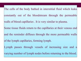
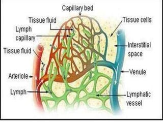
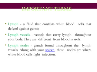
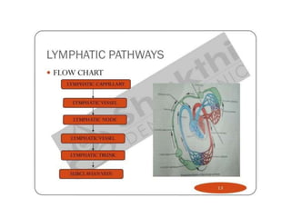
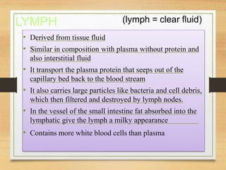
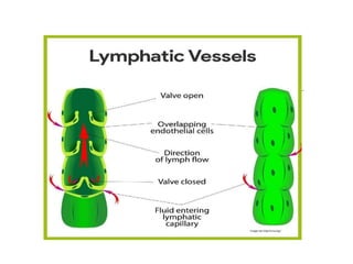
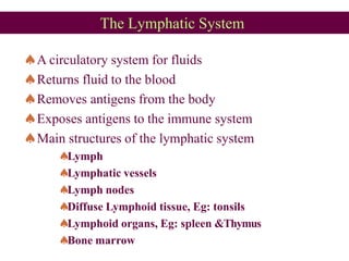
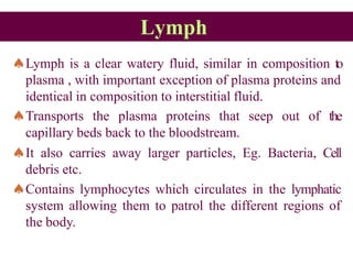
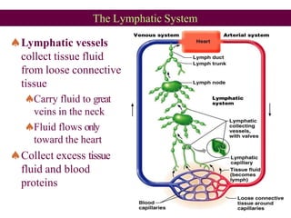
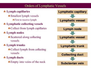
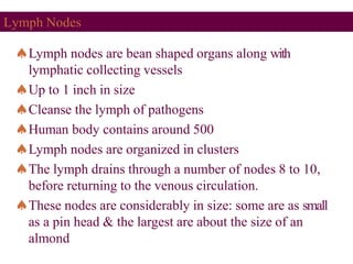
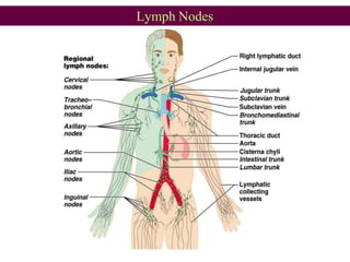
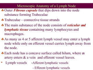
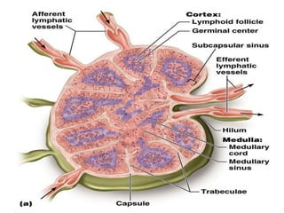
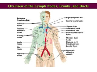
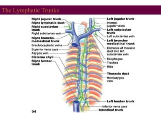
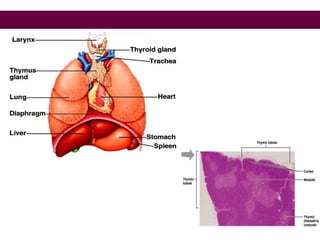


The lymphatic system returns fluid to the bloodstream, removes antigens from tissues, and exposes antigens to the immune system. It consists of lymph, lymphatic vessels, lymph nodes, diffuse lymphoid tissues like tonsils, and lymphoid organs like the spleen and thymus gland. Lymph passes through a series of vessels and lymph nodes before draining into the subclavian veins near the neck.


![ONFH[AVN HIP] -TRIPLE REGIME -A NOVAL SURGICAL CONCEPT .pptx](https://cdn.slidesharecdn.com/ss_thumbnails/onfhavnhip2026koaconcalicutdrgokuldevdrmashraf-260210064517-213ec005-thumbnail.jpg?width=640&height=640&fit=bounds)


![CTEV [ clubfoot] DR ARUN LAL ,DR MOHAMED ASHRAF travancore medical college k...](https://cdn.slidesharecdn.com/ss_thumbnails/ctevclubfootdrarunlaldrmohamedashraftravancoremedicalcollegekollamkeralaindia-260208063247-18fc466c-thumbnail.jpg?width=640&height=640&fit=bounds)

