Download to read offline
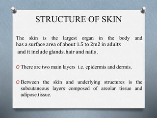
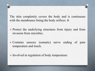
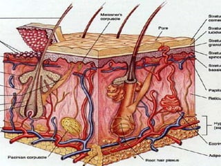

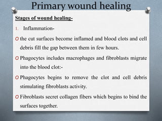
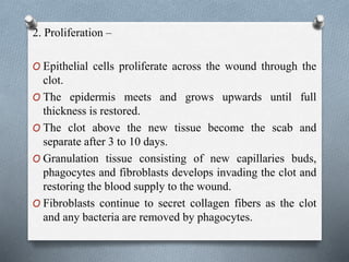
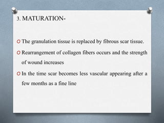
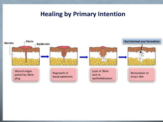

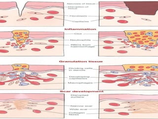


The document provides a comprehensive overview of the skin's structure, including its layers (epidermis and dermis), functions (protection, temperature regulation, sensation), and various components like hair, nails, and glands. It details the processes involved in wound healing, including inflammation, proliferation, and maturation, as well as complications such as infections. Additionally, the document explains the roles of melanin and other factors in skin color and the mechanisms of regulating body temperature.