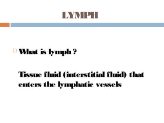
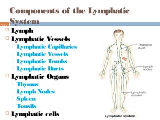
The document provides details on the structure and function of the lymphatic system. It describes lymph, lymphatic vessels, lymph nodes, and other lymphatic organs.
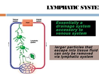
Essentially a
drainage system
accessoryto
venous system
larger particles that
escape into tissue fluid
can only be removed
via lymphatic system
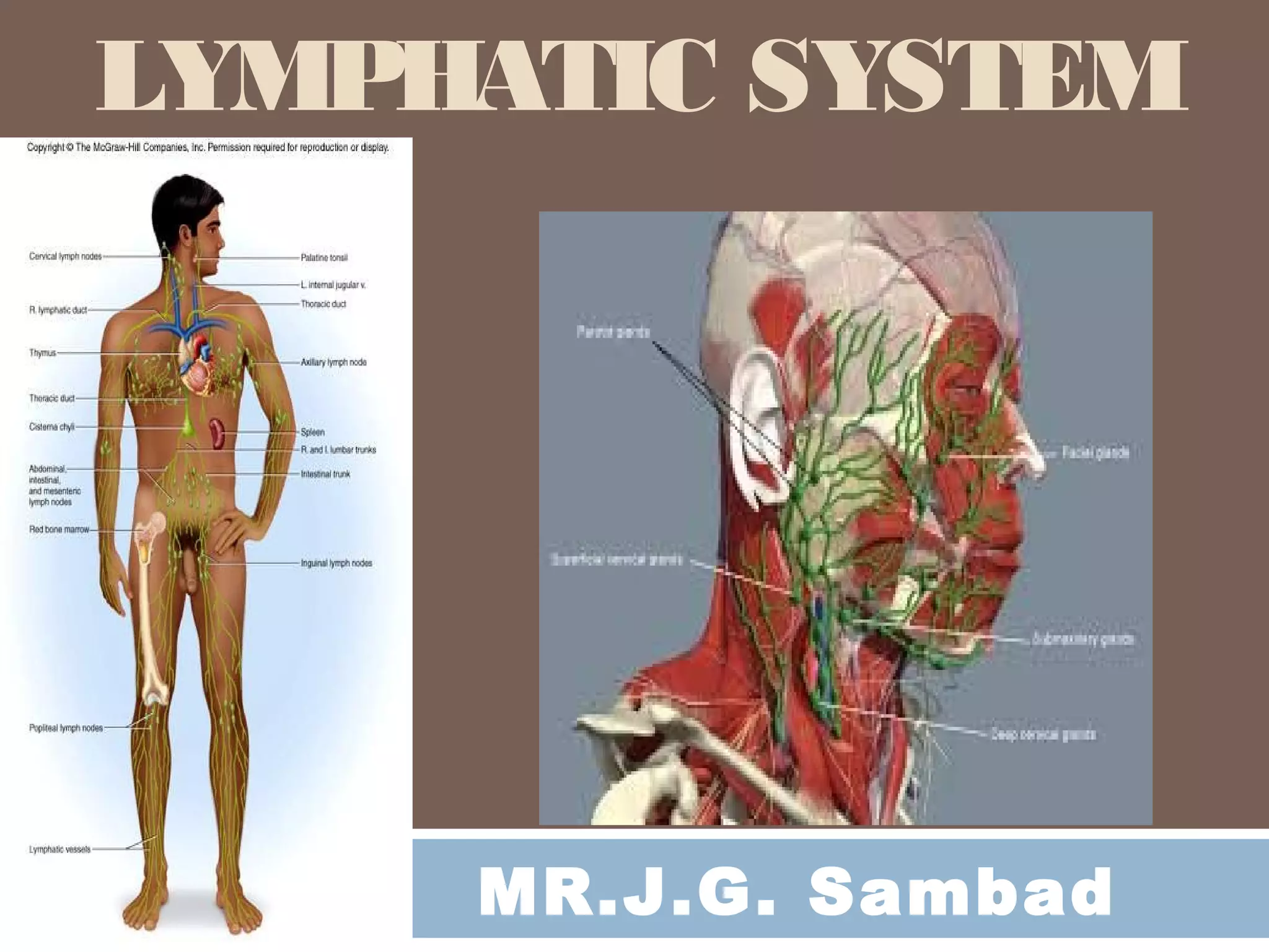
LYMPHATIC SYSTEM
5.
Functions of theLymphatic
System
24-
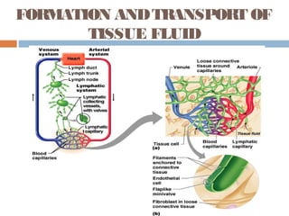
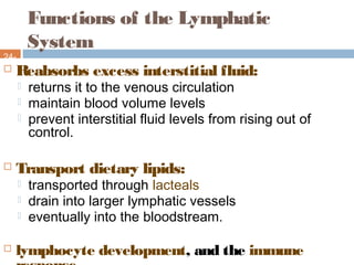
5 Reabsorbs excess interstitial fluid:
returns it to the venous circulation
maintain blood volume levels
prevent interstitial fluid levels from rising out of
control.
Transport dietary lipids:
transported through lacteals
drain into larger lymphatic vessels
eventually into the bloodstream.
lymphocyte development, and the immune
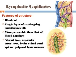
Lymphatic Capillaries
24-
9
Features ofstructure:
Blind end
Single layerof overlapping
endothelial cells
More permeable than that of
blood capillary
Absent fromavascular
structures, brain, spinal cord
splenic pulp and bone marrow
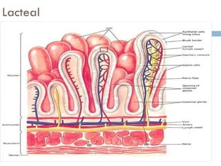
Lymphatic Capillaries –Lacteals
24-
11
The small intestine contains special types
of lymphatic capillaries called lacteals.
Lacteals pickup not only interstitial fluid,
but also dietary lipids and lipid-soluble
vitamins.
The lymph of this area has a milky color
due to the lipid and is also called chyle.
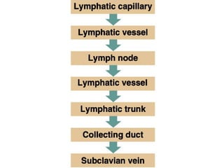
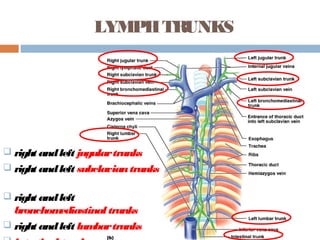
LYMPHTRUNKS
right andleftjugulartrunks
right andleft subclaviantrunks
right andleft
bronchomediastinal trunks
right andleft lumbartrunks
14.
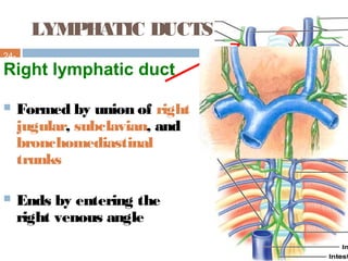
LYMPHATIC DUCTS
24-
14
Right lymphaticduct
Formed by union of right
jugular, subclavian, and
bronchomediastinal
trunks
Ends by entering the
right venous angle
15.
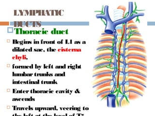
Thoracic duct
Beginsin front of L1 as a
dilated sac, the cisterna
chyli,
formed by left and right
lumbartrunks and
intestinal trunk
Enterthoracic cavity &
ascends
Travels upward, veering to
LYMPHATIC
DUCTS
16.
16
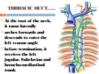
THORACIC DUCT…..
Atthe root of the neck,
it turns laterally
arches forwards and
descends to enterthe
left venous angle
before termination, it
receives the left
jugular, Subclavian and
broncho-mediastinal
trunk
17.
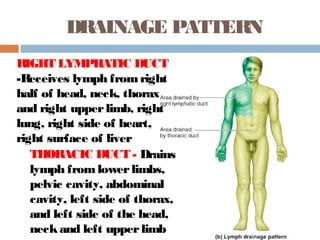
DRAINAGE PATTERN
THORACIC DUCT- Drains
lymph fromlowerlimbs,
pelvic cavity, abdominal
cavity, left side of thorax,
and left side of the head,
neckand left upperlimb
RIGHT LYMPHATIC DUCT
-Receives lymph fromright
half of head, neck, thorax
and right upperlimb, right
lung, right side of heart,
right surface of liver
18.
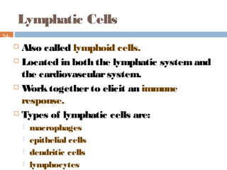
Lymphatic Cells
24-
18
Alsocalled lymphoid cells.
Located in both the lymphatic systemand
the cardiovascularsystem.
Worktogetherto elicit an immune
response.
Types of lymphatic cells are:
macrophages
epithelial cells
dendritic cells
lymphocytes
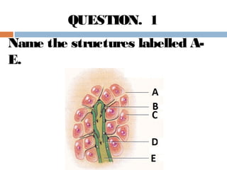
Lymph
Nodes24-
20 Small, roundoroval
located along the
pathways of lymph
vessels.
length from1 - 25
millimeters
Typically found in
clusters
receive lymph frommany
body regions.
Lymph nodes are also
21.
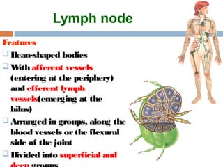
Lymph node
Features
Bean-shapedbodies
With afferent vessels
(entering at the periphery)
and efferent lymph
vessels(emerging at the
hilus)
Arranged in groups, along the
blood vessels orthe flexural
side of the joint
Divided into superficial and
22.
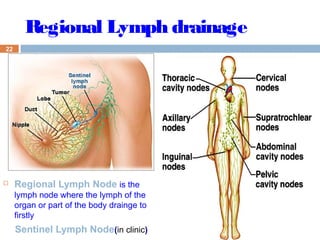
22
Regional LymphNode is the
lymph node where the lymph of the
organ or part of the body drainge to
firstly
Sentinel Lymph Node(in clinic)
Regional Lymph drainage
24
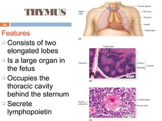
THYMUS
Features
Consists oftwo
elongated lobes
Is a large organ in
the fetus
Occupies the
thoracic cavity
behind the sternum
Secrete
lymphopoietin
25.
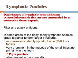
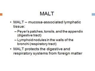
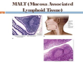
Lymphatic Nodules
24-
25 Ovalclusters of lymphatic cells with some
extracellularmatrix that are not surrounded by a
connective tissue capsule.
Filter and attack antigens.
In some areas of the body, many lymphatic nodules
group together to form larger structures.
mucosa-associated lymphatic tissue (MALT) or
tonsils
very prominent in the mucosa of the small intestine,
primarily in the ileum
Peyerpatches
27.
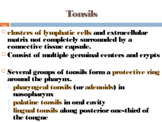
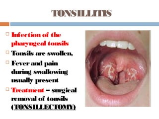
Tonsils
24-
27 clusters oflymphatic cells and extracellular
matrix not completely surrounded by a
connective tissue capsule.
Consist of multiple germinal centers and crypts
Several groups of tonsils forma protective ring
around the pharynx.
pharyngeal tonsils (oradenoids) in
nasopharynx
palatine tonsils in oral cavity
lingual tonsils along posteriorone-third of
the tongue
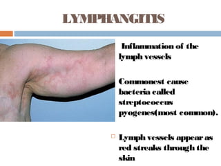
LYMPHANGITIS
Inflammation ofthe
lymph vessels
Commonest cause
bacteria called
streptococcus
pyogenes(most common).
Lymph vessels appearas
red streaks through the
skin
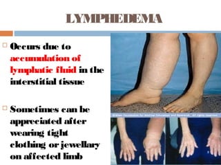
LYMPHEDEMA
Occurs dueto
accumulation of
lymphatic fluid in the
interstitial tissue
Sometimes can be
appreciated after
wearing tight
clothing orjewellary
on affected limb
33.
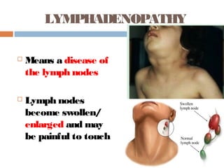
LYMPHADENOPATHY
Means adisease of
the lymph nodes
Lymph nodes
become swollen/
enlarged and may
be painful to touch
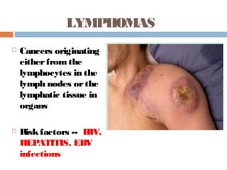
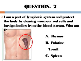
I amapart of lymphatic systemand protect
the body by clearing worn out red cells and
foreign bodies fromthe blood stream. Who am
I?
A. Thymus
B. Palatine
Tonsil
C. Spleen
QUESTION. 2
40.
Which is thecorrect statement about the
Lymphatic system?
A. It Reabsorbs excess interstitial fluid and
returns it to the venous circulation.
B. Transports dietary lipids through lacteals.
C. Helps in lymphocyte development, and the
immune response.
D. All of the above
QUESTION. 3
41.
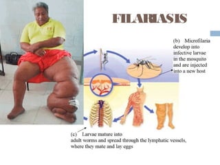
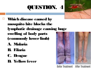
Which diseasecaused by
mosquito bite blocks the
lymphatic drainage causing huge
swelling of body parts
(commonly lowerlimb)
A. Malaria
B. Filaria
C. Dengue
D. Yellow fever
QUESTION. 4
42.
Which ofthese is a primary lymphoid
organ ?
A. Lymph Node
B. Spleen
C. Tonsil
D. Bone Marrow
QUESTION. 5