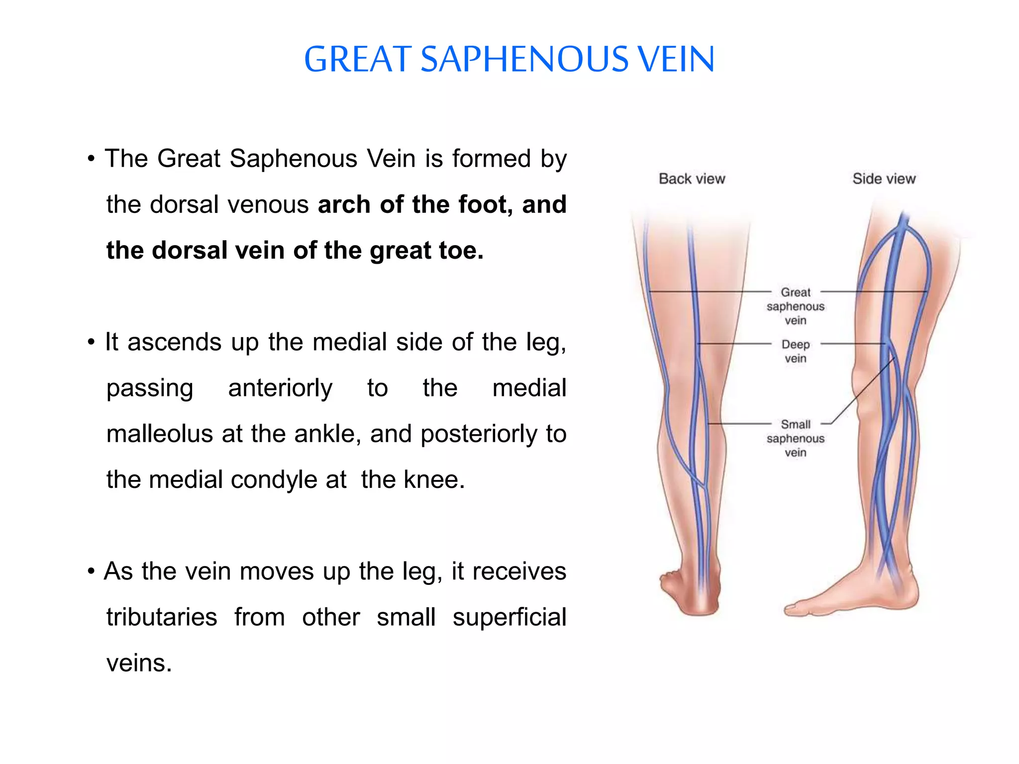
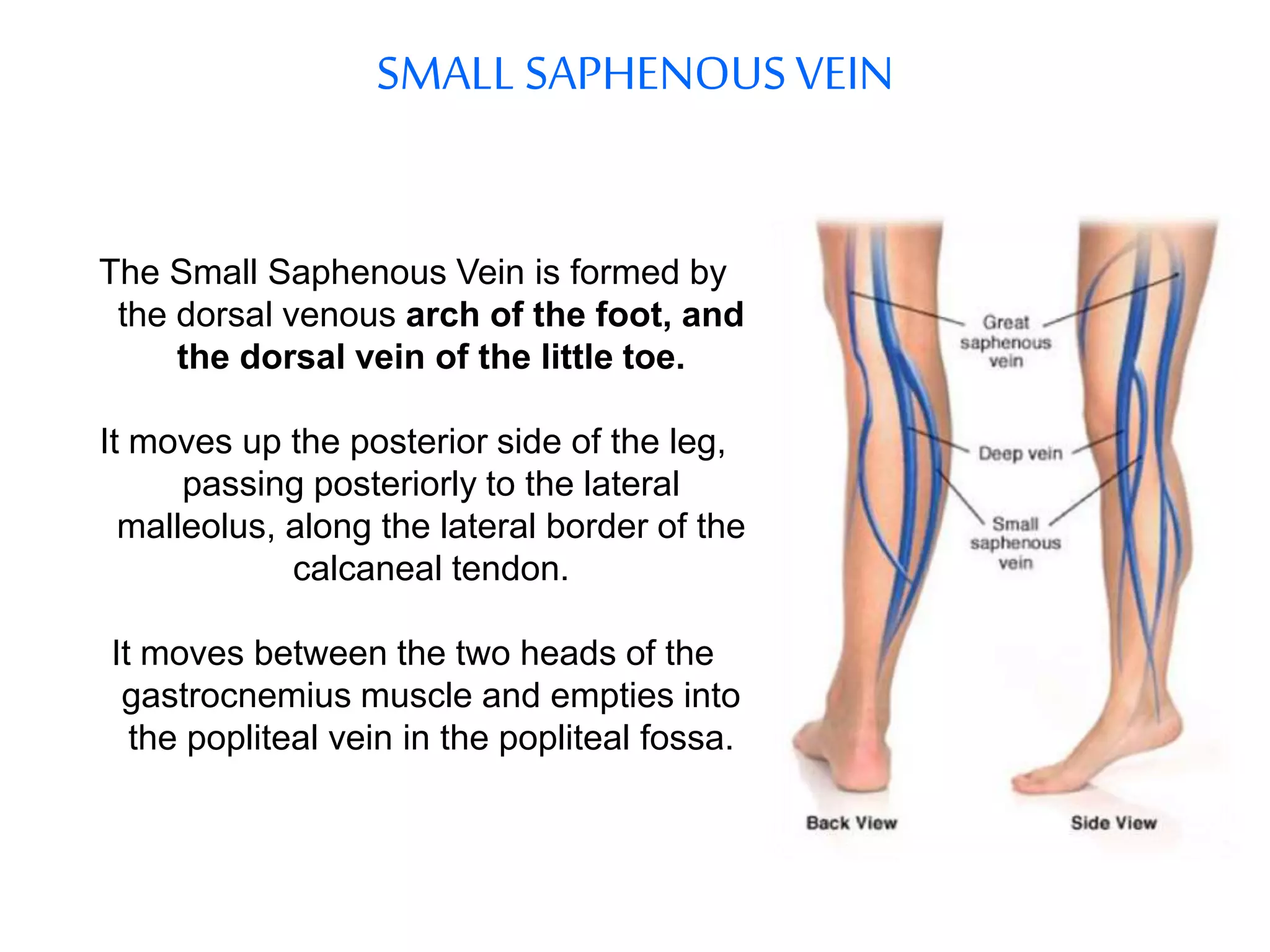
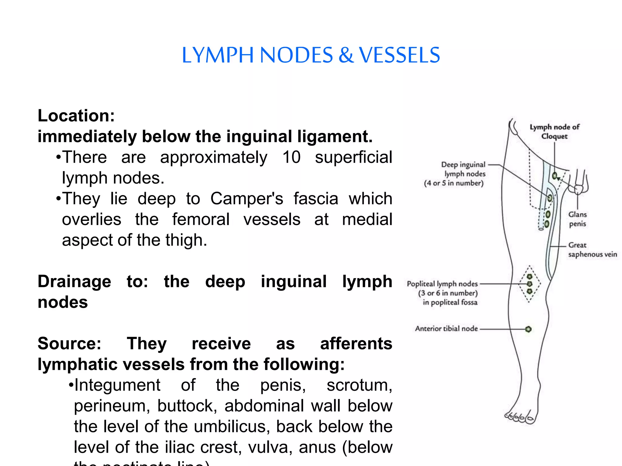
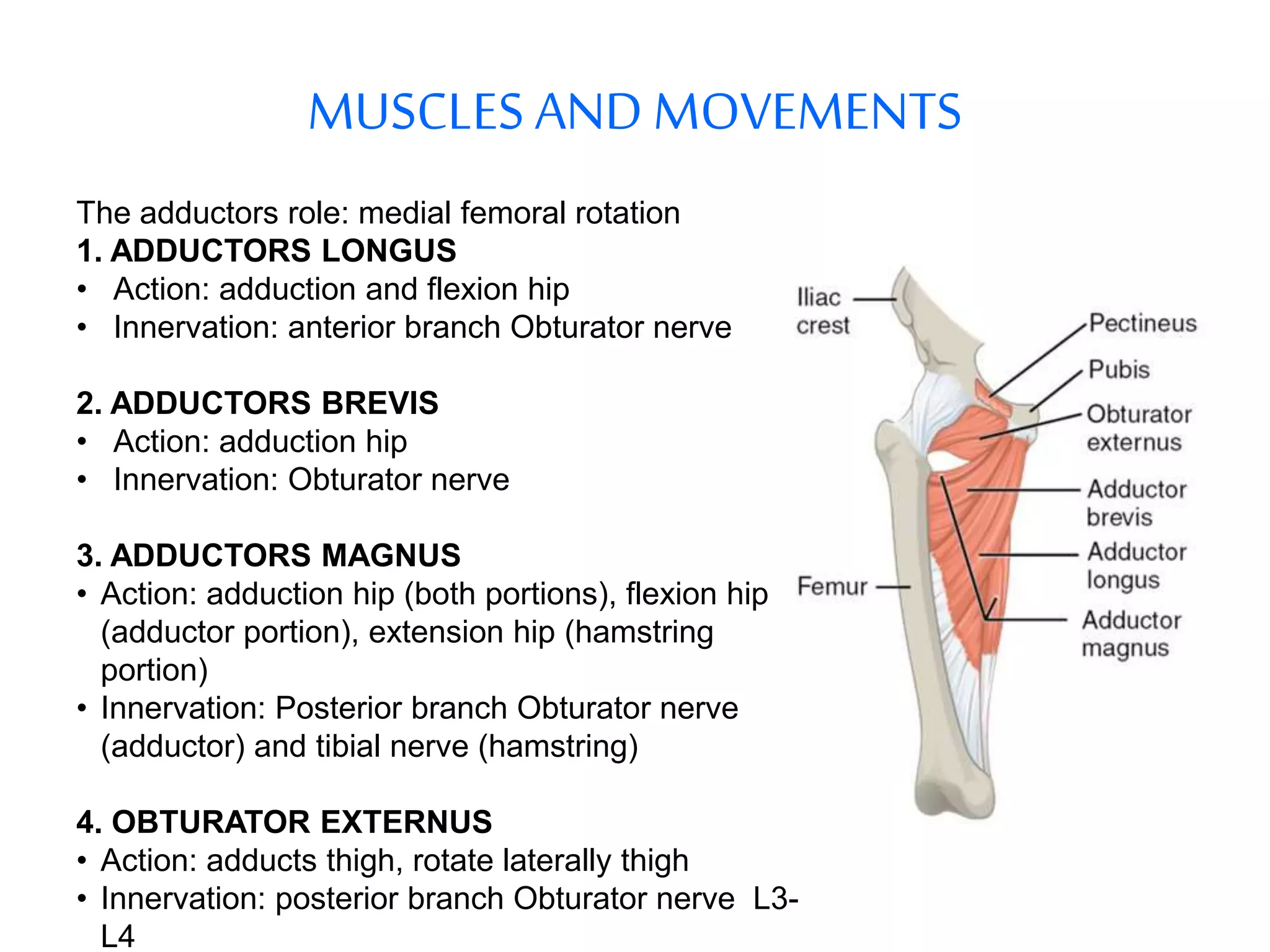
ANATOMY
OF
THIGH
ANTERIOR THIGH ANATOMY
SUPPLY
A. SUPERFICIAL ARTERIES
Cutaneous arteries - Arise from the femoral artery.
B. SUPERFICIAL VEINS
• The superficial veins of the lower limb run in the subcutaneous
tissue.
• There are two major superficial veins:
o The Great Saphenous Vein
o The Small Saphenous Vein.
GREATSAPHENOUSVEIN
• The Great Saphenous Vein is formed by
the dorsal venous arch of the foot, and
the dorsal vein of the great toe.
• It ascends up the medial side of the leg,
passing anteriorly to the medial
malleolus at the ankle, and posteriorly to
the medial condyle at the knee.
• As the vein moves up the leg, it receives
tributaries from other small superficial
veins.
The great saphenous vein then courses anteriorly to lie on the
anterior surface of the thigh before entering an opening in the
fascia lata called the saphenous opening.
It forms an arch, the saphenous arch, to join the common femoral
vein in the region of the femoral triangle at the sapheno-femoral
junction.
SMALL SAPHENOUSVEIN
The Small Saphenous Vein is formed by
the dorsal venous arch of the foot, and
the dorsal vein of the little toe.
It moves up the posterior side of the leg,
passing posteriorly to the lateral
malleolus, along the lateral border of the
calcaneal tendon.
It moves between the two heads of the
gastrocnemius muscle and empties into
the popliteal vein in the popliteal fossa.
LYMPH NODES & VESSELS
Location:
immediately below the inguinal ligament.
•There are approximately 10 superficial
lymph nodes.
•They lie deep to Camper's fascia which
overlies the femoral vessels at medial
aspect of the thigh.
Drainage to: the deep inguinal lymph
nodes
Source: They receive as afferents
lymphatic vessels from the following:
•Integument of the penis, scrotum,
perineum, buttock, abdominal wall below
the level of the umbilicus, back below the
level of the iliac crest, vulva, anus (below
FEMORAL TRIANGLE
• Boundaries: Mnemonic SAIL
• Lateral: medial border of Sartorious.
• Medial: medial border of Adductor longus.
• Superior: Inguinal Ligament.
• Floor: Adductor Longus, lliopsoas, and Pectineus.
• Roof: skin, subcutaneous tissue, a continuation of Scarpa's fascia,
Great Saphenous Vein (joins the femoral vein), superficial lymph
nodes.
CLINICAL SIGNIFICANE OF FEMORAL TRIANGLE
•Access to the Femoral Artery
•Coronary angiography. Here, the femoral artery is catheterised with a
long, thin tube. This tube is navigated up the external iliac artery,
common iliac artery, aorta, and into the coronary vessels.
•Heavy bleeding in the leg can be stopped by applying pressure to
points in the femoral triangle
Contents: Mnemonic NAVEL (From lateral to medial):
 Femoral Nerve: Directly behind sheath
 Femoral Artery and its branches: within sheath
 Femoral Vein, and deep lymph nodes: within sheath
 Femoral canal (Empty space) between vein and LN
 Contains fat and lymph node (Lymph Node of Cloquet)
FEMORALSHEATH
• This oval, funnel-shaped fascia or tube encloses the proximal parts of
the femoral vessels, which lie inferior to the inguinal ligament.
• It is a diverticulum or inferior prolongation of the fasciae lining of the
abdomen: Trasversalis fascia anteriorly and iliac fascia posteriorly.
• It is covered by the fascia lata.
• Its presence allows the femoral artery and vein to glide in and out,
deep to the inguinal ligament, during movements of the hip joint.
Thesheathdoesnotprojectintothe thigh whenthe thigh isfullyflexed,butis drawnfurtherintothe
femoraltrianglewhen the thighis extended.
• Subdivided by two vertical septa into three compartments:
1.Lateral compartment for femoral artery
2.Intermediate compartment for femoral vein
3.Medial compartment or space called femoral canal.
FEMORALCANAL
Bordered: LAMP-FILP
 laterally by the Femoral vein.
 Anteriorly by the Inguinal ligament.
 Medially by the lacunar ligament.
 Posteriorly by the Pectineal ligament
• It contains the lymph node of Cloquet.
• The entrance to the femoral canal is the femoral ring, through which
bowel can sometimes enter, causing a femoral hernia.
MUSCLES AND MOVEMENTS
A. SARTORIUS
• Action: flexion, abduction and lateral
rotation hip, flexion knee
• Innervation: femoral nerve
B. ILIACUS
• Action: flexes and rotates laterally thigh
• Innervation: femoral nerve
C. PSOAS MAJOR
• Action: flexion hip joint
• Innervation: lumbar plexus via ant.
branches L1-L3
D. PECTINEUS
• Action: thigh flexion, adduction
• Innervation: femoral nerve, sometimes
Obturator nerve.
E. QUADRICEPS FEMORIS
(Rectus femoris, Vasti lateralis, intermedius
and medialis)
• Action: knee extension, hip flexion
• Nerve: Femoral
FEMORALARTERY
It is a large artery in the thigh and the
main arterial supply to the lower limb. It
enters the thigh from behind the inguinal
ligament as the common femoral artery, a
continuation of the external iliac artery.
Then lies midway between the anterior
superior iliac spine and the symphysis
pubis.
It enters and passes through the adductor
(subsartorial) canal, and becomes the
Popliteal artery as it passes through an
opening in adductor magnus near the
Descending branch ,junction of the
middle and distal thirds of the thigh.
The common femoral artery gives off the
Profunda femoris artery and becomes the
superficial femoral artery to descend
along the anteromedial part of the thigh in
the femoral triangle.
BRANCHES
• Superficial Epigastric Artery
• Superficial Iliac Circumflex
• Superficial External Pudendal
• Deep External Pudenda!
• Deep Femoral Artery (Profunda
Femoris); Its branches:

Put My Leg Down Please:
P: Profunda Femoris artery
M: Medial Circumflex Femoral artery.
L: Lateral Circumflex Femoral artery.
D: Descending Branch of The Lateral
Circumflex Femoral Artery
P: Perforating Arteries
FEMORALVEIN
The femoral vein is a blood vessel that accompanies the femoral artery
in the femoral sheath. It begins at the adductor canal (also known as
Hunter's canal) and is a continuation of the Popliteal vein.
It ends at the inferior margin of the inguinal ligament, where it becomes
the external iliac vein.
Drainage: Several large veins drain into the femoral vein:
•Popliteal vein
•Profunda femoris vein
•Great saphenous vein
FEMORALNERVE
Derivation:
• From the dorsal divisions of the ventral rami of the L2- L4.
Surface marking:
• Descends through the fibers of the Psoas major muscle, emerging
from the muscle at the lower part of its lateral border, and passes
down between it and the iliacus muscle, behind the iliac fascia.
• It then runs beneath the inguinal ligament, into the thigh, and splits
into an Quadriceps anterior and a posterior division. Under the
inguinal ligament, it is separated from the femoral artery by a portion
of the Psoas major.
CLINICAL SIGNIFICANCE
FEMORAL NERVE INJURY
Causes
• Direct trauma is most common
• Iatrogenic (i.e. Percutaneous proximal interlocking screw placement through
IM nail)
• Compression from tumor or hematoma
Clinical Signs
Motor
• Rectus femoris
• Vastus fateralis
• Vastus medlalis
• Quadriceps wasting and Loss of knee extension and some hip flexion
(iliacus, pectineus)
Sensory
• Loss of sensation over front and medial side of thigh (anterior and
medial cutaneous nerves of the thigh).
• Loss of sensation over medial aspect of lower leg and foot
MEDIAL THIGH ANATOMY
MUSCLES ANDMOVEMENTS
The adductors role: medial femoral rotation
1. ADDUCTORS LONGUS
• Action: adduction and flexion hip
• Innervation: anterior branch Obturator nerve
2. ADDUCTORS BREVIS
• Action: adduction hip
• Innervation: Obturator nerve
3. ADDUCTORS MAGNUS
• Action: adduction hip (both portions), flexion hip
(adductor portion), extension hip (hamstring
portion)
• Innervation: Posterior branch Obturator nerve
(adductor) and tibial nerve (hamstring)
4. OBTURATOR EXTERNUS
• Action: adducts thigh, rotate laterally thigh
• Innervation: posterior branch Obturator nerve L3-
L4
ARTERIES& NERVES
1. THE PROFUNDA FEMORIS
• Source: Femoral artery.
• Branches: My leg Down Please
M: medial circumflex femoral artery
L: lateral circumflex femoral artery
D: descending branch of the lateral circumflex femoral artery
P: perforating arteries
2. OBTURATOR ARTERY
• Source: internal iliac artery
• Branches: ant and post branches
• Supplies: obturator externus muscle, medial compartment thigh, femur.
3. OBTURATOR NERVE
• From: lumbar plexus L2-L4
• To: post branch obturator nerve, Ant branch Obturator
• Innervates: medial compartment Thigh
anatomy of thigh

anatomy of thigh

  • 1.
  • 2.
  • 3.
    SUPPLY A. SUPERFICIAL ARTERIES Cutaneousarteries - Arise from the femoral artery. B. SUPERFICIAL VEINS • The superficial veins of the lower limb run in the subcutaneous tissue. • There are two major superficial veins: o The Great Saphenous Vein o The Small Saphenous Vein.
  • 4.
    GREATSAPHENOUSVEIN • The GreatSaphenous Vein is formed by the dorsal venous arch of the foot, and the dorsal vein of the great toe. • It ascends up the medial side of the leg, passing anteriorly to the medial malleolus at the ankle, and posteriorly to the medial condyle at the knee. • As the vein moves up the leg, it receives tributaries from other small superficial veins.
  • 5.
    The great saphenousvein then courses anteriorly to lie on the anterior surface of the thigh before entering an opening in the fascia lata called the saphenous opening. It forms an arch, the saphenous arch, to join the common femoral vein in the region of the femoral triangle at the sapheno-femoral junction.
  • 6.
    SMALL SAPHENOUSVEIN The SmallSaphenous Vein is formed by the dorsal venous arch of the foot, and the dorsal vein of the little toe. It moves up the posterior side of the leg, passing posteriorly to the lateral malleolus, along the lateral border of the calcaneal tendon. It moves between the two heads of the gastrocnemius muscle and empties into the popliteal vein in the popliteal fossa.
  • 7.
    LYMPH NODES &VESSELS Location: immediately below the inguinal ligament. •There are approximately 10 superficial lymph nodes. •They lie deep to Camper's fascia which overlies the femoral vessels at medial aspect of the thigh. Drainage to: the deep inguinal lymph nodes Source: They receive as afferents lymphatic vessels from the following: •Integument of the penis, scrotum, perineum, buttock, abdominal wall below the level of the umbilicus, back below the level of the iliac crest, vulva, anus (below
  • 8.
    FEMORAL TRIANGLE • Boundaries:Mnemonic SAIL • Lateral: medial border of Sartorious. • Medial: medial border of Adductor longus. • Superior: Inguinal Ligament. • Floor: Adductor Longus, lliopsoas, and Pectineus. • Roof: skin, subcutaneous tissue, a continuation of Scarpa's fascia, Great Saphenous Vein (joins the femoral vein), superficial lymph nodes.
  • 10.
    CLINICAL SIGNIFICANE OFFEMORAL TRIANGLE •Access to the Femoral Artery •Coronary angiography. Here, the femoral artery is catheterised with a long, thin tube. This tube is navigated up the external iliac artery, common iliac artery, aorta, and into the coronary vessels. •Heavy bleeding in the leg can be stopped by applying pressure to points in the femoral triangle
  • 11.
    Contents: Mnemonic NAVEL(From lateral to medial):  Femoral Nerve: Directly behind sheath  Femoral Artery and its branches: within sheath  Femoral Vein, and deep lymph nodes: within sheath  Femoral canal (Empty space) between vein and LN  Contains fat and lymph node (Lymph Node of Cloquet)
  • 12.
    FEMORALSHEATH • This oval,funnel-shaped fascia or tube encloses the proximal parts of the femoral vessels, which lie inferior to the inguinal ligament. • It is a diverticulum or inferior prolongation of the fasciae lining of the abdomen: Trasversalis fascia anteriorly and iliac fascia posteriorly. • It is covered by the fascia lata. • Its presence allows the femoral artery and vein to glide in and out, deep to the inguinal ligament, during movements of the hip joint. Thesheathdoesnotprojectintothe thigh whenthe thigh isfullyflexed,butis drawnfurtherintothe femoraltrianglewhen the thighis extended.
  • 13.
    • Subdivided bytwo vertical septa into three compartments: 1.Lateral compartment for femoral artery 2.Intermediate compartment for femoral vein 3.Medial compartment or space called femoral canal.
  • 14.
    FEMORALCANAL Bordered: LAMP-FILP  laterallyby the Femoral vein.  Anteriorly by the Inguinal ligament.  Medially by the lacunar ligament.  Posteriorly by the Pectineal ligament • It contains the lymph node of Cloquet. • The entrance to the femoral canal is the femoral ring, through which bowel can sometimes enter, causing a femoral hernia.
  • 15.
    MUSCLES AND MOVEMENTS A.SARTORIUS • Action: flexion, abduction and lateral rotation hip, flexion knee • Innervation: femoral nerve B. ILIACUS • Action: flexes and rotates laterally thigh • Innervation: femoral nerve C. PSOAS MAJOR • Action: flexion hip joint • Innervation: lumbar plexus via ant. branches L1-L3 D. PECTINEUS • Action: thigh flexion, adduction • Innervation: femoral nerve, sometimes Obturator nerve. E. QUADRICEPS FEMORIS (Rectus femoris, Vasti lateralis, intermedius and medialis) • Action: knee extension, hip flexion • Nerve: Femoral
  • 16.
    FEMORALARTERY It is alarge artery in the thigh and the main arterial supply to the lower limb. It enters the thigh from behind the inguinal ligament as the common femoral artery, a continuation of the external iliac artery. Then lies midway between the anterior superior iliac spine and the symphysis pubis. It enters and passes through the adductor (subsartorial) canal, and becomes the Popliteal artery as it passes through an opening in adductor magnus near the Descending branch ,junction of the middle and distal thirds of the thigh. The common femoral artery gives off the Profunda femoris artery and becomes the superficial femoral artery to descend along the anteromedial part of the thigh in the femoral triangle.
  • 17.
    BRANCHES • Superficial EpigastricArtery • Superficial Iliac Circumflex • Superficial External Pudendal • Deep External Pudenda! • Deep Femoral Artery (Profunda Femoris); Its branches:  Put My Leg Down Please: P: Profunda Femoris artery M: Medial Circumflex Femoral artery. L: Lateral Circumflex Femoral artery. D: Descending Branch of The Lateral Circumflex Femoral Artery P: Perforating Arteries
  • 18.
    FEMORALVEIN The femoral veinis a blood vessel that accompanies the femoral artery in the femoral sheath. It begins at the adductor canal (also known as Hunter's canal) and is a continuation of the Popliteal vein. It ends at the inferior margin of the inguinal ligament, where it becomes the external iliac vein. Drainage: Several large veins drain into the femoral vein: •Popliteal vein •Profunda femoris vein •Great saphenous vein
  • 19.
    FEMORALNERVE Derivation: • From thedorsal divisions of the ventral rami of the L2- L4. Surface marking: • Descends through the fibers of the Psoas major muscle, emerging from the muscle at the lower part of its lateral border, and passes down between it and the iliacus muscle, behind the iliac fascia. • It then runs beneath the inguinal ligament, into the thigh, and splits into an Quadriceps anterior and a posterior division. Under the inguinal ligament, it is separated from the femoral artery by a portion of the Psoas major.
  • 21.
    CLINICAL SIGNIFICANCE FEMORAL NERVEINJURY Causes • Direct trauma is most common • Iatrogenic (i.e. Percutaneous proximal interlocking screw placement through IM nail) • Compression from tumor or hematoma Clinical Signs Motor • Rectus femoris • Vastus fateralis • Vastus medlalis • Quadriceps wasting and Loss of knee extension and some hip flexion (iliacus, pectineus) Sensory • Loss of sensation over front and medial side of thigh (anterior and medial cutaneous nerves of the thigh). • Loss of sensation over medial aspect of lower leg and foot
  • 22.
  • 23.
    MUSCLES ANDMOVEMENTS The adductorsrole: medial femoral rotation 1. ADDUCTORS LONGUS • Action: adduction and flexion hip • Innervation: anterior branch Obturator nerve 2. ADDUCTORS BREVIS • Action: adduction hip • Innervation: Obturator nerve 3. ADDUCTORS MAGNUS • Action: adduction hip (both portions), flexion hip (adductor portion), extension hip (hamstring portion) • Innervation: Posterior branch Obturator nerve (adductor) and tibial nerve (hamstring) 4. OBTURATOR EXTERNUS • Action: adducts thigh, rotate laterally thigh • Innervation: posterior branch Obturator nerve L3- L4
  • 25.
    ARTERIES& NERVES 1. THEPROFUNDA FEMORIS • Source: Femoral artery. • Branches: My leg Down Please M: medial circumflex femoral artery L: lateral circumflex femoral artery D: descending branch of the lateral circumflex femoral artery P: perforating arteries 2. OBTURATOR ARTERY • Source: internal iliac artery • Branches: ant and post branches • Supplies: obturator externus muscle, medial compartment thigh, femur. 3. OBTURATOR NERVE • From: lumbar plexus L2-L4 • To: post branch obturator nerve, Ant branch Obturator • Innervates: medial compartment Thigh