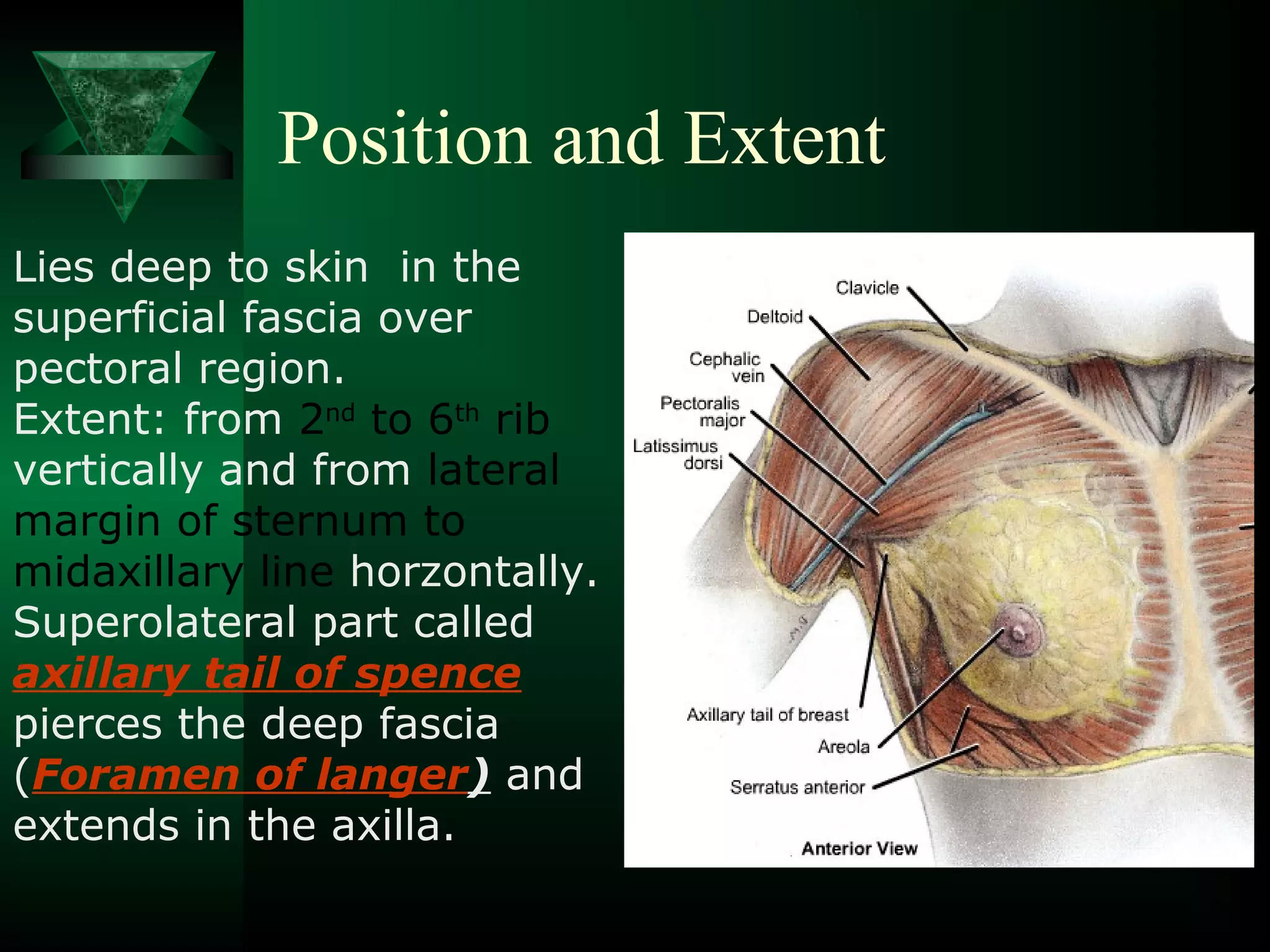
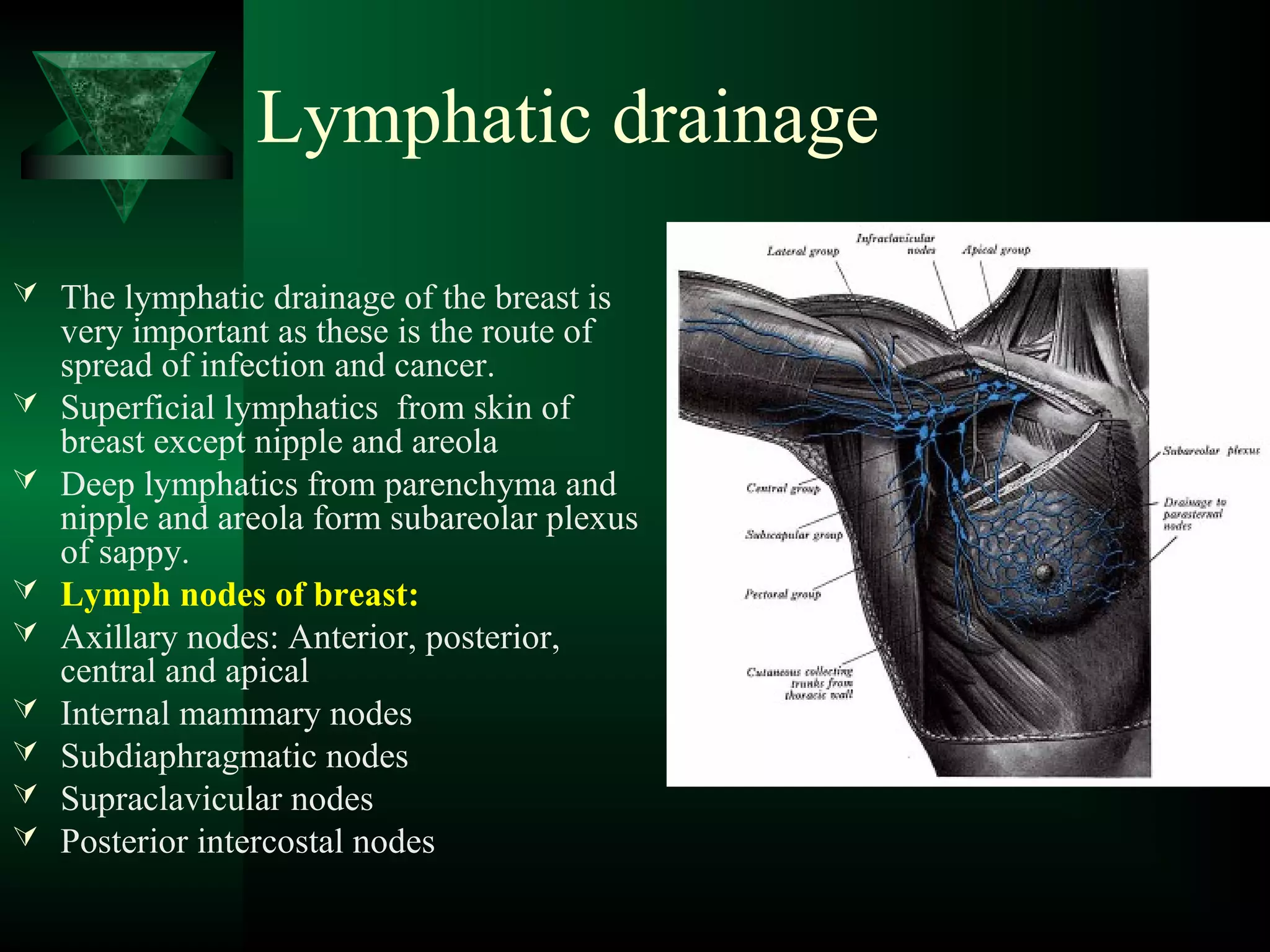
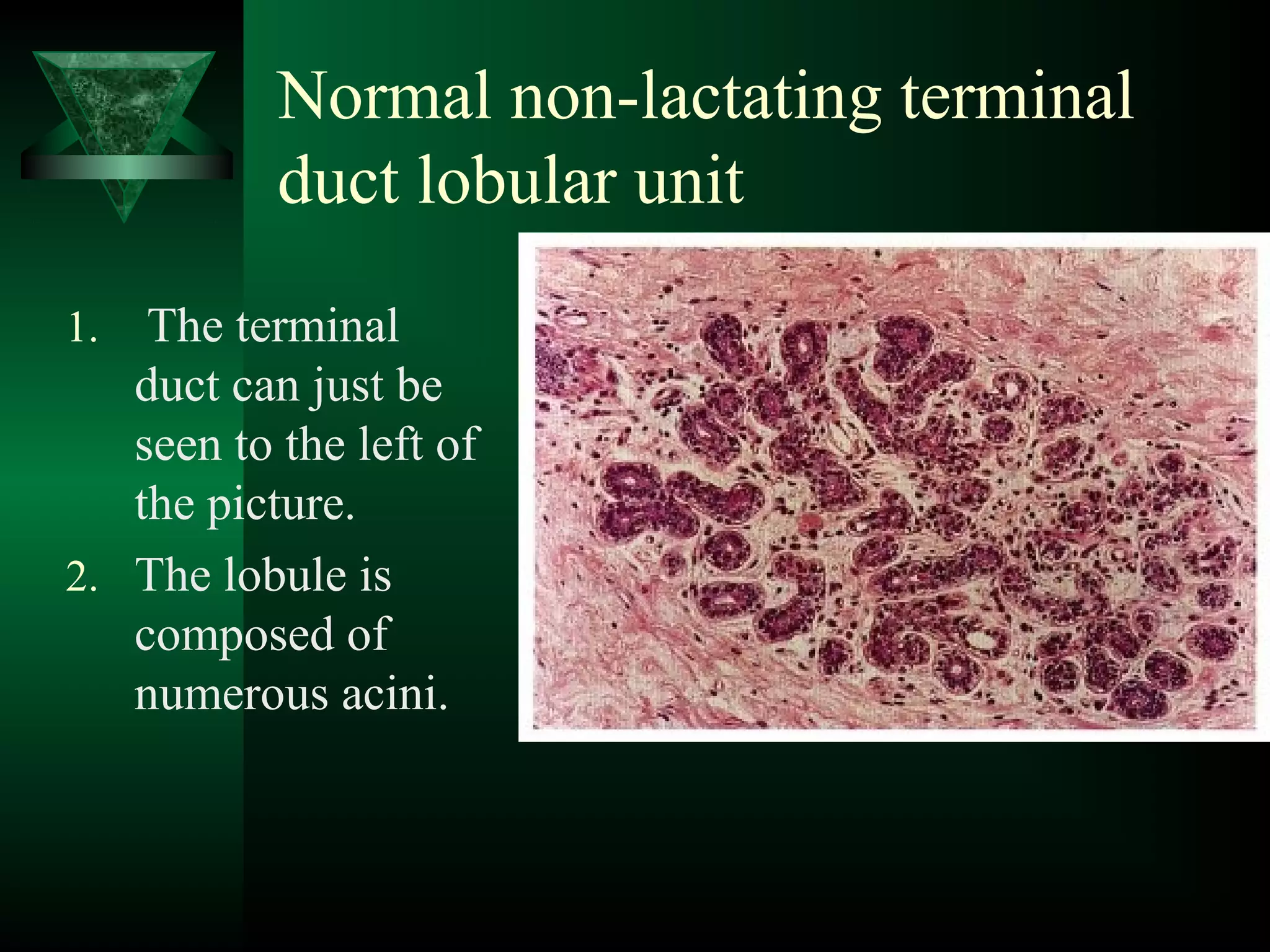
Cynthia Nixon Sheryl Crow 
Christina Applegate 
Olivia Newton John 
What 
important 
fact is 
common 
with these 
females?
Anatomy of Breast 
Dr.B.B.Gosai
Introduction 
Breast found in both sexes but well 
developed in female after puberty. 
Accessory organ of female reproductive 
system. 
Modified sweat gland 
Hemispherical in adult and pendulous in old 
age
Position and Extent 
Lies deep to skin in the 
superficial fascia over 
pectoral region. 
Extent: from 2nd to 6th rib 
vertically and from lateral 
margin of sternum to 
midaxillary line horzontally. 
Superolateral part called 
axillary tail of spence 
pierces the deep fascia 
(Foramen of langer) and 
extends in the axilla.
Deep relations of breast 
Retromammary 
space: loose 
areolar tissue 
Pectoral fascia 
Pectoralis major, 
serratus anterior 
and external 
oblique
Structure of the Breast 
The breast contains: 
 Skin 
 Parenchyma: epithelial glandular tissue 
of the tubulo-alveolar type 
 fibrous connective tissue and fat (stroma) 
surrounding the glandular tissue 

Skin 
 Nipple: Conical projection 
just below the centre of 
breast, pierced by 15-20 
lactiferous ducts and contain 
smooth muscles and sensory 
end organs 
 Areola: Pigmented area 
surrounding the base of nipple 
contain modified sebaceous 
glands which enlarge during 
pregnancy, These enlarged 
glands are known as Tubercles 
of Montgomery for secretion to 
prevent cracking of nipple 
Nipple 
Tubercles of Montgomery
Glandular Tissue (Parenchyma) 
 This consists of branching ducts and 
terminal secretory lobules. 
 In the mature breast 15-20 lobules. Each 
lobule consists of several blind-ending 
branches or expansions, the alveoli 
(acini), converging on an alveolar duct 
 The ducts converge on to the 15–20 
larger lactiferous ducts which open on 
to the apex of the nipple. 
 Breast cancers arise at the junction of 
the lobules and ducts, and as they 
increase in size they lead to fibrous 
tissue formation so that they are hard 
and irregular.
Stroma of the Breast 
 Formed by fibrous tissue and fat 
 Fibrous tissue extend from the ducts to 
the dermis, and these are often well 
developed in the upper part of the breast 
as the suspensory ligaments (of Astley 
Cooper), which assist in the support of 
the breast tissue. 
 Pathologically, these may be contracted 
by fibrosis in carcinoma, causing 
retraction or pitting of the overlying skin. 
Peau de orange
Arterial supply 
 Supplying the female breasts are branches of the axillary 
artery, the internal thoracic artery, and some intercostal 
arteries, as follows: 
 the axillary artery supplies blood to the breast via several 
branches: the superior thoracic, the pectoral branches of 
the thoraco-acromial artery, the lateral thoracic 
 the internal thoracic artery gives perforating branches to 
the anteromedial part of the breast; 
 the second to fourth intercostal arteries give 
perforating branches more laterally in the anterior thorax.
Arteries
Venous drainage 
Around the areola there is a circular venous 
plexus. 
From this and from the glandular tissue, 
blood drains in veins accompanying the 
arterial blood supply, i.e. to the axillary, 
internal thoracic and intercostal veins. 
Great individual variation may occur
Nerve supply 
 The nerve supply of the breast is derived from the 
branches of the fourth to sixth intercostal nerves 
 They carry sensory and sympathetic efferent fibres. 
 The nipple supply is from the T4; 
 this forms an extensive nerve plexus within the 
nipple, its sensory fibres terminating close to the 
epithelium as free endings, Meissner corpuscles and 
Merkel disc endings. 
 These are essential in signalling suckling to the 
central nervous system; however, secretory 
activities of the gland are largely controlled by 
ovarian and hypophyseal hormones (Prolactin) 
rather than by efferent motor fibres.
Lymphatic drainage 
 The lymphatic drainage of the breast is 
very important as these is the route of 
spread of infection and cancer. 
 Superficial lymphatics from skin of 
breast except nipple and areola 
 Deep lymphatics from parenchyma and 
nipple and areola form subareolar plexus 
of sappy. 
 Lymph nodes of breast: 
 Axillary nodes: Anterior, posterior, 
central and apical 
 Internal mammary nodes 
 Subdiaphragmatic nodes 
 Supraclavicular nodes 
 Posterior intercostal nodes
Lymph 
Vessels
Lymphatic drainage by quadrant of breast 
Supraclavicular Nodes
Lymphatic drainage by quadrants 
 From superolateral quadrant: 
– Anterior, posterior axillary nodes 
– Supraclavicular nodes 
 From superomedial quadrant: 
– Internal mammary nodes 
– Supraclavicular nodes 
 From inferomedial quadrant: 
– Intenal mammary nodes 
– Subdiaphragmatic nodes 
 From infrolateral quadrant: 
– Posterior intercostal nodes 
– Subdiaphragmatic nodes
Important characteristics of 
lymphatic drainage 
 the axillary lymph nodes, the predominant site of drainage from the 
breast. (70 %). 
 Internal mammary nodes communicate by lymphatics crossing 
midline and hence one side cancer can spread to opposite breast 
 Route of spread of cancer is according to route of lymphatics 
 Secondary spread of cancer involve axillary nodes, (Axilla) 
supraclavicular nodes (Neck), internal mammary nodes (Opposite 
breast) and subdiaphragmatic nodes (Abdomen-liver, ovary). 
 Nowadays, a simpler nomenclature is generally adopted, based on the relation of 
the nodes to pectoralis minor. 
 Those lying below pectoralis minor are the low nodes (level 1), 
 those behind the muscle are the middle group (level 2), 
 while the nodes between the upper border of pectoralis minor and the lower 
border of the clavicle are the upper or apical nodes (level 3). 
 In addition, between pectoralis minor and major there may be one or two other 
nodes (Rotter's nodes).
Breast Development 
Prenatal Development 
 Epithelial mammary bud 
appearing at a gestational age of 
35 days; by day 37 this has 
become a mammary line 
extending from the axilla through 
to the inguinal region 
 The thoracic mammary bud 
invaginates into the mesenchyme, 
with involution of the remaining 
mammary line 
 Then Nipple is formed , ducts 
develop and get canalized.
•Developmental anomalies of breast 
 Accessory breast tissue may be present in adults 
anywhere along the milk line (polythelia) 
 Accessory breast develop usually in the thoracic region 
(90%) but also occasionally in the axillary (5%) or 
abdominal (5%). 
 In either sex, there may be no breast development 
(amastia) 
 Rarely, the nipple may not develop (athelia) although 
this occurs more commonly in accessory breast tissue. 
 At birth the combination of fetal prolactin and maternal 
oestrogen may give rise to transient hyperplasia and 
secretion of 'witch's milk'.
Polymastia 
Amastia
Normal non-lactating lobule with 
terminal duct. 
1.The duct can be seen 
leading to the lobule 
which is composed of 
multiple, small acini set 
within a loose intralobular 
stroma. 
2. The denser, interlobular 
stroma can be seen 
surrounding the lobule 
and duct. 
3. A small amount of 
adipose tissue is also 
present
Normal non-lactating terminal 
duct lobular unit 
1. The terminal 
duct can just be 
seen to the left of 
the picture. 
2. The lobule is 
composed of 
numerous acini.
High power view of a non-lactating 
lobule 
 . The two cell type lining 
of the acini can be clearly 
seen with the inner 
luminal epithelium and 
outer myoepithelial layer. 
 The eosinophilic basement 
membrane can also be 
discerned. 
 The loose intralobular 
stroma contrasts with the 
denser, interlobular stroma 
surrounding the lobule.
Lactating breast 
 . The lobules are greatly 
expanded. Many more 
acini are present 
 lined by cells showing 
evidence of secretory 
activity. 
 The intralobular stroma 
has largely been 
displaced.
High power view of lactating 
breast 
 . The enlarged lobule, 
composed of multiple 
distended acini, can be 
seen. 
 The vacuolated cytoplasm 
of the secretory epithelium 
is clearly visible. 
 Myoepithelium is 
difficult to see.
Clinical Anatomy of breast 
Cancer of breast: second common cancer in 
female 
Retraction of skin (Peau de orange) 
Retraction of nipple 
Stony hard swelling in breast 
Spread of cancer to nodes of breast 
Distant metastasis 
Early detection can be treated and gives better 
prognosis
Fungating Fulminant cancer 
Surgical removal of 
breast: Mastectomy
Artificial breast for 
cancer patients 
Breast implants for 
cosmetic surgery 
Sarah Jessica Parker
Early detection 
Breast examination 
Mammogram
Other clincal conditions 
Mastitis: Infection of 
breast 
Breast abscess: Incision 
for drainage should be 
radial to avoid injury to 
ducts
Cystic swelling in breast 
due to blockage of ducts 
Gynecomastia: Female like 
breast in male
Learning never ends !!!!

Anatomy of Breast in clinical perspective-Dr.Gosai

  • 1.
    Cynthia Nixon SherylCrow Christina Applegate Olivia Newton John What important fact is common with these females?
  • 2.
    Anatomy of Breast Dr.B.B.Gosai
  • 3.
    Introduction Breast foundin both sexes but well developed in female after puberty. Accessory organ of female reproductive system. Modified sweat gland Hemispherical in adult and pendulous in old age
  • 4.
    Position and Extent Lies deep to skin in the superficial fascia over pectoral region. Extent: from 2nd to 6th rib vertically and from lateral margin of sternum to midaxillary line horzontally. Superolateral part called axillary tail of spence pierces the deep fascia (Foramen of langer) and extends in the axilla.
  • 5.
    Deep relations ofbreast Retromammary space: loose areolar tissue Pectoral fascia Pectoralis major, serratus anterior and external oblique
  • 6.
    Structure of theBreast The breast contains:  Skin  Parenchyma: epithelial glandular tissue of the tubulo-alveolar type  fibrous connective tissue and fat (stroma) surrounding the glandular tissue 
  • 7.
    Skin  Nipple:Conical projection just below the centre of breast, pierced by 15-20 lactiferous ducts and contain smooth muscles and sensory end organs  Areola: Pigmented area surrounding the base of nipple contain modified sebaceous glands which enlarge during pregnancy, These enlarged glands are known as Tubercles of Montgomery for secretion to prevent cracking of nipple Nipple Tubercles of Montgomery
  • 8.
    Glandular Tissue (Parenchyma)  This consists of branching ducts and terminal secretory lobules.  In the mature breast 15-20 lobules. Each lobule consists of several blind-ending branches or expansions, the alveoli (acini), converging on an alveolar duct  The ducts converge on to the 15–20 larger lactiferous ducts which open on to the apex of the nipple.  Breast cancers arise at the junction of the lobules and ducts, and as they increase in size they lead to fibrous tissue formation so that they are hard and irregular.
  • 9.
    Stroma of theBreast  Formed by fibrous tissue and fat  Fibrous tissue extend from the ducts to the dermis, and these are often well developed in the upper part of the breast as the suspensory ligaments (of Astley Cooper), which assist in the support of the breast tissue.  Pathologically, these may be contracted by fibrosis in carcinoma, causing retraction or pitting of the overlying skin. Peau de orange
  • 10.
    Arterial supply Supplying the female breasts are branches of the axillary artery, the internal thoracic artery, and some intercostal arteries, as follows:  the axillary artery supplies blood to the breast via several branches: the superior thoracic, the pectoral branches of the thoraco-acromial artery, the lateral thoracic  the internal thoracic artery gives perforating branches to the anteromedial part of the breast;  the second to fourth intercostal arteries give perforating branches more laterally in the anterior thorax.
  • 11.
  • 12.
    Venous drainage Aroundthe areola there is a circular venous plexus. From this and from the glandular tissue, blood drains in veins accompanying the arterial blood supply, i.e. to the axillary, internal thoracic and intercostal veins. Great individual variation may occur
  • 13.
    Nerve supply The nerve supply of the breast is derived from the branches of the fourth to sixth intercostal nerves  They carry sensory and sympathetic efferent fibres.  The nipple supply is from the T4;  this forms an extensive nerve plexus within the nipple, its sensory fibres terminating close to the epithelium as free endings, Meissner corpuscles and Merkel disc endings.  These are essential in signalling suckling to the central nervous system; however, secretory activities of the gland are largely controlled by ovarian and hypophyseal hormones (Prolactin) rather than by efferent motor fibres.
  • 14.
    Lymphatic drainage The lymphatic drainage of the breast is very important as these is the route of spread of infection and cancer.  Superficial lymphatics from skin of breast except nipple and areola  Deep lymphatics from parenchyma and nipple and areola form subareolar plexus of sappy.  Lymph nodes of breast:  Axillary nodes: Anterior, posterior, central and apical  Internal mammary nodes  Subdiaphragmatic nodes  Supraclavicular nodes  Posterior intercostal nodes
  • 15.
  • 16.
    Lymphatic drainage byquadrant of breast Supraclavicular Nodes
  • 17.
    Lymphatic drainage byquadrants  From superolateral quadrant: – Anterior, posterior axillary nodes – Supraclavicular nodes  From superomedial quadrant: – Internal mammary nodes – Supraclavicular nodes  From inferomedial quadrant: – Intenal mammary nodes – Subdiaphragmatic nodes  From infrolateral quadrant: – Posterior intercostal nodes – Subdiaphragmatic nodes
  • 18.
    Important characteristics of lymphatic drainage  the axillary lymph nodes, the predominant site of drainage from the breast. (70 %).  Internal mammary nodes communicate by lymphatics crossing midline and hence one side cancer can spread to opposite breast  Route of spread of cancer is according to route of lymphatics  Secondary spread of cancer involve axillary nodes, (Axilla) supraclavicular nodes (Neck), internal mammary nodes (Opposite breast) and subdiaphragmatic nodes (Abdomen-liver, ovary).  Nowadays, a simpler nomenclature is generally adopted, based on the relation of the nodes to pectoralis minor.  Those lying below pectoralis minor are the low nodes (level 1),  those behind the muscle are the middle group (level 2),  while the nodes between the upper border of pectoralis minor and the lower border of the clavicle are the upper or apical nodes (level 3).  In addition, between pectoralis minor and major there may be one or two other nodes (Rotter's nodes).
  • 19.
    Breast Development PrenatalDevelopment  Epithelial mammary bud appearing at a gestational age of 35 days; by day 37 this has become a mammary line extending from the axilla through to the inguinal region  The thoracic mammary bud invaginates into the mesenchyme, with involution of the remaining mammary line  Then Nipple is formed , ducts develop and get canalized.
  • 20.
    •Developmental anomalies ofbreast  Accessory breast tissue may be present in adults anywhere along the milk line (polythelia)  Accessory breast develop usually in the thoracic region (90%) but also occasionally in the axillary (5%) or abdominal (5%).  In either sex, there may be no breast development (amastia)  Rarely, the nipple may not develop (athelia) although this occurs more commonly in accessory breast tissue.  At birth the combination of fetal prolactin and maternal oestrogen may give rise to transient hyperplasia and secretion of 'witch's milk'.
  • 21.
  • 22.
    Normal non-lactating lobulewith terminal duct. 1.The duct can be seen leading to the lobule which is composed of multiple, small acini set within a loose intralobular stroma. 2. The denser, interlobular stroma can be seen surrounding the lobule and duct. 3. A small amount of adipose tissue is also present
  • 23.
    Normal non-lactating terminal duct lobular unit 1. The terminal duct can just be seen to the left of the picture. 2. The lobule is composed of numerous acini.
  • 24.
    High power viewof a non-lactating lobule  . The two cell type lining of the acini can be clearly seen with the inner luminal epithelium and outer myoepithelial layer.  The eosinophilic basement membrane can also be discerned.  The loose intralobular stroma contrasts with the denser, interlobular stroma surrounding the lobule.
  • 25.
    Lactating breast . The lobules are greatly expanded. Many more acini are present  lined by cells showing evidence of secretory activity.  The intralobular stroma has largely been displaced.
  • 26.
    High power viewof lactating breast  . The enlarged lobule, composed of multiple distended acini, can be seen.  The vacuolated cytoplasm of the secretory epithelium is clearly visible.  Myoepithelium is difficult to see.
  • 27.
    Clinical Anatomy ofbreast Cancer of breast: second common cancer in female Retraction of skin (Peau de orange) Retraction of nipple Stony hard swelling in breast Spread of cancer to nodes of breast Distant metastasis Early detection can be treated and gives better prognosis
  • 29.
    Fungating Fulminant cancer Surgical removal of breast: Mastectomy
  • 30.
    Artificial breast for cancer patients Breast implants for cosmetic surgery Sarah Jessica Parker
  • 31.
    Early detection Breastexamination Mammogram
  • 32.
    Other clincal conditions Mastitis: Infection of breast Breast abscess: Incision for drainage should be radial to avoid injury to ducts
  • 33.
    Cystic swelling inbreast due to blockage of ducts Gynecomastia: Female like breast in male
  • 34.