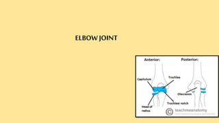
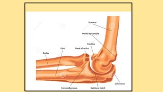
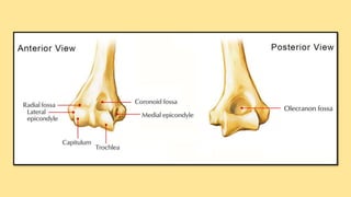
ELBOW JOINT
SURFACE ANATOMY OF ELBOW JOINT
Surface Anatomy: Elbow - Medial and Posterior aspect https://www.youtube.com/watch?v=QLFKWpqyYyk
INTRODUCTION
THE ELBOW JOINT is a synovial joint
found in the upper limb between the arm
and the forearm.
It is the point of articulation of 03 bones:
the humerus of the arm and the radius
and the ulna of the forearm
The elbow joint is classified structurally as a
SYNOVIAL JOINT. It is also classified
structurally as a compound joint, as there are
two articulations in the joint. Synovial joints, also
called diarthroses, are free movable joints. The
articular surfaces of the bones at these joints are
separated from each other by a layer of hyaline
cartilage. Smooth movement at these joints is
provided by a highly viscous synovial fluid, which
acts as a lubricant.
A fibrous capsule encloses the joint, and is lined
internally by a synovial membrane.
Synovial joints can be further categorized based on
function. The elbow joint is functionally a hinge
joint, allowing movement in only one plane
(uniaxial).
SYNOVIAL JOINT
COMPOUND JOINT
DIARTHROSES
HINGE JOINT
UNIAXIAL
Osteology
There are three bones that comprise the elbow joint:
1. The humerus
2. The radius
3. The ulna.
These bones give rise to 02 joints:
HUMEROULNAR JOINT is the joint between the
pulley like trochlea on the medial aspect of the distal
of the humerus and the trochlear notch on the
ulna.
Humeroradial joint is the joint between the
capitulum on the lateral aspect of the distal end of the
humerus with the head of the radius.
The humeroulnar and the humeroradial joints are the
joints that give the elbow its characteristic hinge like
The PROXIMAL RADIOULNAR JOINT is the articulation
between the circumferential head of the radius and a fibro-
osseous ring formed by the radial groove of the ulna and the
annular ligament that hold the head of the radius in this groove.
The proximal radioulnar joint is functionally a PIVOT JOINT,
allowing a rotational movement of the radius on the ulna.
Ligaments
The joint capsule of the elbow is strengthened by ligaments medially and
laterally.
The RADIAL COLLATERAL LIGAMENT is found on the lateral side of
the joint, extending from the lateral epicondyle, and blending with the
annular ligament of the radius (a ligament from the proximal radioulnar
primary restraint to varus and external stress during full arc of elbow
motion
The ULNAR COLLATERAL LIGAMENT originates from the medial
epicondyle, and attaches to the coronoid process and olecranon of the ulna.
MCL provides resistance to valgus and distractive stresses
BURSAE RELATED TO THE ELBOW JOINT
Subtendinous olecranon bursa lies between triceps tendon
and upper surface of the olecranon process.
Subcutaneous olecranon bursa is comparatively a larger
bursa that lies between skin and subcutaneous triangular area on
the posterior surface of the olecranon.
Bicipitoradial bursa is a small bursa separating biceps tendon
from smooth anterior part of the radial tuberosity.
A small bursa separating the biceps tendon from the oblique
cord.
BLOOD SUPPLY OF THE ELBOW JOINT
The blood supply of elbow joints is by arterial
anastomosis around the elbow formed by the branches of
brachial, radial and ulnar arteries.
Movements of the Joint
The orientation of the bones forming the elbow joint produces a
hinge type synovial joint, which allows for extension and flexion of
the forearm:
Extension – triceps brachii and anconeus
Flexion – brachialis, biceps brachii, brachioradialis
Movements at this joint are called pronation and
supination.
These are rotational movements that occur when the distal
end of the radius moves over the distal end of the ulna by
rotating the radius in the pivot joint formed by the circular
head of the radius, the radial groove of the ulna and the
ligament.
OSSIFICATION CENTRES ABOUT THE ELBOW
http://www0.sun.ac.za/ortho/webct-ortho/age/critoe.html
3 View Standard Elbow Radiology Tutorial
https://www.youtube.com/watch?v=wWO8W2CEFwU
https://emedicine.medscape.com/article/389069-overview#a2
Carrying angle is a small degree of cubitus valgus, formed
between the axis of a radially deviated forearm and the axis of the
humerus. It helps the arms to swing without hitting the hips
while walking.
Normally it is 5-15degree away from the body or 165-175degree
towards the body.
A decreased carrying angle can result in the forearm pointing
towards the body, known as gunstock deformity or cubitus varus.
ELBOW JOINT ANATOMY https://www.youtube.com/watch?v=RlR6p3RHsjk
https://www.youtube.com/watch?v=ZUF7tpkVAIYThe Goniometer
Carrying Angle Of The Elbow https://www.youtube.com/watch?v=XIXeTZiCnRk
Anatomy Of The Elbow https://www.youtube.com/watch?v=VEg2rReyM6k
Elbow muscles (anatomy) https://www.youtube.com/watch?v=jKS1Tm8awUA&t=12s
https://www.youtube.com/watch?v=UpmsntJ5rngElbow Anatomy Biomechanics Pathomechanics Independent
Ligaments of the Elbow Stability Of The Elbow https://www.youtube.com/watch?v=KMvKqoPbXTI
https://www.slideshare.net/varuntandra/clinical-examination-
of-elbow-joint
https://www.youtube.com/watch?v=UpmsntJ5rng

Elbow joint anatomy

  • 1.
  • 3.
    SURFACE ANATOMY OFELBOW JOINT
  • 4.
    Surface Anatomy: Elbow- Medial and Posterior aspect https://www.youtube.com/watch?v=QLFKWpqyYyk
  • 5.
    INTRODUCTION THE ELBOW JOINTis a synovial joint found in the upper limb between the arm and the forearm. It is the point of articulation of 03 bones: the humerus of the arm and the radius and the ulna of the forearm
  • 6.
    The elbow jointis classified structurally as a SYNOVIAL JOINT. It is also classified structurally as a compound joint, as there are two articulations in the joint. Synovial joints, also called diarthroses, are free movable joints. The articular surfaces of the bones at these joints are separated from each other by a layer of hyaline cartilage. Smooth movement at these joints is provided by a highly viscous synovial fluid, which acts as a lubricant.
  • 7.
    A fibrous capsuleencloses the joint, and is lined internally by a synovial membrane. Synovial joints can be further categorized based on function. The elbow joint is functionally a hinge joint, allowing movement in only one plane (uniaxial).
  • 8.
  • 9.
    Osteology There are threebones that comprise the elbow joint: 1. The humerus 2. The radius 3. The ulna.
  • 10.
    These bones giverise to 02 joints: HUMEROULNAR JOINT is the joint between the pulley like trochlea on the medial aspect of the distal of the humerus and the trochlear notch on the ulna. Humeroradial joint is the joint between the capitulum on the lateral aspect of the distal end of the humerus with the head of the radius. The humeroulnar and the humeroradial joints are the joints that give the elbow its characteristic hinge like
  • 13.
    The PROXIMAL RADIOULNARJOINT is the articulation between the circumferential head of the radius and a fibro- osseous ring formed by the radial groove of the ulna and the annular ligament that hold the head of the radius in this groove. The proximal radioulnar joint is functionally a PIVOT JOINT, allowing a rotational movement of the radius on the ulna.
  • 14.
    Ligaments The joint capsuleof the elbow is strengthened by ligaments medially and laterally. The RADIAL COLLATERAL LIGAMENT is found on the lateral side of the joint, extending from the lateral epicondyle, and blending with the annular ligament of the radius (a ligament from the proximal radioulnar primary restraint to varus and external stress during full arc of elbow motion The ULNAR COLLATERAL LIGAMENT originates from the medial epicondyle, and attaches to the coronoid process and olecranon of the ulna. MCL provides resistance to valgus and distractive stresses
  • 15.
    BURSAE RELATED TOTHE ELBOW JOINT Subtendinous olecranon bursa lies between triceps tendon and upper surface of the olecranon process. Subcutaneous olecranon bursa is comparatively a larger bursa that lies between skin and subcutaneous triangular area on the posterior surface of the olecranon. Bicipitoradial bursa is a small bursa separating biceps tendon from smooth anterior part of the radial tuberosity. A small bursa separating the biceps tendon from the oblique cord.
  • 16.
    BLOOD SUPPLY OFTHE ELBOW JOINT The blood supply of elbow joints is by arterial anastomosis around the elbow formed by the branches of brachial, radial and ulnar arteries.
  • 17.
    Movements of theJoint The orientation of the bones forming the elbow joint produces a hinge type synovial joint, which allows for extension and flexion of the forearm: Extension – triceps brachii and anconeus Flexion – brachialis, biceps brachii, brachioradialis
  • 18.
    Movements at thisjoint are called pronation and supination. These are rotational movements that occur when the distal end of the radius moves over the distal end of the ulna by rotating the radius in the pivot joint formed by the circular head of the radius, the radial groove of the ulna and the ligament.
  • 20.
    OSSIFICATION CENTRES ABOUTTHE ELBOW http://www0.sun.ac.za/ortho/webct-ortho/age/critoe.html
  • 21.
    3 View StandardElbow Radiology Tutorial https://www.youtube.com/watch?v=wWO8W2CEFwU
  • 25.
  • 26.
    Carrying angle isa small degree of cubitus valgus, formed between the axis of a radially deviated forearm and the axis of the humerus. It helps the arms to swing without hitting the hips while walking. Normally it is 5-15degree away from the body or 165-175degree towards the body. A decreased carrying angle can result in the forearm pointing towards the body, known as gunstock deformity or cubitus varus.
  • 28.
    ELBOW JOINT ANATOMYhttps://www.youtube.com/watch?v=RlR6p3RHsjk https://www.youtube.com/watch?v=ZUF7tpkVAIYThe Goniometer Carrying Angle Of The Elbow https://www.youtube.com/watch?v=XIXeTZiCnRk Anatomy Of The Elbow https://www.youtube.com/watch?v=VEg2rReyM6k Elbow muscles (anatomy) https://www.youtube.com/watch?v=jKS1Tm8awUA&t=12s
  • 29.
    https://www.youtube.com/watch?v=UpmsntJ5rngElbow Anatomy BiomechanicsPathomechanics Independent Ligaments of the Elbow Stability Of The Elbow https://www.youtube.com/watch?v=KMvKqoPbXTI https://www.slideshare.net/varuntandra/clinical-examination- of-elbow-joint https://www.youtube.com/watch?v=UpmsntJ5rng