1. Ojvensha E learning Resources-Prepared by Dr.B.B.Gosai
Spinal Cord
Introduction:
It is lower cylindrical part of central nervous system. It is continuation of medulla
oblongata and gradually tapers in the lower part.
Location: Vertebral canal.
Length: 45 cms.
Extent:
In Adult: From Foramen Magnum to lower border of L1 vertebra or upper border of L2
vertebra.
In Child: From Foramen Magnum to upper border of L3 vertebra
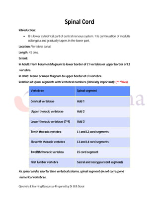
Relation of spinal segments with Vertebral numbers (Clinically important): (***Viva)
Vertebrae Spinal segment
Cervical vertebrae Add 1
Upper thoracic vertebrae Add 2
Lower thoracic vertebrae (7-9) Add 3
Tenth thoracic vertebra L1 and L2 cord segments
Eleventh thoracic vertebra L3 and L4 cord segments
Twelfth thoracic vertebra L5 cord segment
First lumbar vertebra Sacral and coccygeal cord segments
As spinal cord is shorter then vertebral column, spinal segment do not correspond
numerical vertebrae.
2. Ojvensha E learning Resources-Prepared by Dr.B.B.Gosai
(Draw this Diagram for the External Features and Meninges)
3. Ojvensha E learning Resources-Prepared by Dr.B.B.Gosai
(No need to draw this diagram- only for understanding)
Meningeal Coverings of Spinal Cord:
Spinal cord is covered by three layers of Meninges:
1. Dura matter: It is tough outer layer of meninges
2. Arachnoid matter: It is middle layer.
3. Pia matter: It is thin inner layer and closely attached to spinal cord.
4. Ojvensha E learning Resources-Prepared by Dr.B.B.Gosai
Subrachnoid space: Space between arachnoid matter and pia matter is known as subarachnoid
space and it contains Cerebrospinal fluid (CSF). Subarachnoid space extends up to second sacral
vertebra.
Filum Terminale: Cord like structure formed by meninges. Length: 20 cms
Filum Terminale Internum: Continuation of pia matter after termination of spinal cord in
the subarachnoid space. Length: 15 cms
Filum Terminale Externum: Formed by continuation of all meninges together after
second sacral vertebra till first coccygeal vertebra. Length : 5 cms.
Applied Anatomy of Subarachnoid space:
Lumbar Puncture: (*****Important - Viva)
Lumbar puncture is a procedure to tap (withdraw) the cerebrospinal fluid from the
subarachnoid space for diagnostic or therapeutic purpose.
1) Site of puncture: Just above or below fourth lumbar spine.
Explanation: As spinal cord end at L1 and subarachnoid space extend up to second
sacral vertebra, fluid can be tapped without damage to spinal cord.
2) Importance:
Microscopic and bacteriological examination.
Inject drugs for treatment of infection.
Spinal anesthesia for lower abdominal or lower limb surgery.
By attaching manometer-CSF pressure can be measured.
Normal CSF pressure: 60-150 mm of water.
Queckenstedt’s sign: In blockage of subarachnoid space, CSF pressure does not rise
following compression of Internal jugular veins.
5. Ojvensha E learning Resources-Prepared by Dr.B.B.Gosai
(No need to draw this diagram- only for understanding)
(No need to draw this diagram- only for understanding)
6. Ojvensha E learning Resources-Prepared by Dr.B.B.Gosai
External Features of Spinal Cord:
1. Anterior median fissure: Longitudinal fissure contains Anterior spinal artery.
2. Anterolateral sulcus: located lateral to anterior medial fissure and ventral (Anterior)
nerve roots comes out from here.
3. Posterior median sulcus: Longitudinal sulcus posteriorly.
4. Posterolateral sulcus: located lateral to posterior median sulcus. Posterior spinal
arteries are located in it and dorsal (Posterior) Roots enter in spinal cord from here.
5. Conus medullaris: Lower tapering conical part of spinal cord.
6. Enlargement of Spinal cord: Cervical Enlargement (C3-T2) for brachial plexus neurons of
upper limb and Lumbosacral enlargement (L1-S3) for lumbosacral plexus for lower limb.
7. Cauda Equina: Horse tail like appearance formed by obliquely running nerve fibers from
conus medullaris.
(Draw this Diagram for the External Features and Meninges)
7. Ojvensha E learning Resources-Prepared by Dr.B.B.Gosai
(No need to draw this diagram- only for understanding)
Internal Features of Spinal Cord:
Internal Features is divided into:
Gray matter (Inner)
White matter (Outer)
Gray matter of Spinal Cord:
1. “H” shaped.
2. Anterior (Ventral) Horn: Wide, short and do not reach to surface of spinal cord. It is
motor give origin to ventral root. (Contain Medial, lateral and central Nuclei)
3. Posterior (Dorsal) Horn: Narrow, long and reach to surface of spinal cord. It is sensory
and receives dorsal root. (contain Substantia gelatinosa, Nucleus proprius, Clark’s
column)
4. Lateral Horn: Only present at T1 to L2 and S2,3,4 spinal segments. It is responsible for
origin of fibers of Autonomic nervous system.
5. Right and left side is connected by anterior and posterior gray commissure.
6. In the centre of Gray matter is present Central Canal containing Cerebrospinal fliud.
8. Ojvensha E learning Resources-Prepared by Dr.B.B.Gosai
White matter of Spinal Cord:
1. Outer to gray matter.
2. It is divided into three columns.
3. White matter contains ascending and descending tracts of spinal cord.
4. Anterior white column: Located between anterior median fissure and ventral root.
Ascending tracts present in Anterior white column:
a. Anterior spinothalamic tract: Crosses in spinal cord and carries touch and
pressure sensations.
b. Other tracts like spinotectal, spinoolivary, spinovestibular, spinoreticular tracts.
Descending tracts present in Anterior white column:
a. Anterior corticospinal tract: Motor tract (20 % of uncrossed fibers of pyramidal
tract) finally cross at spinal cord.
b. Other tracts like tectospinal, olivospinal, vestibulospinal, reticulospinal tracts.
5. Lateral white column: Located between Ventral root and dorsal root.
Ascending tracts present in Lateral white column:
a. Lateral spinothalamic tract: Crosses in spinal cord and carries pain and
temperature sensations.
b. Anterior and posterior spinocerebellear tracts: carries unconscious joint position
sensation (Kinesthetic) to cerebellum.
Descending tracts present in Anterior white column:
c. Lateral corticospinal tract: Motor tract (80 % of crossed fibers of pyramidal
tract).
d. Rubrospinal tract: motor tract from red nucleus of midbrain to spinal cord.
6. Posterior white column: Located between posterior median sulcus and dorsal root.
Ascending tracts present in Posterior white column:
a. Tractus Gracilis and Tractus Cuneatus (Tract of Gall and Burdach): Remain on
same side in spinal cord but Crosses in medulla oblongata and carries vibration,
tectile discrimination and conscious joint position sensations.
9. Ojvensha E learning Resources-Prepared by Dr.B.B.Gosai
(Draw this diagram- for ascending and descending tracts)
Blood supply of Spinal Cord:
1. Anterior spinal artery : Supply anterior two third of spinal cord.
2. Posterior spinal artery: Supply posterior one third of spinal cord.
3. Blood supply to spinal cord is reinforced by redinacular branches from aorta and largest
branch is known as Artery of Adamkewics
Applied Anatomy of Spinal Cord:
1. Spinal cord may be damaged due to fracture, dislocation of vertebra or by prolapsed
of intervertebral disc.
2. Injuries of spinal cord can lead to complete cord section, Hemisection of cord (Brown
Sequard syndrome), Posterior cord compression, central lesions (Syringomyelia).
3. Complete cord section leads to loss of all motor and sensory activities below the level of
lesion.
10. Ojvensha E learning Resources-Prepared by Dr.B.B.Gosai
4. Hemisection of Spinal cord (Brown Sequard syndrome):
a. On same side below the lesion:
i. Spastic paralysis
ii. Loss of vibration, joint position and tectile discrimination
b. On opposite side below the lesion:
i. Loss of pain, temperature, touch and pressure sensations.
(Draw this Diagram for the short note)
==================X================