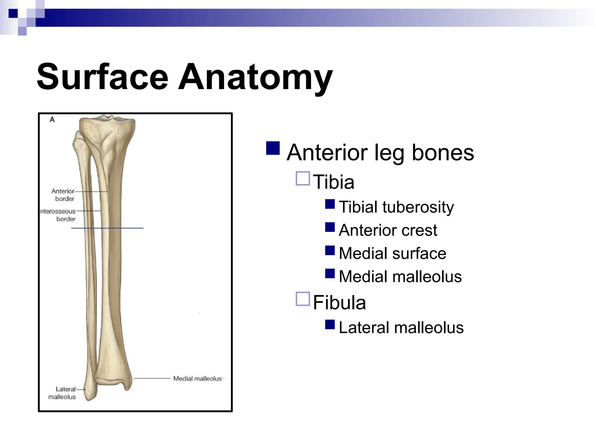
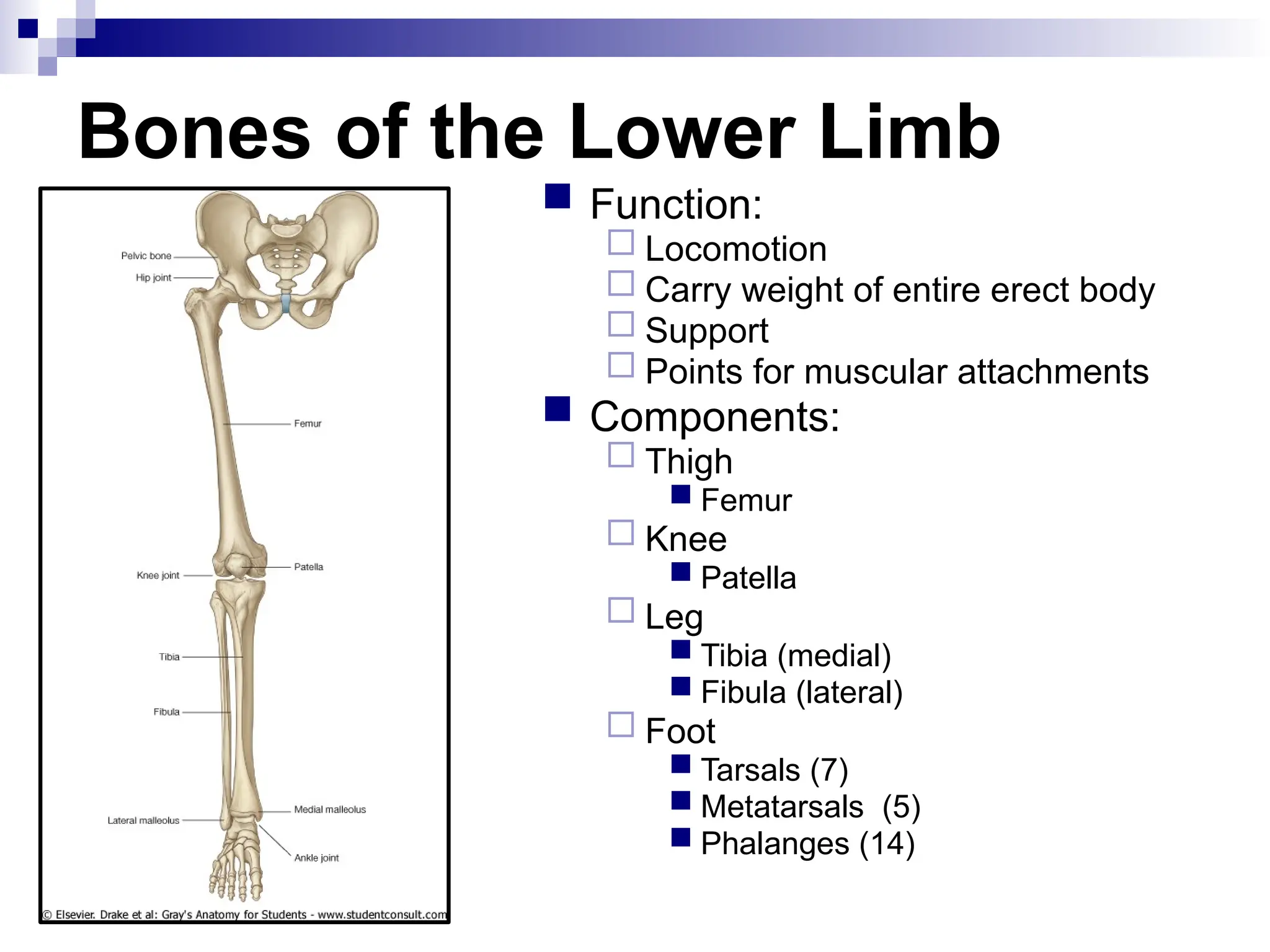
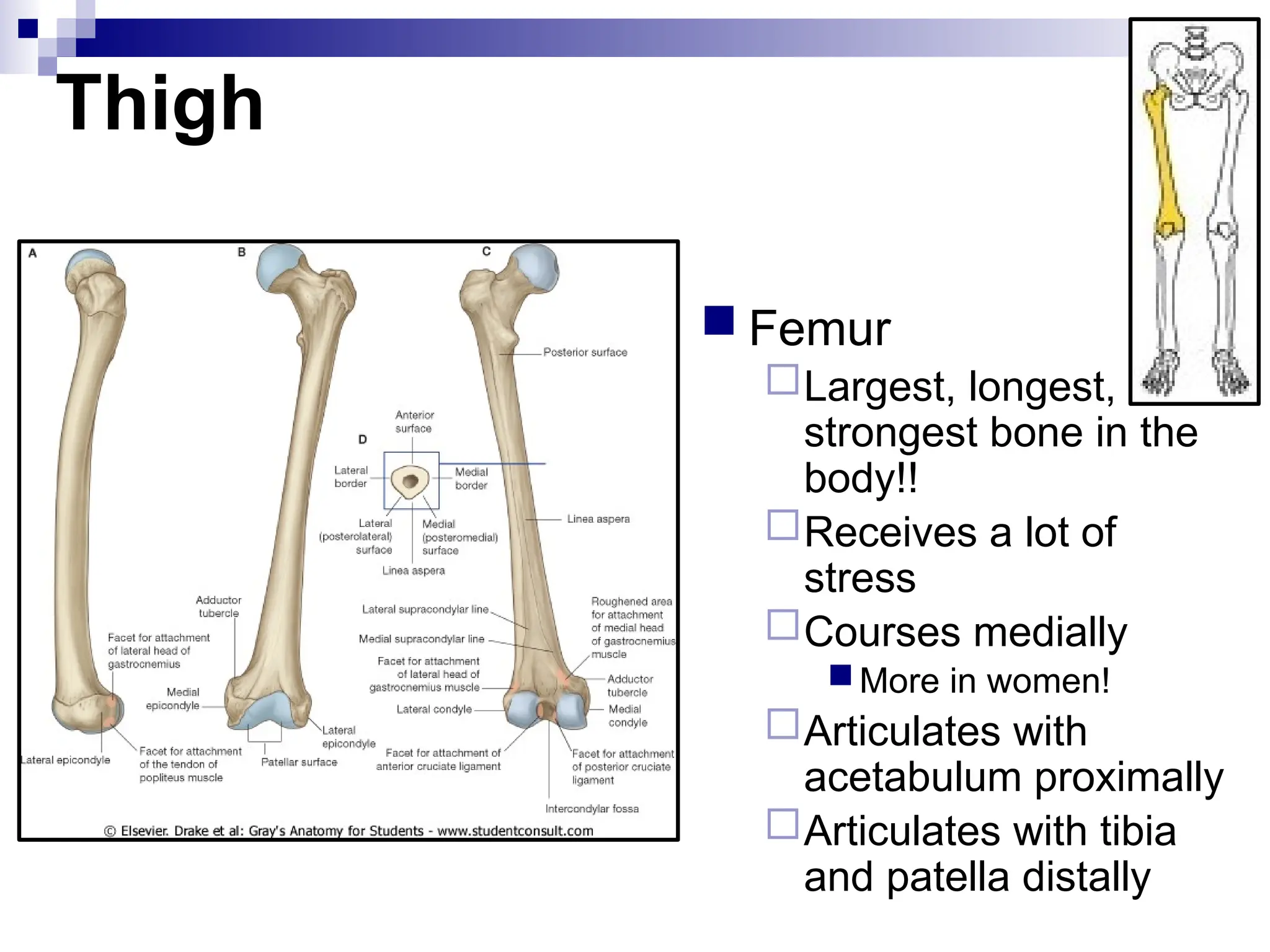
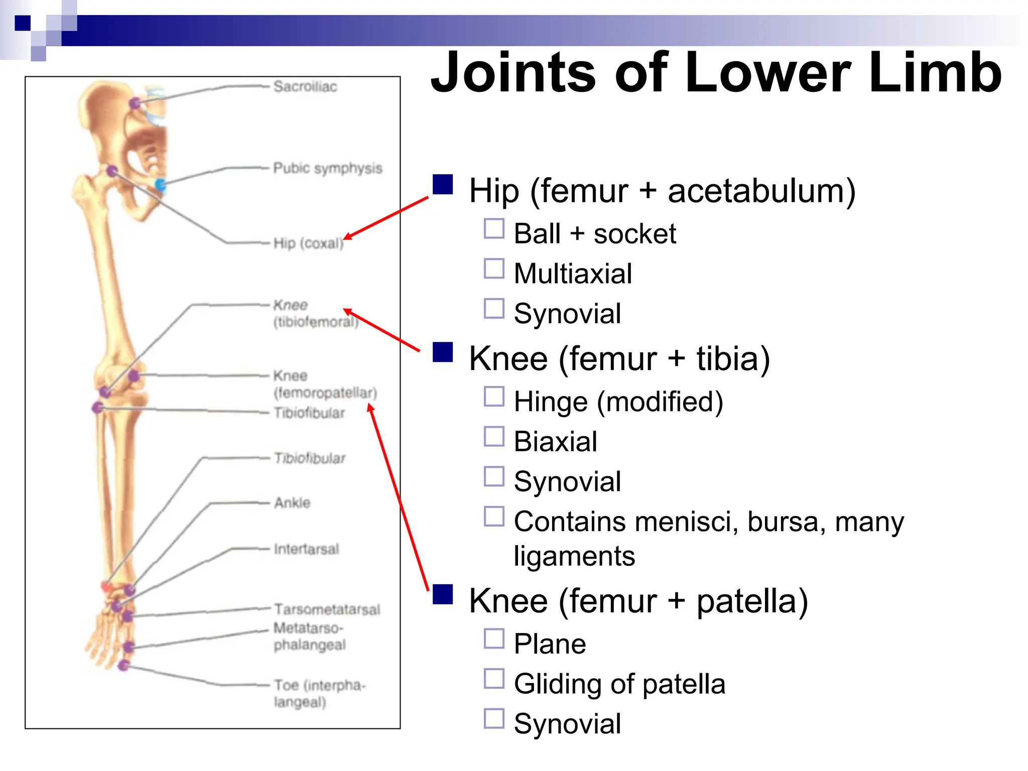
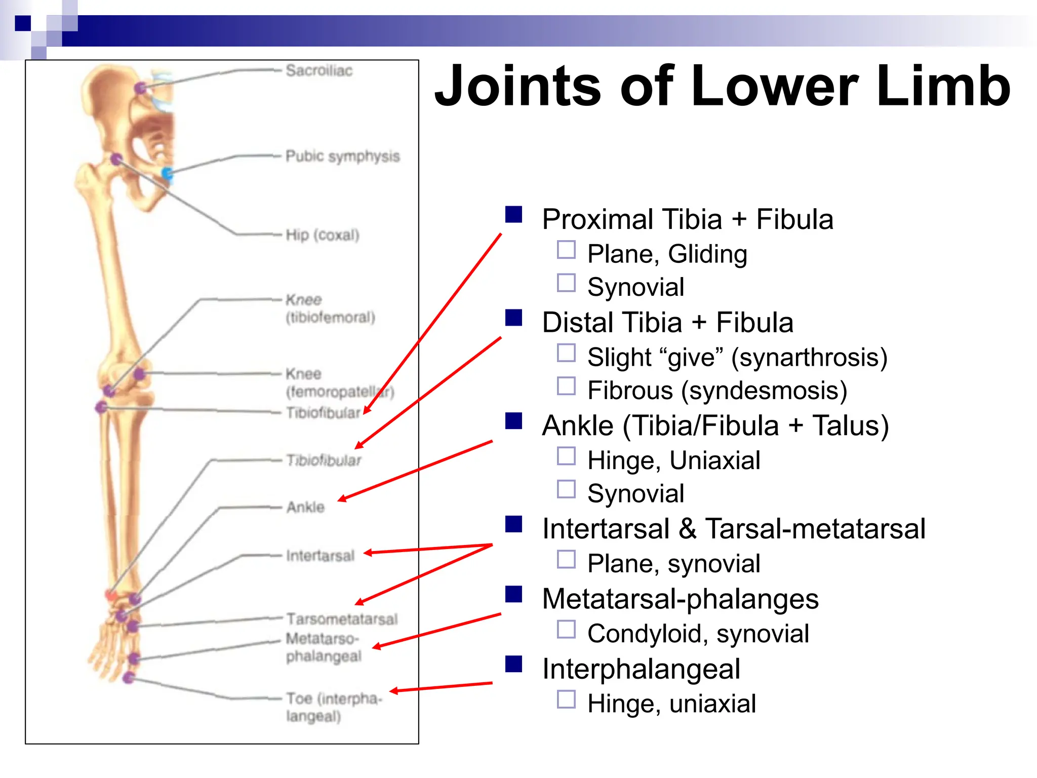
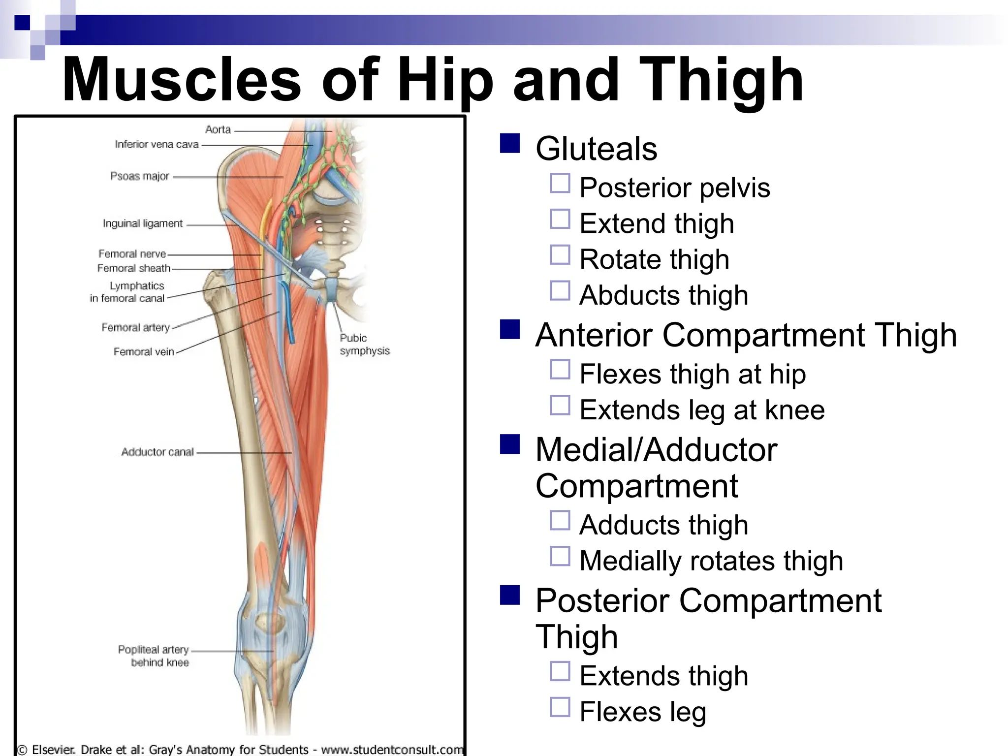
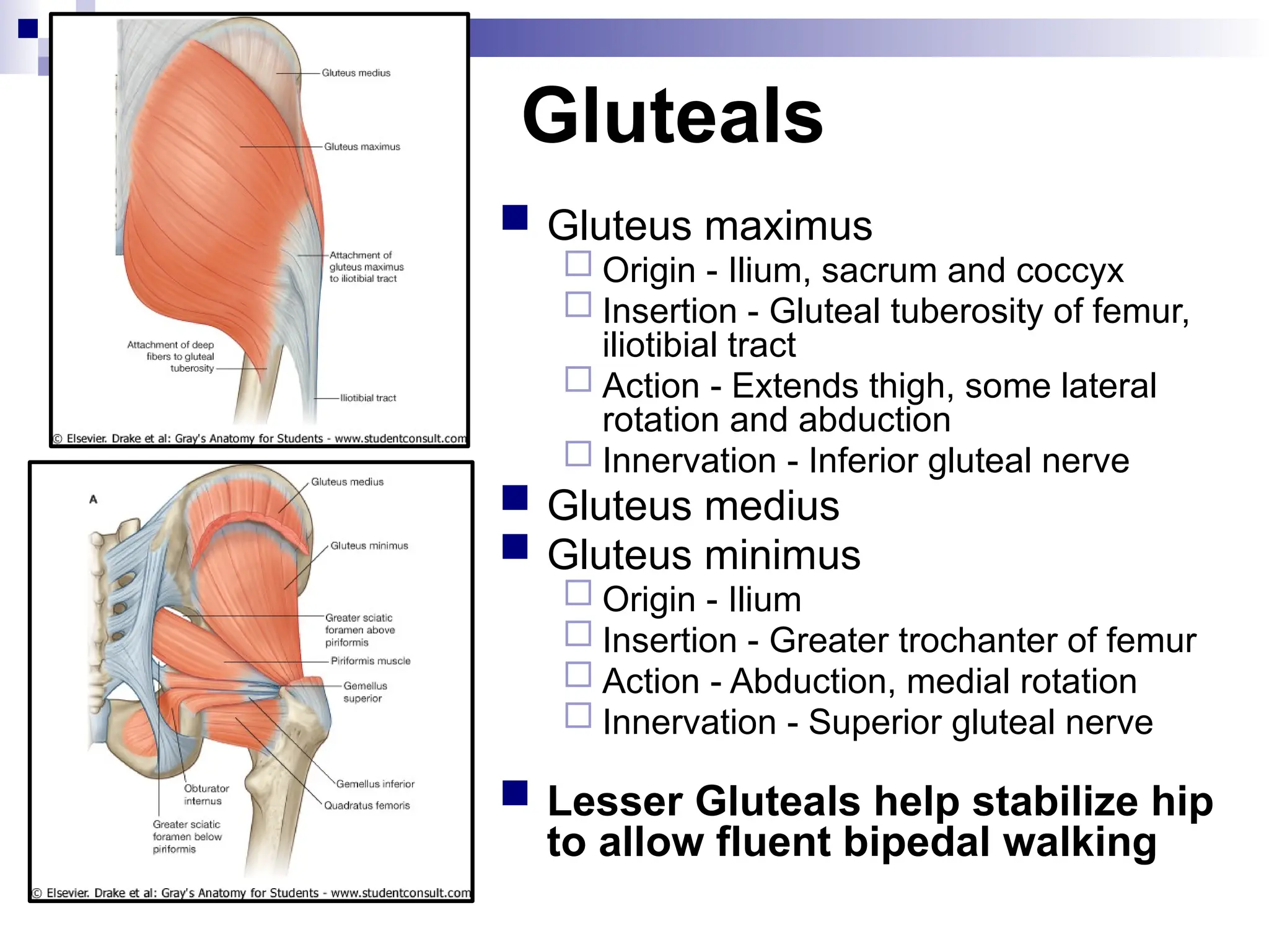
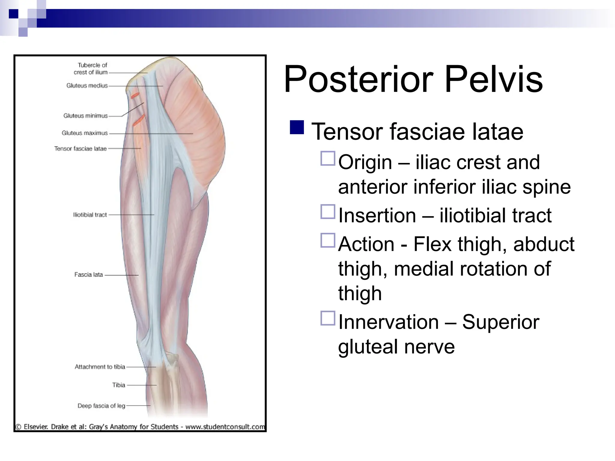
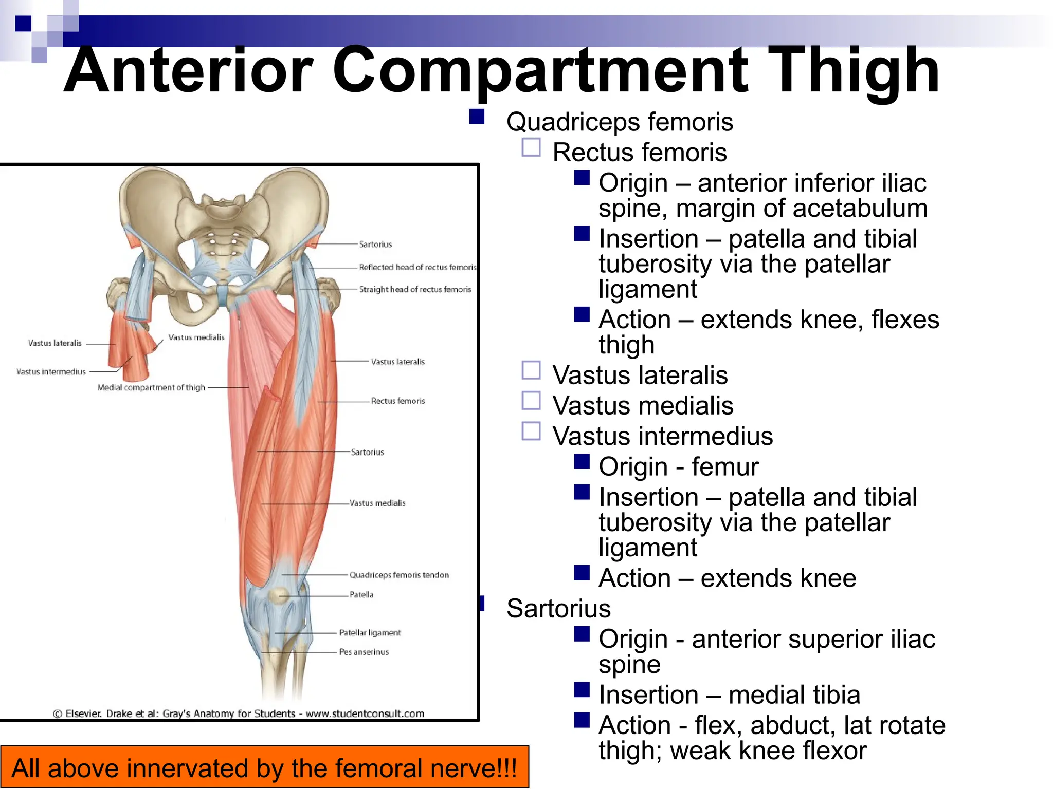
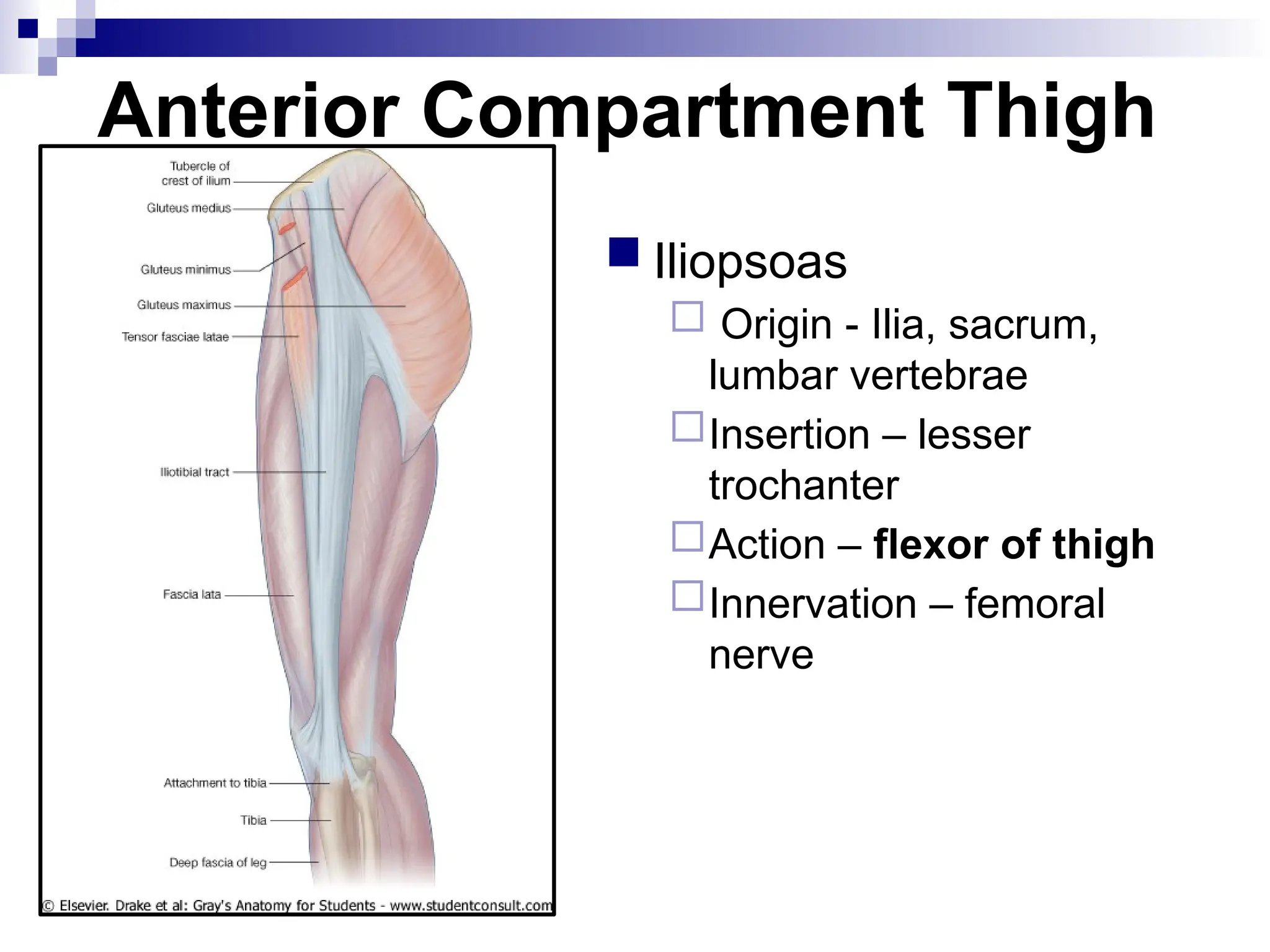
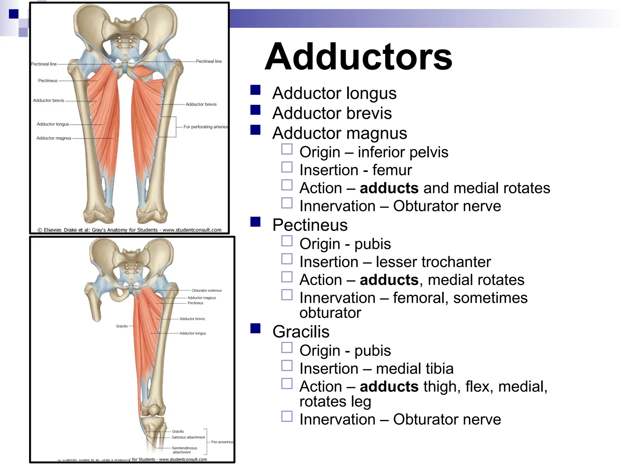
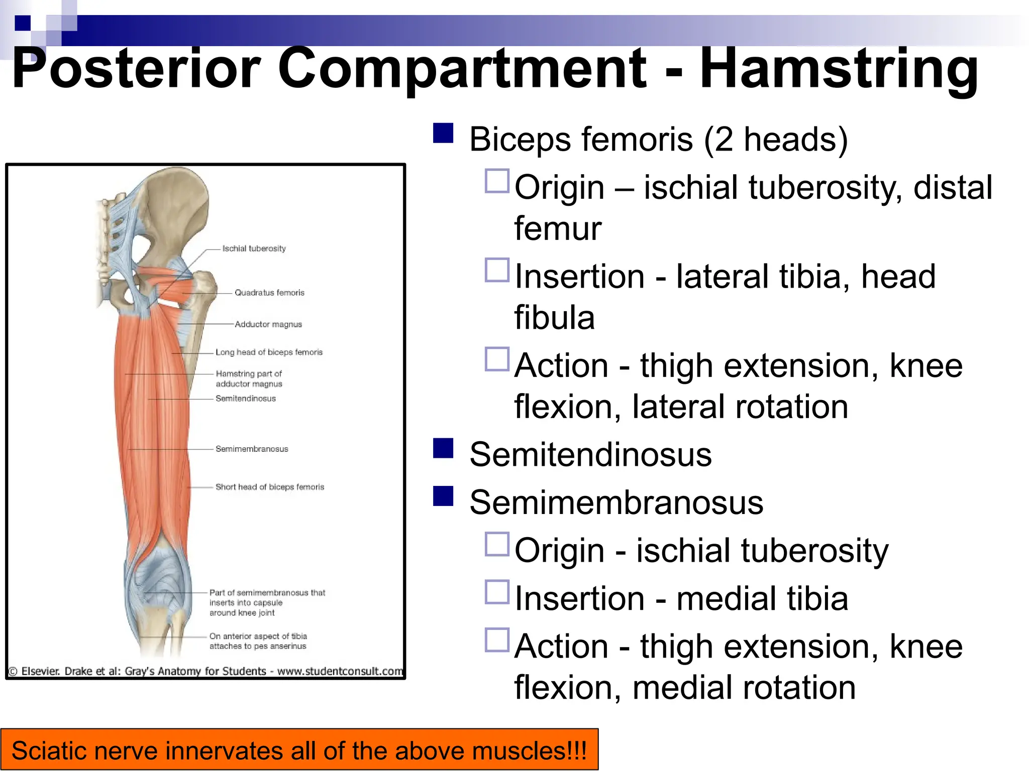
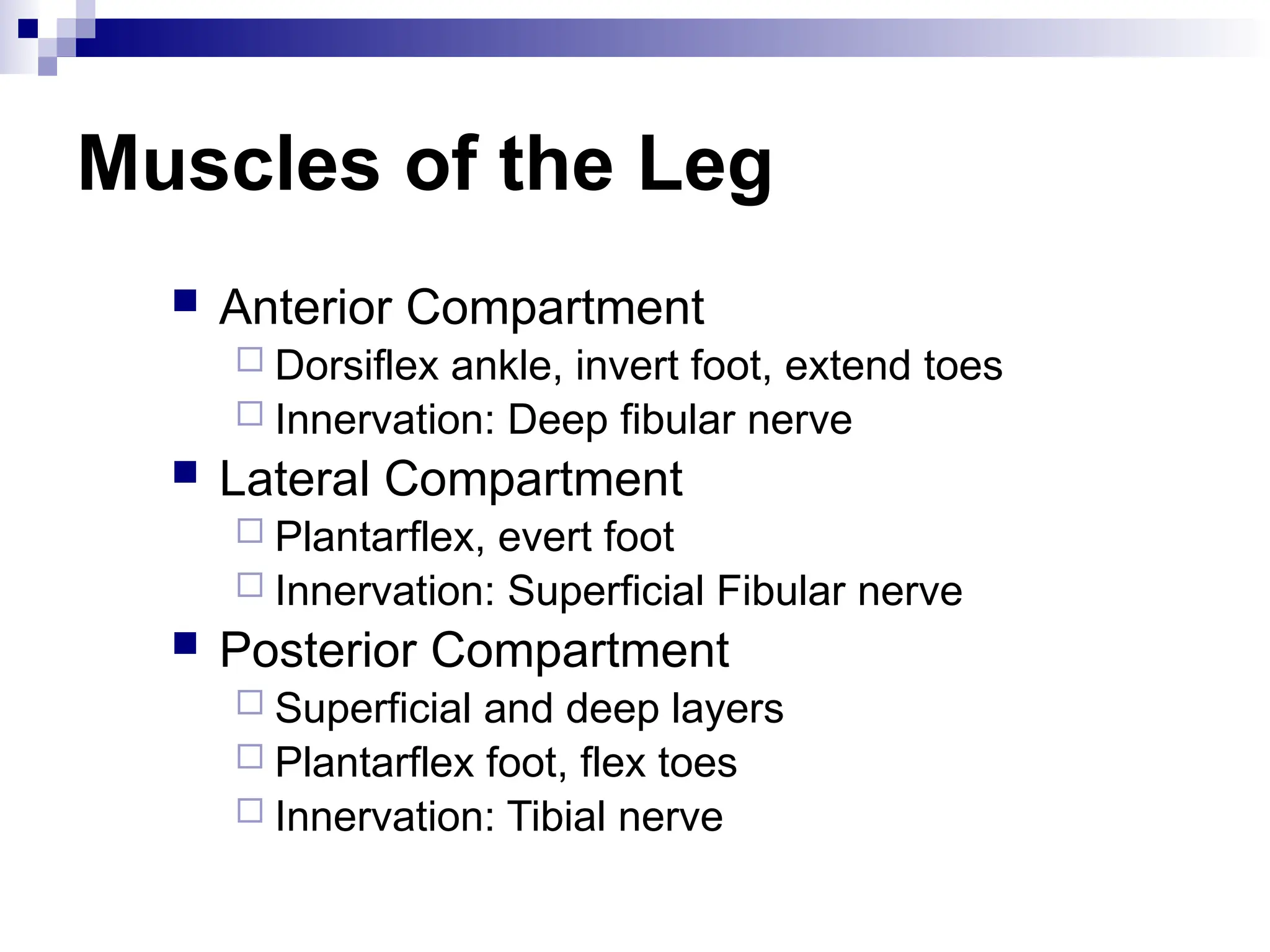
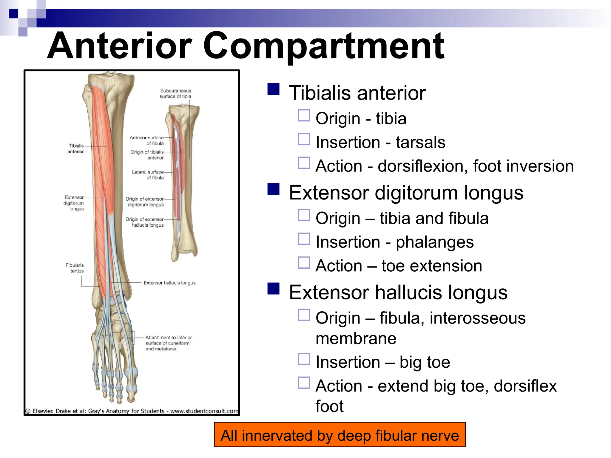
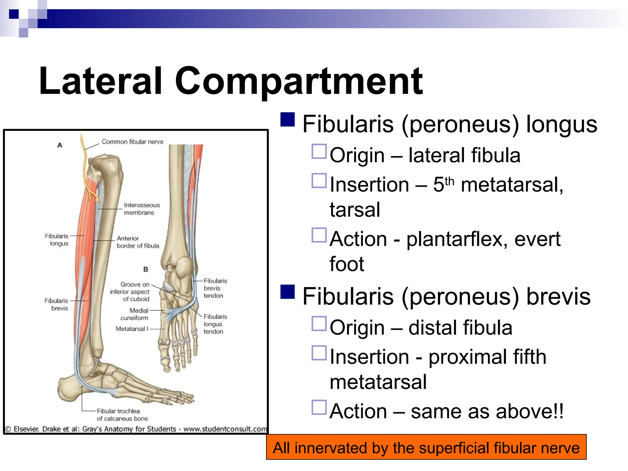
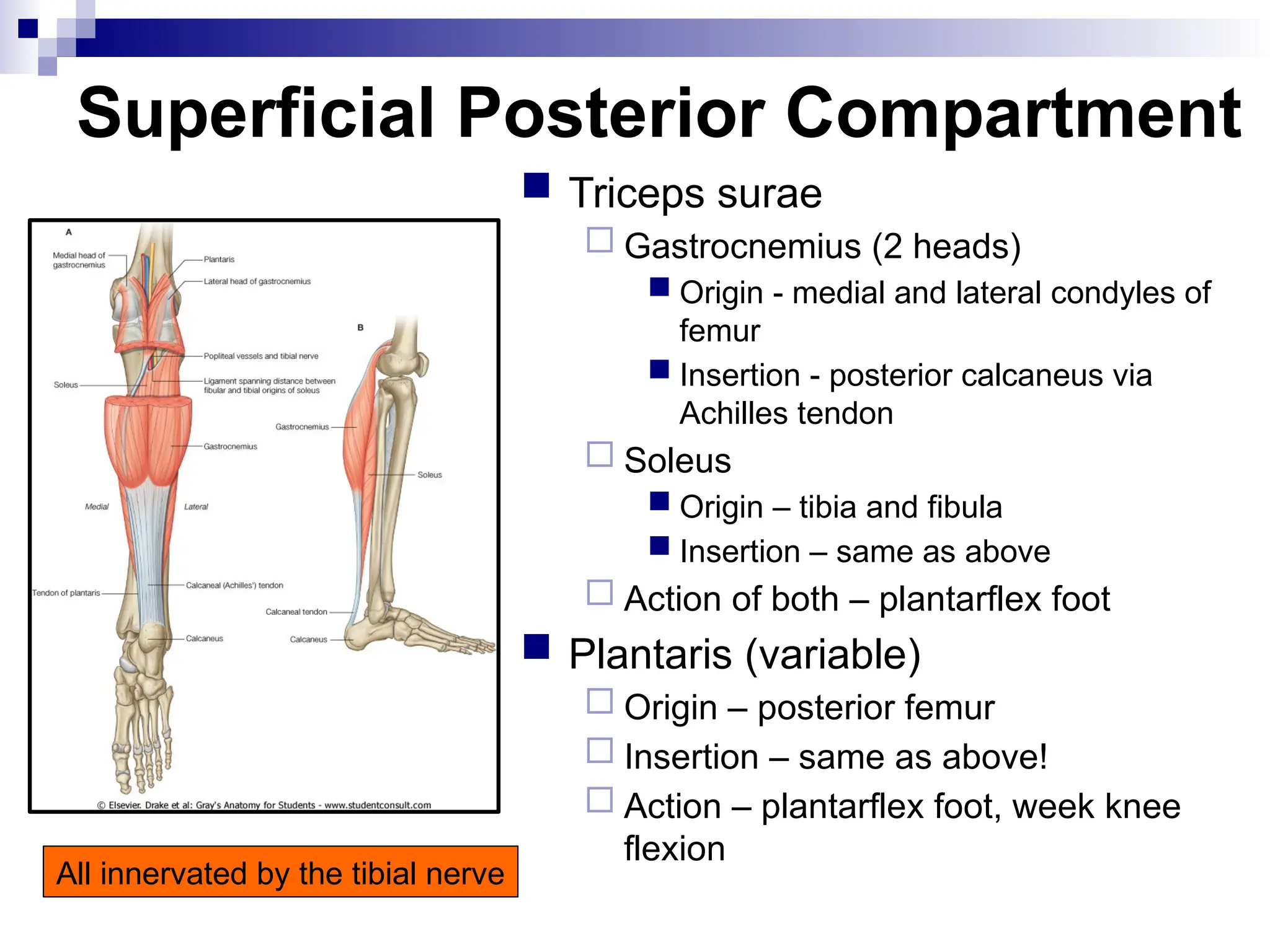
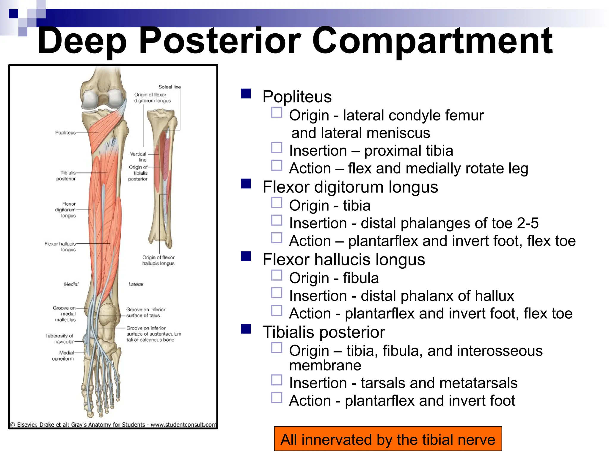
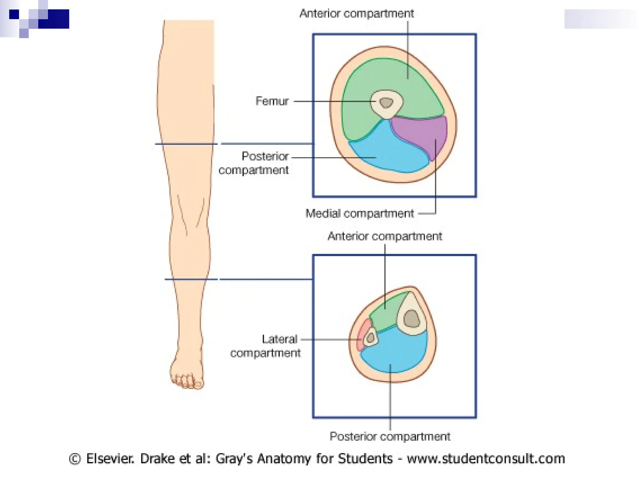
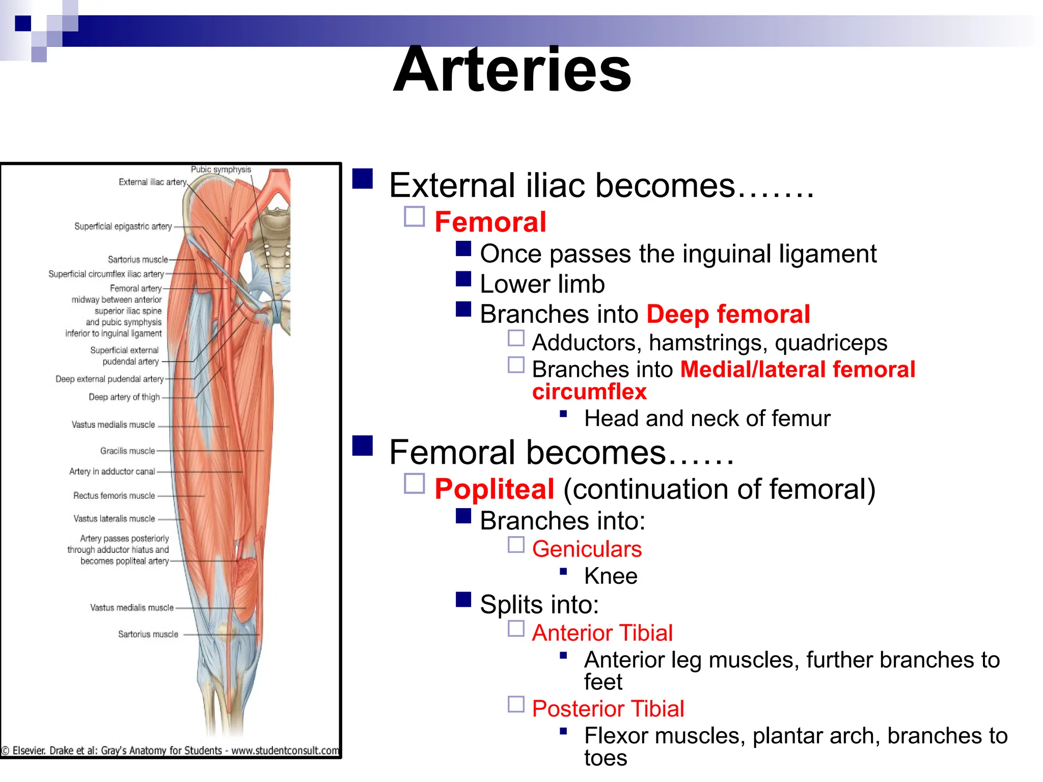
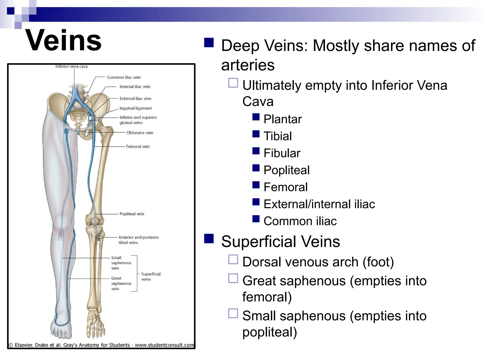
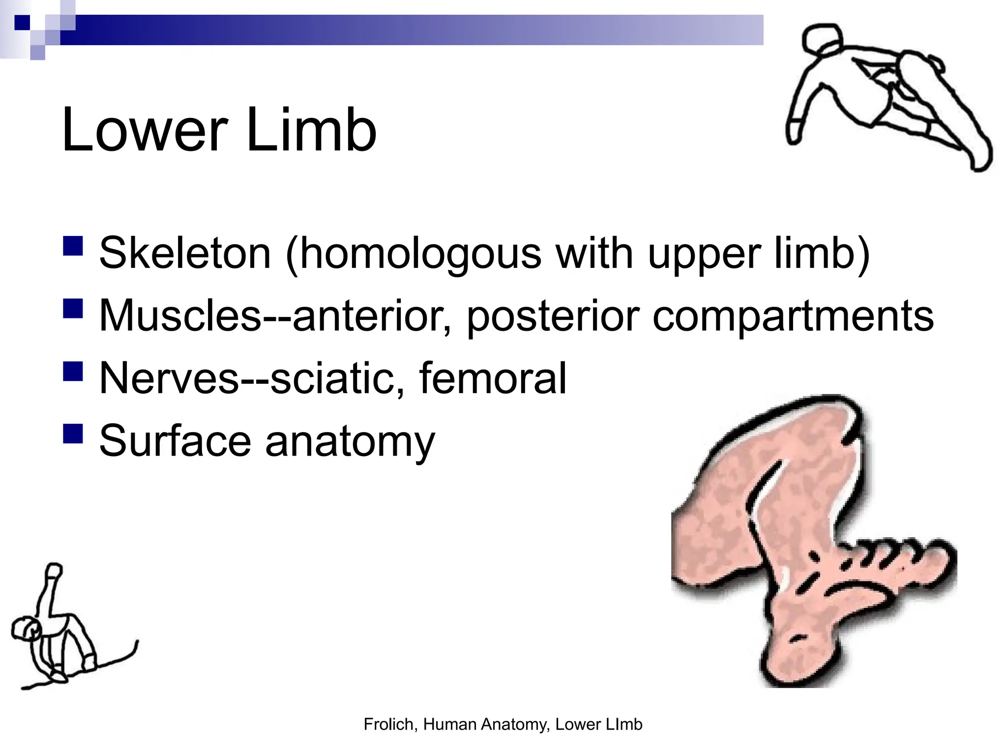
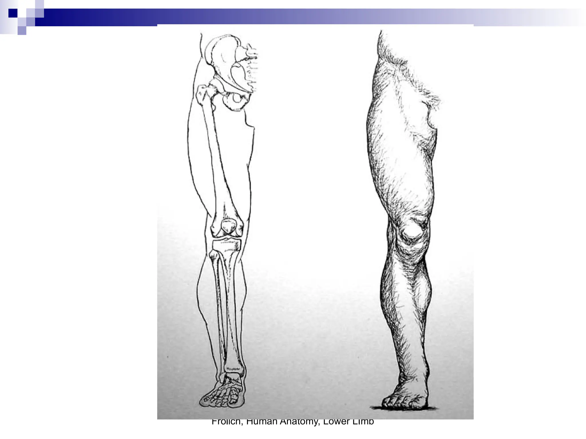
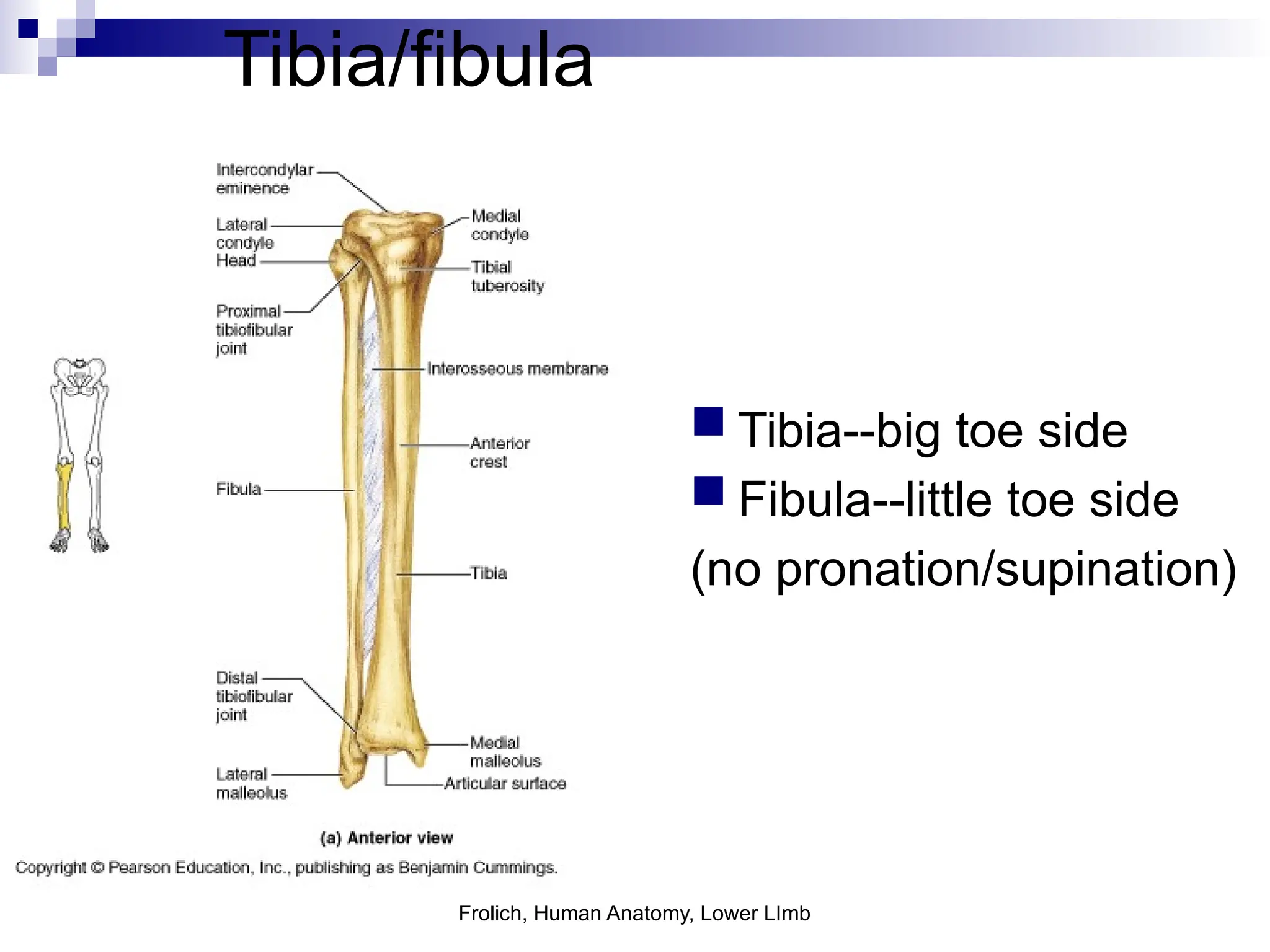
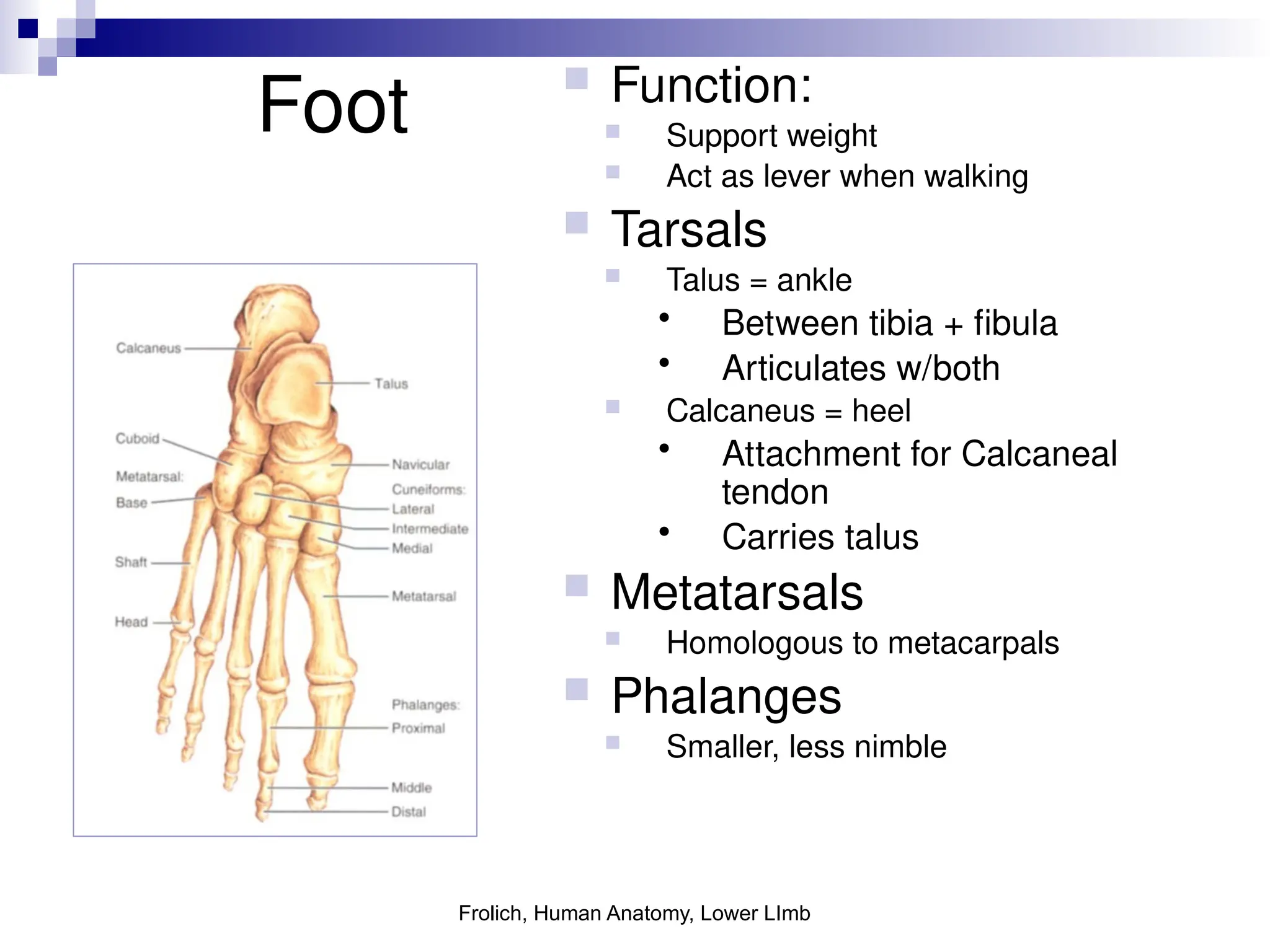
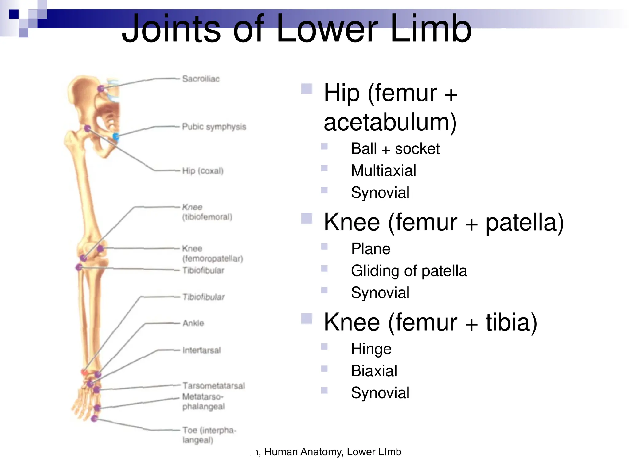
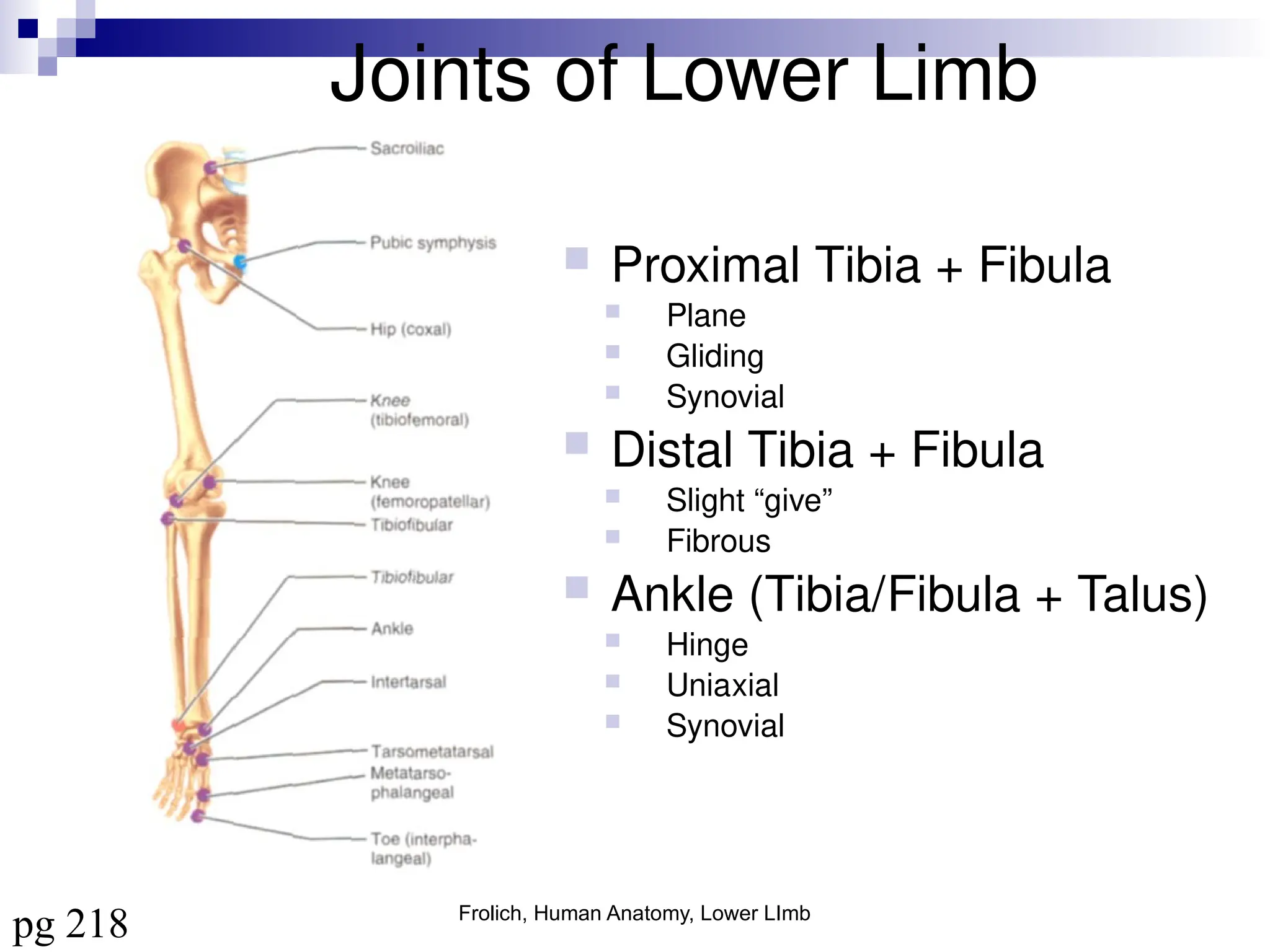
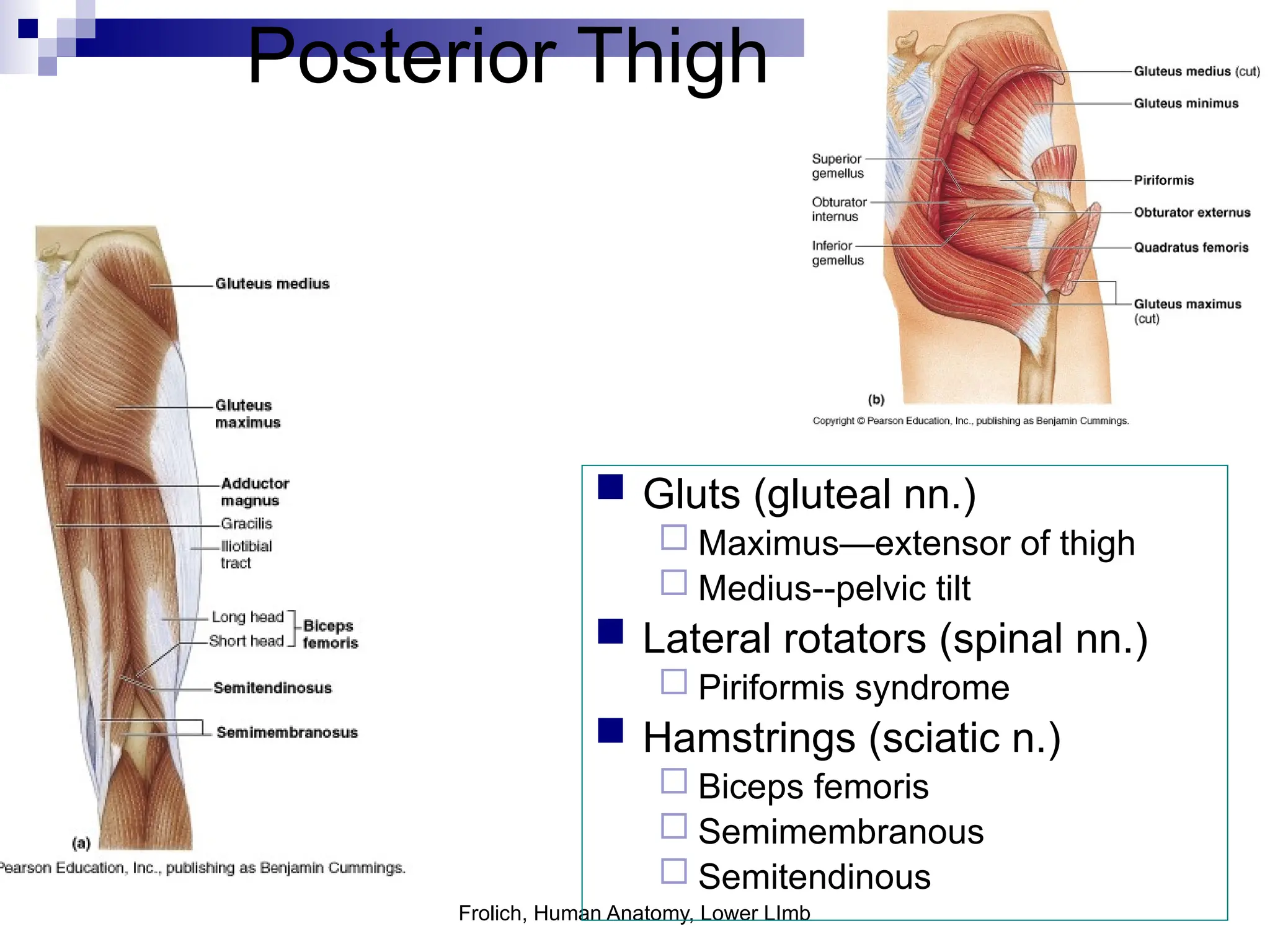
The Lower Limb
An Overview
Surface Anatomy
Surface Anatomy
 Gluteal region /
posterior pelvis
Iliac crest
Gluteus maximus
Cheeks
Natal/gluteal cleft
Vertical midline;
“Crack”
Gluteal folds
Bottom of cheek;
“prominence”
Surface Anatomy
 Anterior thigh and leg
Palpate
Patella
Condyles of femur
Femoral Triangle
Boundaries:
Sartorius (lateral)
Adductor longus (medial)
Inguinal ligament (superior)
Contents:
Femoral artery, vein and
nerve, lymph nodes
Surface Anatomy
 Posterior leg
Popliteal fossa
Diamond-shape fossa
behind knee
Boundaries
Biceps femoris
(superior-lateral)
Semitendinosis and
semimembranosis
(superior-medial)
Gastrocnemius heads
(inferior)
Contents
Popliteal artery and vein
Calcaneal (Achilles)
tendon
Surface Anatomy
 Anterior leg bones
Tibia
Tibial tuberosity
Anterior crest
Medial surface
Medial malleolus
Fibula
Lateral malleolus
Skeletal
Composition
Bones of the Lower Limb
 Function:
 Locomotion
 Carry weight of entire erect body
 Support
 Points for muscular attachments
 Components:
 Thigh
 Femur
 Knee
 Patella
 Leg
 Tibia (medial)
 Fibula (lateral)
 Foot
 Tarsals (7)
 Metatarsals (5)
 Phalanges (14)
Thigh
 Femur
Largest, longest,
strongest bone in the
body!!
Receives a lot of
stress
Courses medially
More in women!
Articulates with
acetabulum proximally
Articulates with tibia
and patella distally
Knee
 Patella
Triangular sesamoid
bone
Protects knee joint
Improves leverage of
thigh muscles acting
across the knee
Contained within
patellar ligament
Leg
 Tibia
 Receives the weight of body
from femur and transmits to foot
 Second to femur in size and
weight
 Articulates with fibula proximally
and distally
 Interosseous membrane
 Fibula
 Does NOT bear weight
 Muscle attachment
 Not part of knee joint
 Stabilize ankle joint
Foot
 Function:
 Supports the weight of the
body
 Act as a lever to propel the
body forward
 Parts:
 Tarsals
 Talus = ankle
 Between tibia and fibula
 Articulates with both
 Calcaneus = heel
 Attachment for Calcaneal
tendon
 Carries talus
 Navicular
 Cuboid
 Medial, lateral and
intermediate cuneiforms
 Metatarsals
 Phalanges
Foot
 3 arches
Medial
Lateral
Transverse
Has tendons that run
inferior to foot bones
Help support arches
of foot
 Function
Recoil after stepping
Longitudinal
Joints of Lower Limb
 Hip (femur + acetabulum)
 Ball + socket
 Multiaxial
 Synovial
 Knee (femur + tibia)
 Hinge (modified)
 Biaxial
 Synovial
 Contains menisci, bursa, many
ligaments
 Knee (femur + patella)
 Plane
 Gliding of patella
 Synovial
Joints of Lower Limb
 Proximal Tibia + Fibula
 Plane, Gliding
 Synovial
 Distal Tibia + Fibula
 Slight “give” (synarthrosis)
 Fibrous (syndesmosis)
 Ankle (Tibia/Fibula + Talus)
 Hinge, Uniaxial
 Synovial
 Intertarsal & Tarsal-metatarsal
 Plane, synovial
 Metatarsal-phalanges
 Condyloid, synovial
 Interphalangeal
 Hinge, uniaxial
Muscles
Muscles of Hip and Thigh
 Gluteals
 Posterior pelvis
 Extend thigh
 Rotate thigh
 Abducts thigh
 Anterior Compartment Thigh
 Flexes thigh at hip
 Extends leg at knee
 Medial/Adductor
Compartment
 Adducts thigh
 Medially rotates thigh
 Posterior Compartment
Thigh
 Extends thigh
 Flexes leg
Gluteals
 Gluteus maximus
 Origin - Ilium, sacrum and coccyx
 Insertion - Gluteal tuberosity of femur,
iliotibial tract
 Action - Extends thigh, some lateral
rotation and abduction
 Innervation - Inferior gluteal nerve
 Gluteus medius
 Gluteus minimus
 Origin - Ilium
 Insertion - Greater trochanter of femur
 Action - Abduction, medial rotation
 Innervation - Superior gluteal nerve
 Lesser Gluteals help stabilize hip
to allow fluent bipedal walking
Posterior Pelvis
 Tensor fasciae latae
Origin – iliac crest and
anterior inferior iliac spine
Insertion – iliotibial tract
Action - Flex thigh, abduct
thigh, medial rotation of
thigh
Innervation – Superior
gluteal nerve
Anterior Compartment Thigh
 Quadriceps femoris
 Rectus femoris
 Origin – anterior inferior iliac
spine, margin of acetabulum
 Insertion – patella and tibial
tuberosity via the patellar
ligament
 Action – extends knee, flexes
thigh
 Vastus lateralis
 Vastus medialis
 Vastus intermedius
 Origin - femur
 Insertion – patella and tibial
tuberosity via the patellar
ligament
 Action – extends knee
 Sartorius
 Origin - anterior superior iliac
spine
 Insertion – medial tibia
 Action - flex, abduct, lat rotate
thigh; weak knee flexor
All above innervated by the femoral nerve!!!
Anterior Compartment Thigh
 Iliopsoas
 Origin - Ilia, sacrum,
lumbar vertebrae
Insertion – lesser
trochanter
Action – flexor of thigh
Innervation – femoral
nerve
Adductors
 Adductor longus
 Adductor brevis
 Adductor magnus
 Origin – inferior pelvis
 Insertion - femur
 Action – adducts and medial rotates
 Innervation – Obturator nerve
 Pectineus
 Origin - pubis
 Insertion – lesser trochanter
 Action – adducts, medial rotates
 Innervation – femoral, sometimes
obturator
 Gracilis
 Origin - pubis
 Insertion – medial tibia
 Action – adducts thigh, flex, medial,
rotates leg
 Innervation – Obturator nerve
Posterior Compartment - Hamstring
 Biceps femoris (2 heads)
Origin – ischial tuberosity, distal
femur
Insertion - lateral tibia, head
fibula
Action - thigh extension, knee
flexion, lateral rotation
 Semitendinosus
 Semimembranosus
Origin - ischial tuberosity
Insertion - medial tibia
Action - thigh extension, knee
flexion, medial rotation
Sciatic nerve innervates all of the above muscles!!!
Muscles of the Leg
 Anterior Compartment
 Dorsiflex ankle, invert foot, extend toes
 Innervation: Deep fibular nerve
 Lateral Compartment
 Plantarflex, evert foot
 Innervation: Superficial Fibular nerve
 Posterior Compartment
 Superficial and deep layers
 Plantarflex foot, flex toes
 Innervation: Tibial nerve
Anterior Compartment
 Tibialis anterior
 Origin - tibia
 Insertion - tarsals
 Action - dorsiflexion, foot inversion
 Extensor digitorum longus
 Origin – tibia and fibula
 Insertion - phalanges
 Action – toe extension
 Extensor hallucis longus
 Origin – fibula, interosseous
membrane
 Insertion – big toe
 Action - extend big toe, dorsiflex
foot
All innervated by deep fibular nerve
Lateral Compartment
 Fibularis (peroneus) longus
Origin – lateral fibula
Insertion – 5th
metatarsal,
tarsal
Action - plantarflex, evert
foot
 Fibularis (peroneus) brevis
Origin – distal fibula
Insertion - proximal fifth
metatarsal
Action – same as above!!
All innervated by the superficial fibular nerve
Superficial Posterior Compartment
 Triceps surae
 Gastrocnemius (2 heads)
 Origin - medial and lateral condyles of
femur
 Insertion - posterior calcaneus via
Achilles tendon
 Soleus
 Origin – tibia and fibula
 Insertion – same as above
 Action of both – plantarflex foot
 Plantaris (variable)
 Origin – posterior femur
 Insertion – same as above!
 Action – plantarflex foot, week knee
flexion
All innervated by the tibial nerve
Deep Posterior Compartment
 Popliteus
 Origin - lateral condyle femur
and lateral meniscus
 Insertion – proximal tibia
 Action – flex and medially rotate leg
 Flexor digitorum longus
 Origin - tibia
 Insertion - distal phalanges of toe 2-5
 Action – plantarflex and invert foot, flex toe
 Flexor hallucis longus
 Origin - fibula
 Insertion - distal phalanx of hallux
 Action - plantarflex and invert foot, flex toe
 Tibialis posterior
 Origin – tibia, fibula, and interosseous
membrane
 Insertion - tarsals and metatarsals
 Action - plantarflex and invert foot
All innervated by the tibial nerve
Innervation
Plexuses of the Lower Limb
 “Lumbosacral plexus”
 Lumbar Plexus
 Arises from L1-L4
 Lies within the psoas major
muscle
 Mostly anterior structures
 Sacral Plexus
 Arises from spinal nerve L4-S4
 Lies caudal to the lumbar
plexus
 Mostly posterior structures
Lumbar Plexus
 Femoral nerve
 Cutaneous branches
 Thigh, leg, foot (e.g. saphenous nerve)
 Motor branches
 Anterior thigh muscles (e.g. quadriceps,
sartorius, iliopsoas)
 Obturator nerve
 Sensory
 Skin medial thigh; hip, knee joints
 Motor
 Adductor muscles
 Lateral femoral cutaneous
 Sensory
 Skin lateral thigh
 Genitofemoral
 Sensory
 Skin scrotum, labia major, anterior thigh
 Motor
 Cremaster muscle
Sacral Plexus
 Sciatic
 Motor:
 Hamstring
 Branches into:
 Tibial nerve
 Cutaneous
 Posterior leg and sole of foot
 Motor
 Posterior leg, foot
 Common fibular (peroneal) nerve
 Cutaneous
 Anterior and lateral leg, dorsum foot
 Motor
 Lateral compartment, tibialis anterior,
toe extensors
 Superior gluteal nerve
 Motor
 Gluteus medius and minimus, tensor
fasciae latae
Sacral Plexus (continued)
 Inferior gluteal nerve
Motor
Gluteus maximus
 Posterior femoral
cutaneous nerve
Sensory
Inferior buttocks, posterior
thigh, popliteal fossa
 Pudendal nerve
Sensory
External genitalia, anus
Motor
Muscles of perineum
Vasculature
Arteries
 Common iliac (from
aorta) branches into:
Internal iliac
Supplies pelvic organs
External iliac
Supplies lower limb
Arteries
 Internal iliac branches into:
Cranial and Caudal Gluteals
(Superior and Inferior)
Gluteals
Internal Pudendal
Perineum, external genitalia
Obturator
Adductor muscles
Other branches supply rectum,
bladder, uterus, vagina, male
reproductive glands
Arteries
 External iliac becomes…….
 Femoral
 Once passes the inguinal ligament
 Lower limb
 Branches into Deep femoral
 Adductors, hamstrings, quadriceps
 Branches into Medial/lateral femoral
circumflex
 Head and neck of femur
 Femoral becomes……
 Popliteal (continuation of femoral)
 Branches into:
 Geniculars
 Knee
 Splits into:
 Anterior Tibial
 Anterior leg muscles, further branches to
feet
 Posterior Tibial
 Flexor muscles, plantar arch, branches to
toes
Veins  Deep Veins: Mostly share names of
arteries
 Ultimately empty into Inferior Vena
Cava
 Plantar
 Tibial
 Fibular
 Popliteal
 Femoral
 External/internal iliac
 Common iliac
 Superficial Veins
 Dorsal venous arch (foot)
 Great saphenous (empties into
femoral)
 Small saphenous (empties into
popliteal)
Frolich, Human Anatomy, Lower LImb
Lower Limb
 Skeleton (homologous with upper limb)
 Muscles--anterior, posterior compartments
 Nerves--sciatic, femoral
 Surface anatomy
Frolich, Human Anatomy, Lower LImb
Tibia/fibula
 Tibia--big toe side
 Fibula--little toe side
(no pronation/supination)
Frolich, Human Anatomy, Lower LImb
Ankle
 Tarsus--forms ankle
joint
 Calcaneus--forms
heel
Frolich, Human Anatomy, Lower LImb
Foot
 Function:
 Support weight
 Act as lever when walking
 Tarsals
 Talus = ankle
• Between tibia + fibula
• Articulates w/both
 Calcaneus = heel
• Attachment for Calcaneal
tendon
• Carries talus
 Metatarsals
 Homologous to metacarpals
 Phalanges
 Smaller, less nimble
Frolich, Human Anatomy, Lower LImb
Joints of Lower Limb
 Hip (femur +
acetabulum)
 Ball + socket
 Multiaxial
 Synovial
 Knee (femur + patella)
 Plane
 Gliding of patella
 Synovial
 Knee (femur + tibia)
 Hinge
 Biaxial
 Synovial
Frolich, Human Anatomy, Lower LImb
Joints of Lower Limb
 Proximal Tibia + Fibula
 Plane
 Gliding
 Synovial
 Distal Tibia + Fibula
 Slight “give”
 Fibrous
 Ankle (Tibia/Fibula + Talus)
 Hinge
 Uniaxial
 Synovial
pg 218
Frolich, Human Anatomy, Lower LImb
Lower Limb Movements
 Hip
Flexion/extension
Abduction/adduction
Lateral/medial rotation
 Knee
Flexion/extension
 Ankle
Dorsiflexion/plantarflexion
Inversion/eversion
 Toes
Flexion/extension
 Bending on posterior side
is flexion (except hip)
 Bending on anterior sided
is extension (except hip)
Frolich, Human Anatomy,
Lower LImb
Anterior/Posterior compartments
ANTERIOR
COMPARTMENT
POSTERIOR
COMPARTMENT
MOVEMENT Extension Flexion
MUSCLES Quads
Shin
Hamstrings
Gastrocs
NERVES Femoral n.
(lumbar plexus)
Sciatic n.
(sacral plexus)
Frolich, Human Anatomy, Lower LImb
Thigh movements by
compartment
Frolich, Human Anatomy, Lower LImb
Posterior Thigh
 Gluts (gluteal nn.)
 Maximus—extensor of thigh
 Medius--pelvic tilt
 Lateral rotators (spinal nn.)
 Piriformis syndrome
 Hamstrings (sciatic n.)
 Biceps femoris
 Semimembranous
 Semitendinous
Frolich, Human Anatomy,
Lower LImb
Anterior thigh (femoral n.)
 Sartorius (Tailor’s
muscle)
 Quads (four)
 Rectus femoris
(crosses hip)
 3 vastus mm. (vast--
big)
Frolich, Human Anatomy, Lower LImb
Medial compartment (obturator n.)
 Adductor muscles
Gracilis
Adductor
Magnus
Longus
brevis
Frolich, Human Anatomy, Lower LImb
Leg movements by
compartment (in leg all nn are branches of sciatic)
Frolich, Human Anatomy,
Lower LImb
Anterior Leg (deep fibular n.)
Frolich, Human Anatomy,
Lower LImb
Lateral Leg (superficial fibular n.)
 Fibularis
brevis/longus
Frolich, Human Anatomy, Lower LImb
Posterior Leg (tibial n.)
Frolich, Human Anatomy, Lower LImb
Intrinsics of foot
Frolich, Human Anatomy, Lower LImb
Dermatomes show twisting of
leg in development
Frolich, Human Anatomy, Lower LImb
supply to
lower limb
Frolich, Human Anatomy, Lower LImb
Surface Anatomy:
Posterior Pelvis
 Iliac crest
 Gluteus maximus = cheeks
 Natal/gluteal cleft = crack
 Gluteal folds = bottom of cheek
pg 789
Frolich, Human Anatomy, Lower LImb
Surface Anatomy:
Anterior Thigh + Leg
 Palpate
Patella
Condyles of femur
 Femoral Triangle
Sartorius (lateral)
Adductor longus (medial)
Inguinal ligament
(superior)
Femoral a + v, lymph
nodes
pg 785
pg 792
Frolich, Human Anatomy, Lower LImb
Surface Anatomy: Posterior Leg
 Popliteal fossa
Diamond-shape fossa
behind knee
 Boundaries
Biceps femoris (sup-lat)
Semitendinosis +
semimembranosis (sup-
med)
Gastrocnemius heads (inf)
 Contents
Popliteal a + v
 Calcaneal (Achilles)
tendon
pg 793

01Anatomy Overview of Lower Extremity.ppt A

  • 1.
  • 2.
  • 3.
    Surface Anatomy  Glutealregion / posterior pelvis Iliac crest Gluteus maximus Cheeks Natal/gluteal cleft Vertical midline; “Crack” Gluteal folds Bottom of cheek; “prominence”
  • 4.
    Surface Anatomy  Anteriorthigh and leg Palpate Patella Condyles of femur Femoral Triangle Boundaries: Sartorius (lateral) Adductor longus (medial) Inguinal ligament (superior) Contents: Femoral artery, vein and nerve, lymph nodes
  • 5.
    Surface Anatomy  Posteriorleg Popliteal fossa Diamond-shape fossa behind knee Boundaries Biceps femoris (superior-lateral) Semitendinosis and semimembranosis (superior-medial) Gastrocnemius heads (inferior) Contents Popliteal artery and vein Calcaneal (Achilles) tendon
  • 6.
    Surface Anatomy  Anteriorleg bones Tibia Tibial tuberosity Anterior crest Medial surface Medial malleolus Fibula Lateral malleolus
  • 7.
  • 8.
    Bones of theLower Limb  Function:  Locomotion  Carry weight of entire erect body  Support  Points for muscular attachments  Components:  Thigh  Femur  Knee  Patella  Leg  Tibia (medial)  Fibula (lateral)  Foot  Tarsals (7)  Metatarsals (5)  Phalanges (14)
  • 9.
    Thigh  Femur Largest, longest, strongestbone in the body!! Receives a lot of stress Courses medially More in women! Articulates with acetabulum proximally Articulates with tibia and patella distally
  • 10.
    Knee  Patella Triangular sesamoid bone Protectsknee joint Improves leverage of thigh muscles acting across the knee Contained within patellar ligament
  • 11.
    Leg  Tibia  Receivesthe weight of body from femur and transmits to foot  Second to femur in size and weight  Articulates with fibula proximally and distally  Interosseous membrane  Fibula  Does NOT bear weight  Muscle attachment  Not part of knee joint  Stabilize ankle joint
  • 12.
    Foot  Function:  Supportsthe weight of the body  Act as a lever to propel the body forward  Parts:  Tarsals  Talus = ankle  Between tibia and fibula  Articulates with both  Calcaneus = heel  Attachment for Calcaneal tendon  Carries talus  Navicular  Cuboid  Medial, lateral and intermediate cuneiforms  Metatarsals  Phalanges
  • 13.
    Foot  3 arches Medial Lateral Transverse Hastendons that run inferior to foot bones Help support arches of foot  Function Recoil after stepping Longitudinal
  • 14.
    Joints of LowerLimb  Hip (femur + acetabulum)  Ball + socket  Multiaxial  Synovial  Knee (femur + tibia)  Hinge (modified)  Biaxial  Synovial  Contains menisci, bursa, many ligaments  Knee (femur + patella)  Plane  Gliding of patella  Synovial
  • 15.
    Joints of LowerLimb  Proximal Tibia + Fibula  Plane, Gliding  Synovial  Distal Tibia + Fibula  Slight “give” (synarthrosis)  Fibrous (syndesmosis)  Ankle (Tibia/Fibula + Talus)  Hinge, Uniaxial  Synovial  Intertarsal & Tarsal-metatarsal  Plane, synovial  Metatarsal-phalanges  Condyloid, synovial  Interphalangeal  Hinge, uniaxial
  • 16.
  • 17.
    Muscles of Hipand Thigh  Gluteals  Posterior pelvis  Extend thigh  Rotate thigh  Abducts thigh  Anterior Compartment Thigh  Flexes thigh at hip  Extends leg at knee  Medial/Adductor Compartment  Adducts thigh  Medially rotates thigh  Posterior Compartment Thigh  Extends thigh  Flexes leg
  • 18.
    Gluteals  Gluteus maximus Origin - Ilium, sacrum and coccyx  Insertion - Gluteal tuberosity of femur, iliotibial tract  Action - Extends thigh, some lateral rotation and abduction  Innervation - Inferior gluteal nerve  Gluteus medius  Gluteus minimus  Origin - Ilium  Insertion - Greater trochanter of femur  Action - Abduction, medial rotation  Innervation - Superior gluteal nerve  Lesser Gluteals help stabilize hip to allow fluent bipedal walking
  • 19.
    Posterior Pelvis  Tensorfasciae latae Origin – iliac crest and anterior inferior iliac spine Insertion – iliotibial tract Action - Flex thigh, abduct thigh, medial rotation of thigh Innervation – Superior gluteal nerve
  • 20.
    Anterior Compartment Thigh Quadriceps femoris  Rectus femoris  Origin – anterior inferior iliac spine, margin of acetabulum  Insertion – patella and tibial tuberosity via the patellar ligament  Action – extends knee, flexes thigh  Vastus lateralis  Vastus medialis  Vastus intermedius  Origin - femur  Insertion – patella and tibial tuberosity via the patellar ligament  Action – extends knee  Sartorius  Origin - anterior superior iliac spine  Insertion – medial tibia  Action - flex, abduct, lat rotate thigh; weak knee flexor All above innervated by the femoral nerve!!!
  • 21.
    Anterior Compartment Thigh Iliopsoas  Origin - Ilia, sacrum, lumbar vertebrae Insertion – lesser trochanter Action – flexor of thigh Innervation – femoral nerve
  • 22.
    Adductors  Adductor longus Adductor brevis  Adductor magnus  Origin – inferior pelvis  Insertion - femur  Action – adducts and medial rotates  Innervation – Obturator nerve  Pectineus  Origin - pubis  Insertion – lesser trochanter  Action – adducts, medial rotates  Innervation – femoral, sometimes obturator  Gracilis  Origin - pubis  Insertion – medial tibia  Action – adducts thigh, flex, medial, rotates leg  Innervation – Obturator nerve
  • 23.
    Posterior Compartment -Hamstring  Biceps femoris (2 heads) Origin – ischial tuberosity, distal femur Insertion - lateral tibia, head fibula Action - thigh extension, knee flexion, lateral rotation  Semitendinosus  Semimembranosus Origin - ischial tuberosity Insertion - medial tibia Action - thigh extension, knee flexion, medial rotation Sciatic nerve innervates all of the above muscles!!!
  • 24.
    Muscles of theLeg  Anterior Compartment  Dorsiflex ankle, invert foot, extend toes  Innervation: Deep fibular nerve  Lateral Compartment  Plantarflex, evert foot  Innervation: Superficial Fibular nerve  Posterior Compartment  Superficial and deep layers  Plantarflex foot, flex toes  Innervation: Tibial nerve
  • 25.
    Anterior Compartment  Tibialisanterior  Origin - tibia  Insertion - tarsals  Action - dorsiflexion, foot inversion  Extensor digitorum longus  Origin – tibia and fibula  Insertion - phalanges  Action – toe extension  Extensor hallucis longus  Origin – fibula, interosseous membrane  Insertion – big toe  Action - extend big toe, dorsiflex foot All innervated by deep fibular nerve
  • 26.
    Lateral Compartment  Fibularis(peroneus) longus Origin – lateral fibula Insertion – 5th metatarsal, tarsal Action - plantarflex, evert foot  Fibularis (peroneus) brevis Origin – distal fibula Insertion - proximal fifth metatarsal Action – same as above!! All innervated by the superficial fibular nerve
  • 27.
    Superficial Posterior Compartment Triceps surae  Gastrocnemius (2 heads)  Origin - medial and lateral condyles of femur  Insertion - posterior calcaneus via Achilles tendon  Soleus  Origin – tibia and fibula  Insertion – same as above  Action of both – plantarflex foot  Plantaris (variable)  Origin – posterior femur  Insertion – same as above!  Action – plantarflex foot, week knee flexion All innervated by the tibial nerve
  • 28.
    Deep Posterior Compartment Popliteus  Origin - lateral condyle femur and lateral meniscus  Insertion – proximal tibia  Action – flex and medially rotate leg  Flexor digitorum longus  Origin - tibia  Insertion - distal phalanges of toe 2-5  Action – plantarflex and invert foot, flex toe  Flexor hallucis longus  Origin - fibula  Insertion - distal phalanx of hallux  Action - plantarflex and invert foot, flex toe  Tibialis posterior  Origin – tibia, fibula, and interosseous membrane  Insertion - tarsals and metatarsals  Action - plantarflex and invert foot All innervated by the tibial nerve
  • 30.
  • 31.
    Plexuses of theLower Limb  “Lumbosacral plexus”  Lumbar Plexus  Arises from L1-L4  Lies within the psoas major muscle  Mostly anterior structures  Sacral Plexus  Arises from spinal nerve L4-S4  Lies caudal to the lumbar plexus  Mostly posterior structures
  • 32.
    Lumbar Plexus  Femoralnerve  Cutaneous branches  Thigh, leg, foot (e.g. saphenous nerve)  Motor branches  Anterior thigh muscles (e.g. quadriceps, sartorius, iliopsoas)  Obturator nerve  Sensory  Skin medial thigh; hip, knee joints  Motor  Adductor muscles  Lateral femoral cutaneous  Sensory  Skin lateral thigh  Genitofemoral  Sensory  Skin scrotum, labia major, anterior thigh  Motor  Cremaster muscle
  • 33.
    Sacral Plexus  Sciatic Motor:  Hamstring  Branches into:  Tibial nerve  Cutaneous  Posterior leg and sole of foot  Motor  Posterior leg, foot  Common fibular (peroneal) nerve  Cutaneous  Anterior and lateral leg, dorsum foot  Motor  Lateral compartment, tibialis anterior, toe extensors  Superior gluteal nerve  Motor  Gluteus medius and minimus, tensor fasciae latae
  • 34.
    Sacral Plexus (continued) Inferior gluteal nerve Motor Gluteus maximus  Posterior femoral cutaneous nerve Sensory Inferior buttocks, posterior thigh, popliteal fossa  Pudendal nerve Sensory External genitalia, anus Motor Muscles of perineum
  • 35.
  • 36.
    Arteries  Common iliac(from aorta) branches into: Internal iliac Supplies pelvic organs External iliac Supplies lower limb
  • 37.
    Arteries  Internal iliacbranches into: Cranial and Caudal Gluteals (Superior and Inferior) Gluteals Internal Pudendal Perineum, external genitalia Obturator Adductor muscles Other branches supply rectum, bladder, uterus, vagina, male reproductive glands
  • 38.
    Arteries  External iliacbecomes…….  Femoral  Once passes the inguinal ligament  Lower limb  Branches into Deep femoral  Adductors, hamstrings, quadriceps  Branches into Medial/lateral femoral circumflex  Head and neck of femur  Femoral becomes……  Popliteal (continuation of femoral)  Branches into:  Geniculars  Knee  Splits into:  Anterior Tibial  Anterior leg muscles, further branches to feet  Posterior Tibial  Flexor muscles, plantar arch, branches to toes
  • 39.
    Veins  DeepVeins: Mostly share names of arteries  Ultimately empty into Inferior Vena Cava  Plantar  Tibial  Fibular  Popliteal  Femoral  External/internal iliac  Common iliac  Superficial Veins  Dorsal venous arch (foot)  Great saphenous (empties into femoral)  Small saphenous (empties into popliteal)
  • 41.
    Frolich, Human Anatomy,Lower LImb Lower Limb  Skeleton (homologous with upper limb)  Muscles--anterior, posterior compartments  Nerves--sciatic, femoral  Surface anatomy
  • 42.
  • 43.
    Frolich, Human Anatomy,Lower LImb Tibia/fibula  Tibia--big toe side  Fibula--little toe side (no pronation/supination)
  • 44.
    Frolich, Human Anatomy,Lower LImb Ankle  Tarsus--forms ankle joint  Calcaneus--forms heel
  • 45.
    Frolich, Human Anatomy,Lower LImb Foot  Function:  Support weight  Act as lever when walking  Tarsals  Talus = ankle • Between tibia + fibula • Articulates w/both  Calcaneus = heel • Attachment for Calcaneal tendon • Carries talus  Metatarsals  Homologous to metacarpals  Phalanges  Smaller, less nimble
  • 46.
    Frolich, Human Anatomy,Lower LImb Joints of Lower Limb  Hip (femur + acetabulum)  Ball + socket  Multiaxial  Synovial  Knee (femur + patella)  Plane  Gliding of patella  Synovial  Knee (femur + tibia)  Hinge  Biaxial  Synovial
  • 47.
    Frolich, Human Anatomy,Lower LImb Joints of Lower Limb  Proximal Tibia + Fibula  Plane  Gliding  Synovial  Distal Tibia + Fibula  Slight “give”  Fibrous  Ankle (Tibia/Fibula + Talus)  Hinge  Uniaxial  Synovial pg 218
  • 48.
    Frolich, Human Anatomy,Lower LImb Lower Limb Movements  Hip Flexion/extension Abduction/adduction Lateral/medial rotation  Knee Flexion/extension  Ankle Dorsiflexion/plantarflexion Inversion/eversion  Toes Flexion/extension  Bending on posterior side is flexion (except hip)  Bending on anterior sided is extension (except hip)
  • 49.
    Frolich, Human Anatomy, LowerLImb Anterior/Posterior compartments ANTERIOR COMPARTMENT POSTERIOR COMPARTMENT MOVEMENT Extension Flexion MUSCLES Quads Shin Hamstrings Gastrocs NERVES Femoral n. (lumbar plexus) Sciatic n. (sacral plexus)
  • 50.
    Frolich, Human Anatomy,Lower LImb Thigh movements by compartment
  • 51.
    Frolich, Human Anatomy,Lower LImb Posterior Thigh  Gluts (gluteal nn.)  Maximus—extensor of thigh  Medius--pelvic tilt  Lateral rotators (spinal nn.)  Piriformis syndrome  Hamstrings (sciatic n.)  Biceps femoris  Semimembranous  Semitendinous
  • 52.
    Frolich, Human Anatomy, LowerLImb Anterior thigh (femoral n.)  Sartorius (Tailor’s muscle)  Quads (four)  Rectus femoris (crosses hip)  3 vastus mm. (vast-- big)
  • 53.
    Frolich, Human Anatomy,Lower LImb Medial compartment (obturator n.)  Adductor muscles Gracilis Adductor Magnus Longus brevis
  • 54.
    Frolich, Human Anatomy,Lower LImb Leg movements by compartment (in leg all nn are branches of sciatic)
  • 55.
    Frolich, Human Anatomy, LowerLImb Anterior Leg (deep fibular n.)
  • 56.
    Frolich, Human Anatomy, LowerLImb Lateral Leg (superficial fibular n.)  Fibularis brevis/longus
  • 57.
    Frolich, Human Anatomy,Lower LImb Posterior Leg (tibial n.)
  • 58.
    Frolich, Human Anatomy,Lower LImb Intrinsics of foot
  • 59.
  • 60.
    Frolich, Human Anatomy,Lower LImb Dermatomes show twisting of leg in development
  • 61.
    Frolich, Human Anatomy,Lower LImb supply to lower limb
  • 62.
    Frolich, Human Anatomy,Lower LImb Surface Anatomy: Posterior Pelvis  Iliac crest  Gluteus maximus = cheeks  Natal/gluteal cleft = crack  Gluteal folds = bottom of cheek pg 789
  • 63.
    Frolich, Human Anatomy,Lower LImb Surface Anatomy: Anterior Thigh + Leg  Palpate Patella Condyles of femur  Femoral Triangle Sartorius (lateral) Adductor longus (medial) Inguinal ligament (superior) Femoral a + v, lymph nodes pg 785 pg 792
  • 64.
    Frolich, Human Anatomy,Lower LImb Surface Anatomy: Posterior Leg  Popliteal fossa Diamond-shape fossa behind knee  Boundaries Biceps femoris (sup-lat) Semitendinosis + semimembranosis (sup- med) Gastrocnemius heads (inf)  Contents Popliteal a + v  Calcaneal (Achilles) tendon pg 793

Editor's Notes