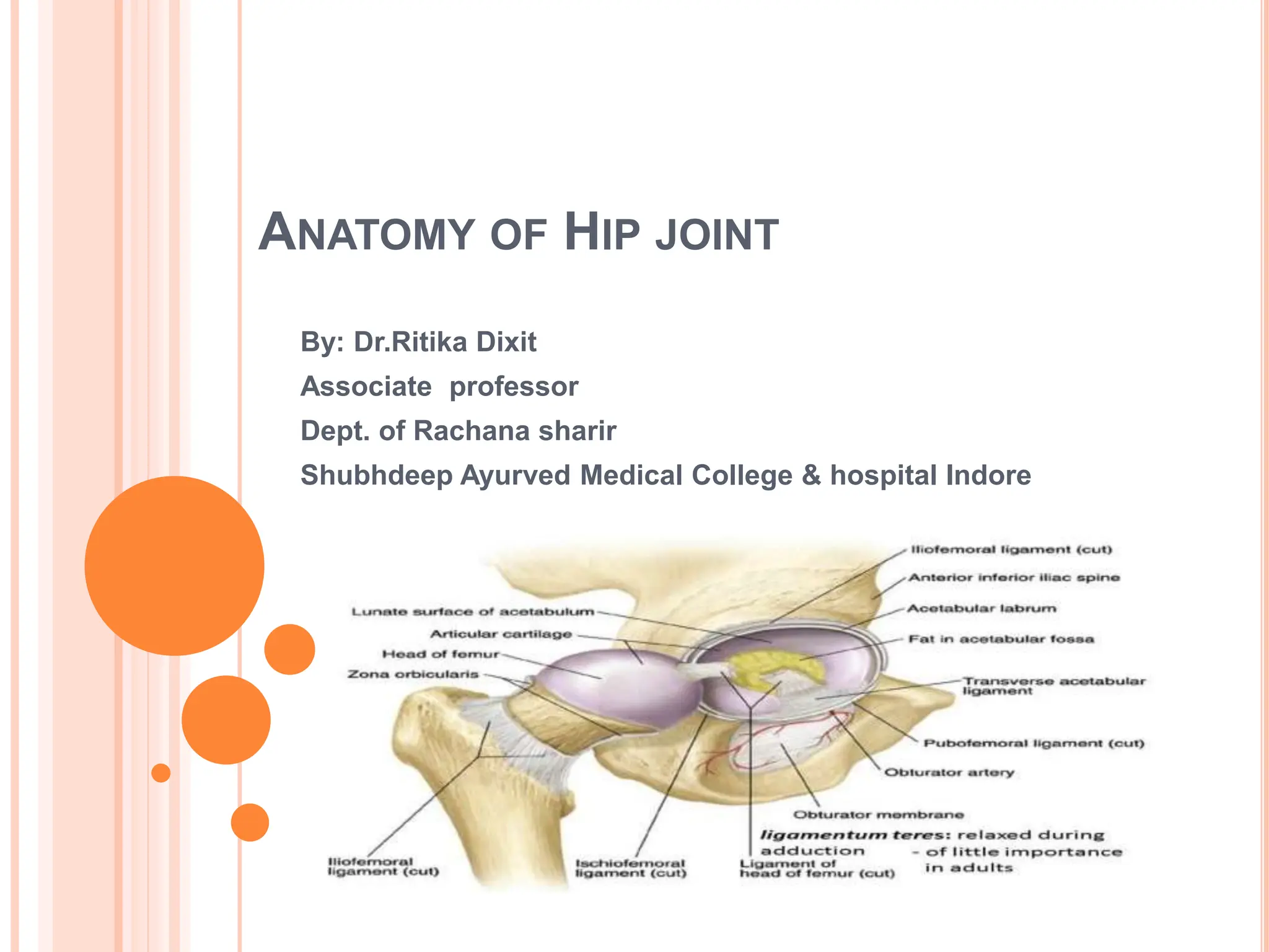
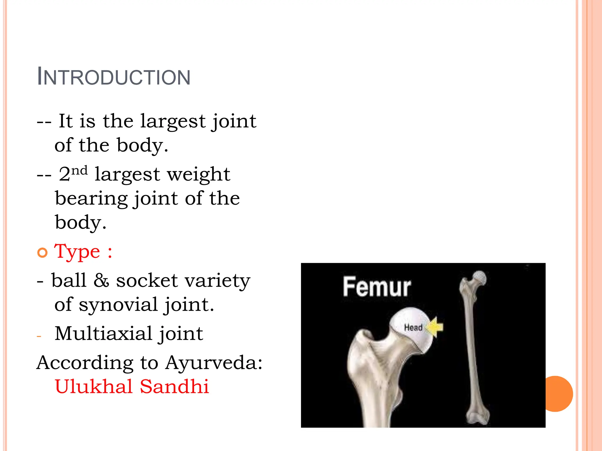
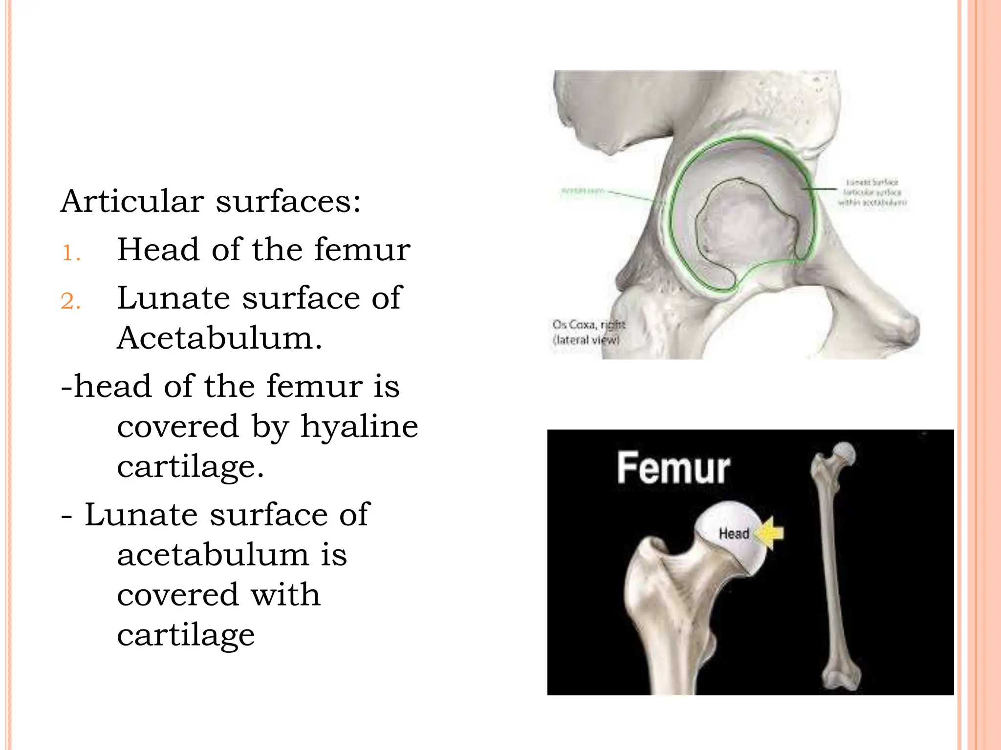
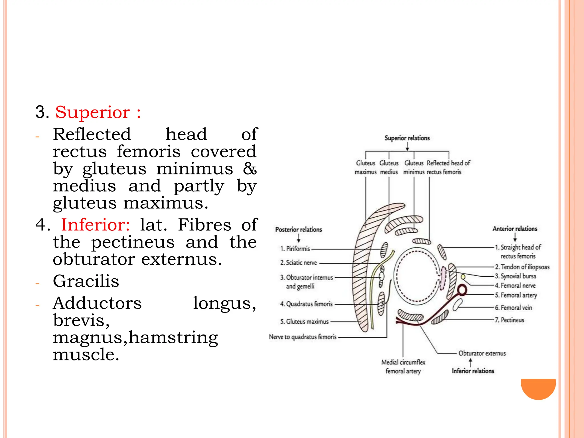
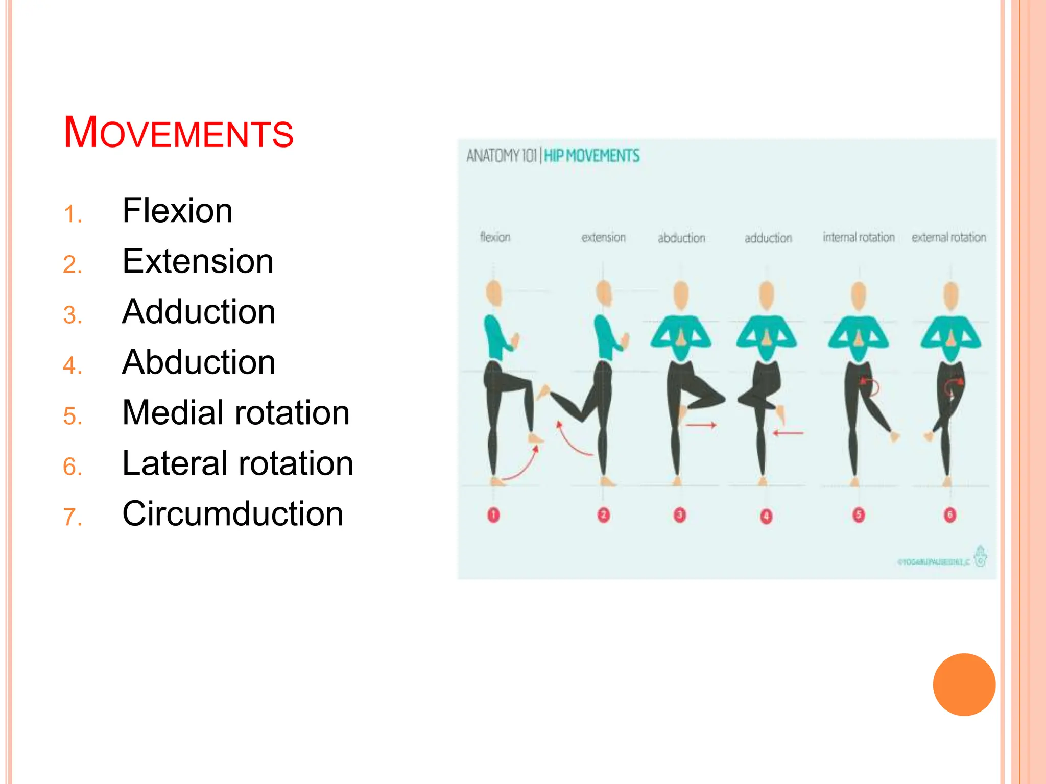
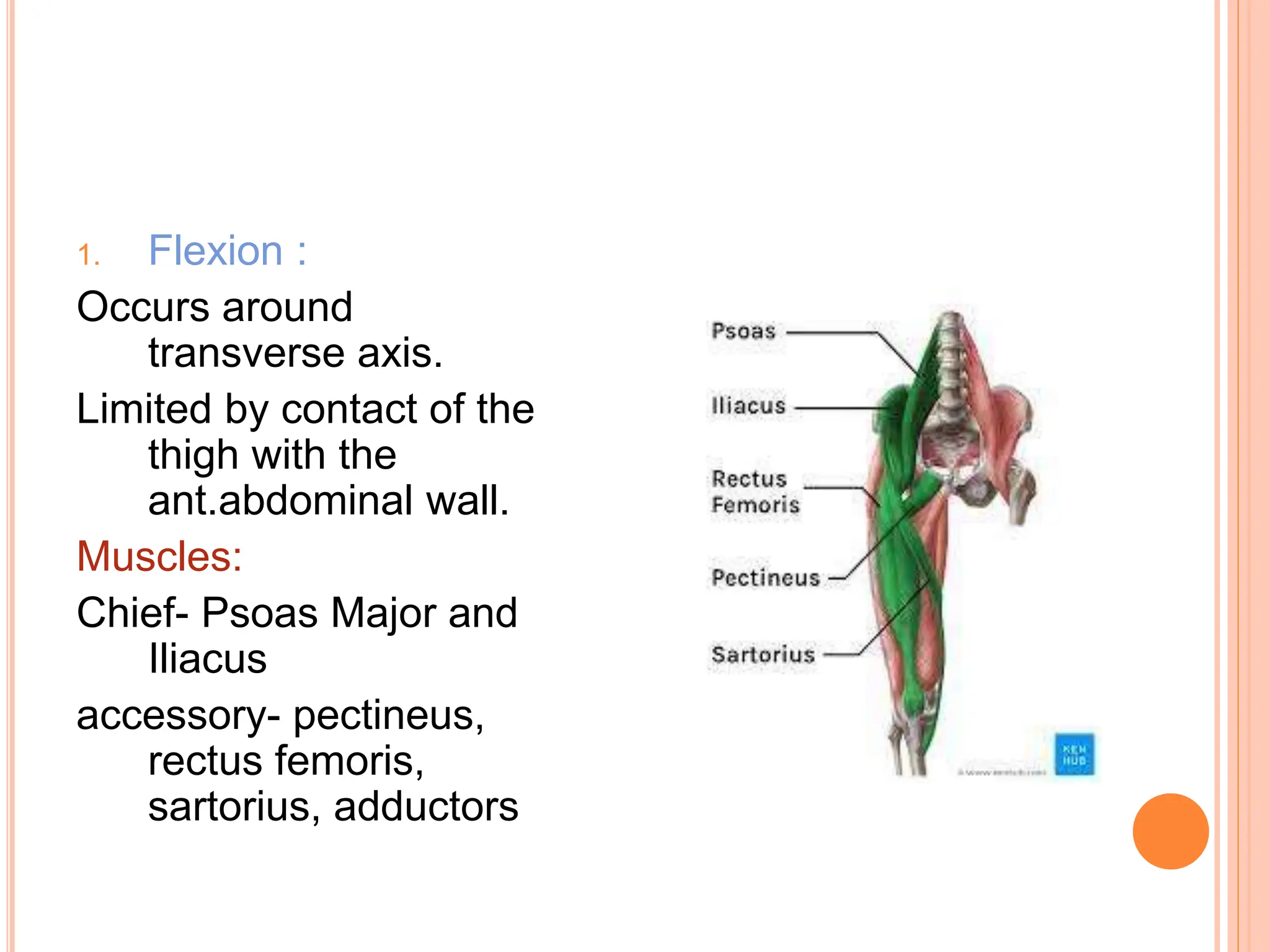
ANATOMY OF HIP JOINT
By: Dr.Ritika Dixit
Associate professor
Dept. of Rachana sharir
Shubhdeep Ayurved Medical College & hospital Indore
INTRODUCTION
-- It is the largest joint
of the body.
-- 2nd largest weight
bearing joint of the
body.
 Type :
- ball & socket variety
of synovial joint.
- Multiaxial joint
According to Ayurveda:
Ulukhal Sandhi
Articular surfaces:
1. Head of the femur
2. Lunate surface of
Acetabulum.
-head of the femur is
covered by hyaline
cartilage.
- Lunate surface of
acetabulum is
covered with
cartilage
Hip joint is designed for stability over a wide
range of movements.
Stability and strength depends upon:
a. Depth of acetabulum and narrowing of its
mouth by acetabular labrum.
b. Tension and strength of ligaments.
c. Strength of surrounding muscles.
d. Length & obliquity of neck of the femur.
e. Atmospheric pressure.
LIGAMENTS
1. The fibrous capsule
2. The iliofemoral ligament
3. The pubofemoral lig.
4. The ischiofemoral lig.
5. The lig. Of the head of the femur
6. The acetabular labrum
7. Transverse acetabular ligament.
1. FIBROUS CAPSULE
Attachment :
-on the hip bone -- Acetabular
labrum, transverse acetabular
lig. and to bone above &
behind acetabulam.
- On femur – to the
intertrochanteric line in front,
& intertrochenteric crest
behind.
- Anterosuperiorly, the capsule
is thick, firmly attached.
- Poster-inferiorly, capsule is
thin & loosely attached.
- Femoral neck intracapsular
- Greater and lesser trochanter
extracapsular.
2. THE ILIOFEMORAL LIGAMENT(BIGELOW)
- Lies anteriorly
- One of the strongest
lig. of the body.
- Prevents trunk from
falling backwards, in
standing posture.
- Triangular in shape.
Attachment :
- Apex is attached to the
lower half of AIIS.
- Base is attached to
base of
intertrochenteric line.
- Upper oblique and
lower vertical fibres
form thick and strong
bands.
- Middle fibres are thin
and weak.
3. THE PUBOFEMORAL LIGAMENT
- It supports the joint
inferomedially.
- Triangular in shape.
- Attachment- superiorly
to the iliopubic
eminence, obturator
crest and the obturator
membrane.
- Inferiorly – merges
with the anteroinferior
part of the capsule &
the with the lower
band of iliofemoral
ligament.
4. THE ISCHIOFEMORAL LIGAMENT
 Comparatively weak
ligament.
 It covers the joint
posteriorly.
 Fibres are twisted
and extend from
ischium to
acetabulum.
 It’s fibers form the
zona orbicularis.
Some attached to
greater trochanter.
 Fibers of all three ligaments are oriented in a
spiral fashion around the hip joint so that the
become taught when joint is extended.
 This stabilizes the joint and reduces the
amount of muscle energy required to maintain
a standing position.
5. THE LIG. OF THE HEAD OF FEMUR(ROUND LIG.
OR LIGAMENTUM TERES)
- Flat & triangular lig.
- Apex attached to
fovea capitis.
- Base attached to
transverse lig. &
margins of
acetabular notch.
- It transmit arteries
to the head of the
femur.
6. THE ACETABULAR LABRUM
It is a fibrocartilaginous rim .
Attached to the margins of acetabulam.
This helps in holding the head of femur in
position.
7. THE TRANSVERSE LIGAMENT
 It is a part of acetabular labrum.
 It bridges the acetabular notch.the notch is
thus converted into foramen.
RELATIONS OF THE JOINT
1. Anterior relation:
- Tendon of iliopsoas
- Femoral vein
- Femoral artery
- Femoral nerve
2. Posterior : from below
upwards –
- Tendon of obturator
externusobturator
internus
- Gamelli
- Piriformis
- Sciatic nerve
- Gluteus maximus muscle.
3. Superior :
- Reflected head of
rectus femoris covered
by gluteus minimus &
medius and partly by
gluteus maximus.
4. Inferior: lat. Fibres of
the pectineus and the
obturator externus.
- Gracilis
- Adductors longus,
brevis,
magnus,hamstring
muscle.
Blood Supply
- Obturator artery
- 2 circumflex femoral arteries.
- 2 gluteal arteries.
Nerve supply
- Femoral nerve
- Ant. Division of obturator nerve
- The nerve of the quadratus femoris
- Sup. Gluteal nerve.
MOVEMENTS
1. Flexion
2. Extension
3. Adduction
4. Abduction
5. Medial rotation
6. Lateral rotation
7. Circumduction
1. Flexion :
Occurs around
transverse axis.
Limited by contact of the
thigh with the
ant.abdominal wall.
Muscles:
Chief- Psoas Major and
Iliacus
accessory- pectineus,
rectus femoris,
sartorius, adductors
2. Extension:
- Occurs around transverse axis.
Muscles–
Gluteus maximus & hamstrings.
3. Adduction :
- Occurs around anteroposterior axis.
Muscles :
Chief – adductor longus, brevis magnus.
Accessory – pectineus gracilis

Anatomy of Hip joint.pptx

  • 1.
    ANATOMY OF HIPJOINT By: Dr.Ritika Dixit Associate professor Dept. of Rachana sharir Shubhdeep Ayurved Medical College & hospital Indore
  • 2.
    INTRODUCTION -- It isthe largest joint of the body. -- 2nd largest weight bearing joint of the body.  Type : - ball & socket variety of synovial joint. - Multiaxial joint According to Ayurveda: Ulukhal Sandhi
  • 3.
    Articular surfaces: 1. Headof the femur 2. Lunate surface of Acetabulum. -head of the femur is covered by hyaline cartilage. - Lunate surface of acetabulum is covered with cartilage
  • 4.
    Hip joint isdesigned for stability over a wide range of movements. Stability and strength depends upon: a. Depth of acetabulum and narrowing of its mouth by acetabular labrum. b. Tension and strength of ligaments. c. Strength of surrounding muscles. d. Length & obliquity of neck of the femur. e. Atmospheric pressure.
  • 5.
    LIGAMENTS 1. The fibrouscapsule 2. The iliofemoral ligament 3. The pubofemoral lig. 4. The ischiofemoral lig. 5. The lig. Of the head of the femur 6. The acetabular labrum 7. Transverse acetabular ligament.
  • 6.
    1. FIBROUS CAPSULE Attachment: -on the hip bone -- Acetabular labrum, transverse acetabular lig. and to bone above & behind acetabulam. - On femur – to the intertrochanteric line in front, & intertrochenteric crest behind. - Anterosuperiorly, the capsule is thick, firmly attached. - Poster-inferiorly, capsule is thin & loosely attached. - Femoral neck intracapsular - Greater and lesser trochanter extracapsular.
  • 7.
    2. THE ILIOFEMORALLIGAMENT(BIGELOW) - Lies anteriorly - One of the strongest lig. of the body. - Prevents trunk from falling backwards, in standing posture. - Triangular in shape.
  • 8.
    Attachment : - Apexis attached to the lower half of AIIS. - Base is attached to base of intertrochenteric line. - Upper oblique and lower vertical fibres form thick and strong bands. - Middle fibres are thin and weak.
  • 9.
    3. THE PUBOFEMORALLIGAMENT - It supports the joint inferomedially. - Triangular in shape. - Attachment- superiorly to the iliopubic eminence, obturator crest and the obturator membrane. - Inferiorly – merges with the anteroinferior part of the capsule & the with the lower band of iliofemoral ligament.
  • 10.
    4. THE ISCHIOFEMORALLIGAMENT  Comparatively weak ligament.  It covers the joint posteriorly.  Fibres are twisted and extend from ischium to acetabulum.  It’s fibers form the zona orbicularis. Some attached to greater trochanter.
  • 11.
     Fibers ofall three ligaments are oriented in a spiral fashion around the hip joint so that the become taught when joint is extended.  This stabilizes the joint and reduces the amount of muscle energy required to maintain a standing position.
  • 12.
    5. THE LIG.OF THE HEAD OF FEMUR(ROUND LIG. OR LIGAMENTUM TERES) - Flat & triangular lig. - Apex attached to fovea capitis. - Base attached to transverse lig. & margins of acetabular notch. - It transmit arteries to the head of the femur.
  • 13.
    6. THE ACETABULARLABRUM It is a fibrocartilaginous rim . Attached to the margins of acetabulam. This helps in holding the head of femur in position.
  • 14.
    7. THE TRANSVERSELIGAMENT  It is a part of acetabular labrum.  It bridges the acetabular notch.the notch is thus converted into foramen.
  • 15.
    RELATIONS OF THEJOINT 1. Anterior relation: - Tendon of iliopsoas - Femoral vein - Femoral artery - Femoral nerve 2. Posterior : from below upwards – - Tendon of obturator externusobturator internus - Gamelli - Piriformis - Sciatic nerve - Gluteus maximus muscle.
  • 16.
    3. Superior : -Reflected head of rectus femoris covered by gluteus minimus & medius and partly by gluteus maximus. 4. Inferior: lat. Fibres of the pectineus and the obturator externus. - Gracilis - Adductors longus, brevis, magnus,hamstring muscle.
  • 17.
    Blood Supply - Obturatorartery - 2 circumflex femoral arteries. - 2 gluteal arteries. Nerve supply - Femoral nerve - Ant. Division of obturator nerve - The nerve of the quadratus femoris - Sup. Gluteal nerve.
  • 18.
    MOVEMENTS 1. Flexion 2. Extension 3.Adduction 4. Abduction 5. Medial rotation 6. Lateral rotation 7. Circumduction
  • 19.
    1. Flexion : Occursaround transverse axis. Limited by contact of the thigh with the ant.abdominal wall. Muscles: Chief- Psoas Major and Iliacus accessory- pectineus, rectus femoris, sartorius, adductors
  • 20.
    2. Extension: - Occursaround transverse axis. Muscles– Gluteus maximus & hamstrings. 3. Adduction : - Occurs around anteroposterior axis. Muscles : Chief – adductor longus, brevis magnus. Accessory – pectineus gracilis