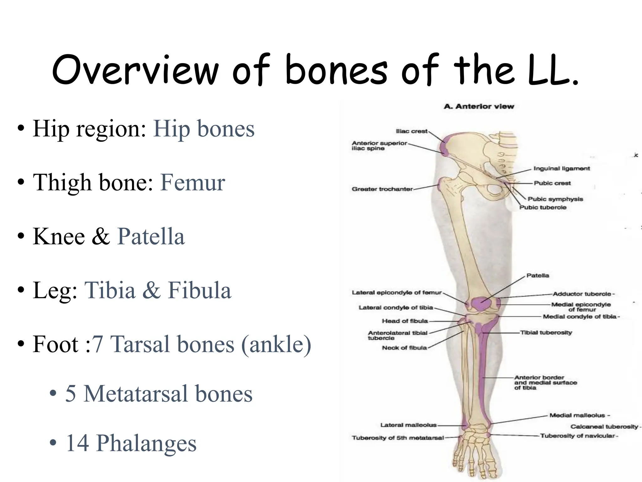
SlideShare a Scribd company logo
Anatomy of the lower
limb
By Solomon D
Introduction
Regions of the LL
• Gluteal region: buttocks and
hip region
• Thigh or femoral region
• Knee region
• Leg
• Ankle
• Foot
Overview of bones of the LL.
• Hip region: Hip bones
• Thigh bone: Femur
• Knee & Patella
• Leg: Tibia & Fibula
• Foot :7 Tarsal bones (ankle)
• 5 Metatarsal bones
• 14 Phalanges
Femoral bone
• The longest, heaviest &
strongest bone in the
body
Proximal end
• Head
• rounded end,
articulates with
acetabulum
• is directed medially,
upward, and slightly
forward
• Possesses a medial
pit, fovea capitis for
ligament of head
Femur
• Neck
• Trapezoidal in shape
• Is narrower near the
head
• Forms an angle of about
126 degrees with the
shaft, so it is a common
site of fractures.
• The angle of inclination
(angle measured b/n the
long axis of neck and
shaft) is less in females
because of wider pelvic
diameter
Clinical
• Normal: angle of
inclination between 120
and 135
• Coxa valga: if the angle
of inclination >135
degrees.
• coxa vara: if the angle of
inclination <120 degrees.
Trochanter
• Greater trochanter
• blunt elevation, projects
upward from the junction of
the neck with the shaft.
• Provides an insertion for
many muscles.
• Lesser trochanter
• Projects posteriomedially
near the angle between the
neck and the shaft.
Body
• Shaft
• Is smooth anteriorly
• posses linea aspera;
double edge ridge on
posterior aspect
Distal end
• has medial & lateral
condyles
• intercondylar fossa
• Epicondyles:
• projections on upper part
of condyle
• Medial epicondyle is more
prominent
• patellar surface for
articulation with the
patella anteriorly
Patella (knee cap)
• Largest sesamoid bone
• located within the quariceps
tendon
• Triangular in shape
• Base – thicker superior
border
• Apex – pointed inferiorly
Tibia
• Larger and weight
bearing bone on the
anteromedial side of the
leg
• Articulates
• proximally with femur
• distally with the talus
• Join with fibula at:
• fibular articular facet
proximally, and
• fibular notch distally
Proximal features
• Proximal end
• has medial & lateral
tibial condyles
• Interconylar eminence
• Is an elevation b/n the
two condyles
• Separates the flat
articular surfaces of
condyles (tibial plateau)
• Is formed by two
intercondylar tubercles
(medial and lateral)
Cont. …
• Anteriorly, below
the condyles is tibial
tuberosity
• Distal end
• Medial malleolus -
inferiorly directed
projection from
medial side of distal
end
Fibula
• Long, slender bone on the
lateral side of leg
• is firmly attached to tibia by:
• interosseous membrane
• Serves mainly for muscle
attachment than weight
bearing
• is a common site of
bone grafting
• Distal end:
• Is enlarged to form lateral
malleolus
Ankle joint (tarsal bones )
• There are seven tarsal bones
Cuboid
Calcaneus
Talus
Navicular
Medial
Cuneiform
Intermediate
Cuneiform
Lateral Cuneiform
Thigh region muscles, membrane
and neurovascular structure
• Fascia lata
Is the deep fascia
investing the thigh
• Iliotibial tract
• is dense (thickened) lateral
part of fascia lata
• extends from iliac tubercle
to tibial tubercle
• Stabilizes the hip and
knee in standing.
Saphenous opening
• A gap in the fascia lata
inferior to medial part of
inguinal ligament
• Great saphenous vein and
some lymphatics pass
through it
Fascia of the leg
• Continuous with fascia
lata, and is thick
proximally
• Attaches to anterior
and medial borders of
tibia
• Forms extensor
retinacula at distal end
Superficial veins of the
lower limb
Great saphenous
vein
• Origin
• By the union of:
• dorsal digital vein
of great toe and
• Medial end of
dorsal venous
arch on the
dorsum of the
foot
Course on the leg
• runs anterior to the
medial malleolus
• crosses the lower 1/3 of
the medial surface of
tibia
• lies a hand breadth
posterior to the medial
border of patella at the
knee
• Course on the thigh
• Ascends on the medial
aspect of thigh
• passes through the
saphenous opening &
• joins femoral vein, 4 cm
inferolateral to the pubic
tubercle
Tributaries -
• 3 superficial veins near the
inguinal ligament.
• Superficial epigastric
• Superficial circumflex
iliac
• Superficial external
pudendal vein
2. Small saphenous Vein
• Begins- lateral end of
venous arch + dorsal
digital V of little toe
• Course below and
posterior to lateral
malleolus
• Ascends along lateral
border of achiles tendon
• After a course b/n the
two heads of
gastrocnemius, → pierce
popliteal fascia & joins
popliteal Vein
Perforating (communicating) veins
• arise from superficial veins
and drains to deep veins
perforating the fascias
• Have valves that allow
blood flow only toward
deep veins.
• Valve damage → reverse the
direction of flow → superficial
veins becomes tortous, dilated
and prominent = varicose vein
Lymphatic drainage of lower
limb
Major cutaneous nerves of
the lower limb
• Posterior cutaneous
nerve of the thigh
• The sural nerve (calf
of the leg)
• Lateral femoral
cutaneous nerve
• Anterior cutaneous
branches of the
femoral nerve
• Saphenous nerve
• Superficial fibular
nerve
Muscles Crossing the Hip and
Knee
• Muscles crossing the hip and knee can be grouped into six.
• Medial hip muscles (2-Hip flexors)
• Gluteal muscles (3)
• Medial compartment (5 adductors)
• Anterior compartment (5 knee extensors)
• Posterior compartment (3 knee flexors)
• Pelvi-trochanteric muscles (6 Lateral rotators )
Hip flexors
• Cross at the medial
aspect of the hip
region
• Include:
• Psoas Major & Iliacus
Gluteal muscles
1. Gluteus maximus
• O - posterior gluteal line,
sacrum and coccyx
• I – gluteal tuberosity of
femur
• A – strong hip extensor;
• adduction & lateral
rotation (lower fibers)
• N.S: inferior gluteal N
Gluteal muscles
2. Gluteus medius
O – b/n anterior and
posterior gluteal lines
I - lateral part of greater
trochanter of femur
A - abducts and medially
rotates hip, Stablizes
pelvis or hip during gait
N.S: Superior gluteal N
Gluteal muscles
Gluteus minimus
O - between anterior and
inferior gluteal lines
I – anterior border of greater
trochanter of femur
A - abducts and medially
rotates hip
- with gluteus medius,
stabilizes the hip
N.S: superior gluteal N
Lateral rotators (deep gluteal
muscles)
• These sets of muscles
Laterally rotate extended
thigh and abduct flexed
thigh; steady femoral head
in acetabulum
Piriformis clinical importance
Structures emerging via
greater sciatic
foramen
Above piriformis-
• superior gluteal N and VS
Below piriformis-
• Sciatic N,
• Pudendal N,
• Nerve to Quadratus
femoris and obturator
internus,
• inferior gluteal & internal
pudendal VS
Note
Sacral plexus
Thigh
• Thigh muscles are organized
into 3 compartments by
intermuscular septa
• Anterior or extensor
• Medial or adductor
• Posterior or flexor
Anterior thigh muscles
• Include:
1. Sartorius
2. Quadriceps femoris (
four muscles)
• Vastus lateralis
• Vastus medialis
• Vastus intermedius
• Rectus femoris
• Action: Flexors of hip
and extensors of knee
• The nerve supply- femoral
N
Medial thigh muscles
(adductor group)
• Constitute the medial
compartment
• Attach from the pubis and
ischium to the linea aspera
• Adductor muscles are
• Adductor magnus
• Adductor longus
• Adductor brevis
• Pectineus
• Gracilis
• Obturator externus
(optional)
Adductor muscles
• Most muscles are
supplied by obturator
nerve except
• pectineus supplied by
femoral nerve
• hamstring part of
adductor magnus by
sciatic nerve
• Arranged in to 3-layers
• Superficial - pectineus,
gracilis and adductor
longus
• Middle - adductor brevis
• Deep - adductor magnus
& obturator externus
The Femoral Triangle…
Contents of the femoral
triangle:
1. Femoral nerve and its
branches
2. Femoral sheath & its
contents, like;
 femoral artery and its
branch
 femoral vein and its
tributaries
 deep inguinal lymph nodes
LVAN
Posterior thigh muscles
• Also called Hamstring
muscles
• Are 3 in number
• Semitendinosus
• Semimembranosus
• Biceps femoris (long head
only)
NB: Biceps femoris has two
heads
• Short head & long head
• The short head is not
hamstring as it crosses only
knee joint & innervated by
fibular division of sciatic
nerve
Hamstring muscles
• Arise from ischial
tuberosity
• Insert into tibia &
fibula
• Innervated by the
tibial division of
sciatic nerve
Don’t forget nerve supply
Obturator nerve
• Formed by anterior
division of anterior rami
of L2-4
• Descends Passes
through the obturator
canal to supply adductor
muscles
• Divides into anterior
and posterior divisions
as it pass the obturator
canal
• Its branch to gracilis
becomes the only
cutaneous branch
• Anterior division: gives
branch to;
• Adductor longus
• Adductor brevis
• Gracilis
• Articular branch to hip joint
• Cutaneous branch to skin
overlying adductor canal
• Posterior division: branches
to;
• Obturator externus
• Adductor magnus (adductor
part)
Femoral nerve
• Formed by posterior
division of anterior rami
of L2-4
• Lies in front of the
lower part of iliacus;
where innervates it
• Passes deep to the
inguinal ligament
/iliopectineal arch/ to
the femoral triangle
• Breaks up into motor
and sensory branches
• 2 of its branches pass
into the adductor canal:
• nerve to vastus medialis
(motor)
• saphenous nerve
(sensory)
Branches of femoral nerve
• Muscular
• To quadriceps, sartorius,
iliacus & pectineus
• Cutaneous
• anterior cutaneous
branches which supplies
the anterior and lower
part of medial aspect of
thigh.
• saphenous nerve
• supply the skin of the
medial side of the leg,
ankle and foot to the great
toe
Sacral plexus
• Formed by the anterior
rami of L4, L5, S1–S4
• L4 is shared by lumbar
plexus, a branch from it
joins L5 to form the
lumbosacral trunk
• The sacral nerves
emerge from the
anterior sacral foramina
and unite in front of
piriformis where they
are joined by the
lumbosacral trunk
sacral plexus
• The deep gluteal nerves
are -8:
• the superior gluteal nerve
• inferior gluteal nerve
• sciatic nerve
• Nerve to quadratus femoris
• posterior cutaneous nerve
of the thigh
• Nerve to obturator
internus.
• Pudendal nerve
• Nerve to piriformis
• All of these nerves are
branches of the sacral
plexus
Course of sciatic nerve
• Emerges through the
greater sciatic foramen
inferior to the
piriformis
• Runs inferolaterally
under cover of the
gluteus maximus
• descends deep to biceps
femoris
• Bifurcates into tibial
and common peroneal
nerves at popliteal fossa
Assignment
Blood vessels of the thigh
• Femoral artery and vein
• Femoral pulse
• Trendelenburg gait
(gluteal gait)
• Safe area of gluteal IM
(intramuscular injection)
• Piriformis syndrome
• Hamstring injuries
Muscles of Legs
• Has 3-compartments
• Anterior compartment
• posterior compartment
• lateral (fibular or
peroneal) compartment
Anterior compartment of leg
• Is Dorsiflexor
• Located anterior to the
interosseous membrane, b/n the
tibial and fibular shaft
• Inferiorly, two band-like
thickenings of the deep fascia
form retinacula
Muscles of anterior compartment
• Consists of 4- muscles
1. Tibialis anterior
2. Extensor digitorum
longus
3. Extensor hallucis longus
4. Peroneus tertius
• Nerve supply: deep fibular
nerve to all muscles
Muscles of lateral compartment
• Contains two muscles,
that pass posterior to
the lateral malleolus:
• Fibularis longus (peroneus
longus)
• Fibularis (peroneus)
brevis = deep
• Actions: Plantar flexor
and evertor of the foot.
• Nerve supply –
superficial fibular nerve
Posterior compartment of leg
• Is also called plantarflexor compartment, is the largest of
the three leg compartments
• divided into two subcompartments (groups) by the
transverse intermuscular septum
• Neurovascular structures for both groups of posterior
compartment (calf muscles) are:
• Tibial nerve (for all muscles of both group)
• Posterior tibial artery & vein
• Fibular artery & vein
NB: all these neurovascular structures lie just deep to the
transverse intermuscular septum
Muscles of posterior compartment
1. Superficial group has 3
muscles:
• Gastrocnemius
• Soleus
• Plantaris
• The gastrocnemius and soleus
share a common tendon.
Therefore, these 2 muscles are
collectively called triceps surae
Gastrocnemius
• Its tendon together with the
tendon of soleus is called tendo
calcaneus (Achilles' tendon)
which is the largest tendon in the
body
• Origin:
• lateral head: from lateral epicondyle
of femur
• medial head: above the medial
epicondyle of femur
• Insertion
• tendo calcaneus inserts to center of
calcaneal tuberosity
Plantaris
• is a small muscle with a short
belly and a long tendon
• lies b/n the gastrocnemius and
the soleus
• Origin - posterior femur above
lateral condyle;
• Insertion – calcaneus
• Action – weak plantar flexion,
knee flexion
• Acts as organ of proprioception
Muscles of posterior compartment
2. Deep group contains 4
muscles
• Popliteus
• Flexor hallucis longus
• Flexor digitorum longus
• Tibialis posterior
Popliteal fossa
• The popliteal fossa is a fat-
filled diamond-shaped space
posterior to the knee
• All important vessels and
nerves from the thigh to the
leg pass through this fossa
• Distal continuation of the
adductor canal
Popliteal fossa
• Boundaries
• Laterally & inferiorly – lateral
head of gastrocnemius
• Medially & inferiorly- medial
head of gastrocnemius
• Laterally & superiorly - biceps
femoris
• Medially & superiorly –
semimembranosus
Cont. ..
• Contents
• popliteal vessels
(artery is deep to the
vein)
• Tibial nerve (superficial
to the popliteal vein)
• common fibular nerves-
closer to biceps
femoris
• small saphenous vein
(superficial content)
• popliteal lymph nodes &
vessels
Popliteal artery
• Continuation of the
femoral artery
• Begins where the
femoral artery passes
through the adductor
hiatus
• Ends at the inferior
border of the popliteus
muscle by dividing into
anterior and posterior
tibial arteries
• The deepest structure in
the popliteal fossa
Popliteal pulse
• Because the popliteal
artery is deep in the
popliteal fossa, it may
be difficult to feel the
popliteal pulse
• Palpation of this pulse is
commonly performed by
placing the person in the
prone position with the
knee flexed to relax the
popliteal fascia and
hamstrings
Popliteal vein
• Formed at distal end of
popliteus as an assembly
of venae comitantes
below the knee
• Terminate in the
adductor hiatus
Nerves in popliteal fossa
•The sciatic nerve
usually ends at the
superior angle of the
popliteal fossa by
dividing into:
•tibial nerve and
•common fibular
nerves
Tibial nerve
• The medial, larger
terminal branch of the
sciatic nerve
• In the fossa, the
tibial nerve gives
branches to the
soleus, gastrocnemius,
plantaris, and
popliteus muscles
• The most superficial
of the three main
central components of
the popliteal fossa.
Common fibular nerve
• The lateral, smaller terminal
branch of the sciatic nerve
• Descends closely to the
medial border of biceps
femoris
• Leaves the fossa by passing
superficial to the lateral
head of the gastrocnemius
• Winds around the fibular
neck, where it is vulnerable
to injury
• Here it divides into the
superficial and deep fibular
Nerves of the leg
For your further reading
• Saphenous
• Sural
• Tibial
• Common fibular
• Superficial fibular
• Deep fibular
The Foot
• Foot is a region of lower
limb distal to ankle
• Provides platform for
supporting the weight of
the body
• Compartments of the
sole
• Vertical intermuscular
septa extend superiorly
from the margins of the
plantar aponeurosis toward
the 1st and 5th
metatarsals, forming three
compartments of the sole:
Medial compartment of the sole
• covered superficially by
medial plantar fascia
• contains
• abductor hallucis
• flexor hallucis brevis
• tendon of the flexor
hallucis longus
• medial plantar nerve and
vessels
2. Central compartment of the sole
• Is deep to the plantar
aponeurosis
• contains
– flexor digitorum brevis
– flexor digitorum longus
tendons
– quadratus plantae
– lumbricals
– adductor hallucis
– lateral plantar nerve and
vessels
3. Lateral compartment of the sole
• covered by the thinner
lateral plantar fascia
• contains abductor and
flexor digiti minimi
brevis
Dorsum of the foot
• Two Muscles
• Extensor
digitorum brevis
• Extensor hallucis
brevis (medial slip
of the extensor
digitorum brevis)
• Nerve supply –
tibial nerve
Plantar muscles
• Are organized in to 4
layers
• First layer: 3 muscles
• Abductor digiti minimi
• Flexor digitorium brevis
• Abductor hallucis
Second layer:
• 2 muscles and 2 tendons
• The two tendons are:
• Flexor digitorum longus
tendon &
• Flexor hallucis longus
tendon
• Two muscles are:
1. Quadratus plantae
2. Lumbricals
Third layer:
• 3 muscles
1. Adductor halluces
2. Fexor halluces brevis
3. Flexor digiti minimi
Fourth layer:
• 2 muscles and 2 tendons
• The 2 tendons are:
• Tibialis posterior
tendon
• Peroneus (fibularis)
longus tendon
Fourth layer
2 muscle groups
• 7 interossei
• 4 dorsal and
• 3 plantar
lower limb for public health students.pdf

More Related Content

Similar to lower limb for public health students.pdf

Anterior cmpartment of thigh
Anterior  cmpartment of thighAnterior  cmpartment of thigh
Anterior cmpartment of thigh
AtifRaza11
 
HipJoint attachment and action of muscles
HipJoint attachment and action of musclesHipJoint attachment and action of muscles
HipJoint attachment and action of muscles
HarmanSaini54
 
glutealregion-140107113927-phpapp01.pdf
glutealregion-140107113927-phpapp01.pdfglutealregion-140107113927-phpapp01.pdf
glutealregion-140107113927-phpapp01.pdf
UmaMaheshwariJ3
 
02 THE GLUTEAL REGION-AN OVERVIEW OF KURIA SERIES.pptx
02 THE GLUTEAL REGION-AN OVERVIEW OF KURIA SERIES.pptx02 THE GLUTEAL REGION-AN OVERVIEW OF KURIA SERIES.pptx
02 THE GLUTEAL REGION-AN OVERVIEW OF KURIA SERIES.pptx
VivianMwamuye
 
Anatomy of the leg
Anatomy of the legAnatomy of the leg
Anatomy of the leg
Randolph Tulsie
 
back of thigh and popleteal fossa.pptx
back of thigh and popleteal fossa.pptxback of thigh and popleteal fossa.pptx
back of thigh and popleteal fossa.pptx
manishajha49
 
Joints of Lower Limb_ BSC 2013.pptx
Joints of Lower Limb_ BSC 2013.pptxJoints of Lower Limb_ BSC 2013.pptx
Joints of Lower Limb_ BSC 2013.pptx
AimanArifin2
 
Anatomy of Gluteal Region
Anatomy of Gluteal RegionAnatomy of Gluteal Region
Anatomy of Gluteal Region
Dr. Seyed Morteza Mahmoudi
 
joints & Major joints By M Thiru murugan.pptx
joints & Major joints By M Thiru murugan.pptxjoints & Major joints By M Thiru murugan.pptx
joints & Major joints By M Thiru murugan.pptx
thiru murugan
 
Lower limb anatomy.pptx
Lower limb anatomy.pptxLower limb anatomy.pptx
Lower limb anatomy.pptx
FarhanFarhan310858
 
Knee joint
Knee jointKnee joint
Knee joint
RajnishKumar566
 
Anatomy of ankle and foot
Anatomy of ankle and footAnatomy of ankle and foot
Anatomy of ankle and foot
Dr VAIBHAV KALYANJI VIRA
 
Muscles Bones of Lower Limb Lower Limb Bones of LL HUMAN
Muscles  Bones of Lower Limb Lower Limb Bones of LL HUMANMuscles  Bones of Lower Limb Lower Limb Bones of LL HUMAN
Muscles Bones of Lower Limb Lower Limb Bones of LL HUMAN
nidhi sharma
 
knee ..pptx
knee ..pptxknee ..pptx
knee ..pptx
Vivekchanda4
 
Muscles of thigh
Muscles of thighMuscles of thigh
Muscles of thigh
Idris Siddiqui
 
The femur
The femurThe femur
The femur
Idris Siddiqui
 
thefemur-180421095413.pdf
thefemur-180421095413.pdfthefemur-180421095413.pdf
thefemur-180421095413.pdf
UmaMaheshwariJ3
 
02 osteologyLL.ppt
02 osteologyLL.ppt02 osteologyLL.ppt
02 osteologyLL.ppt
ELLYROTSPA
 
Anatomy of the GLUTEAL REGION........ By Shapi.pdf
Anatomy of the GLUTEAL REGION........ By Shapi.pdfAnatomy of the GLUTEAL REGION........ By Shapi.pdf
Anatomy of the GLUTEAL REGION........ By Shapi.pdf
Shapi. MD
 
Bony anatomy of human body medical anatomy
Bony anatomy of human body medical anatomyBony anatomy of human body medical anatomy
Bony anatomy of human body medical anatomy
nidhi sharma
 

Similar to lower limb for public health students.pdf (20)

Anterior cmpartment of thigh
Anterior  cmpartment of thighAnterior  cmpartment of thigh
Anterior cmpartment of thigh
 
HipJoint attachment and action of muscles
HipJoint attachment and action of musclesHipJoint attachment and action of muscles
HipJoint attachment and action of muscles
 
glutealregion-140107113927-phpapp01.pdf
glutealregion-140107113927-phpapp01.pdfglutealregion-140107113927-phpapp01.pdf
glutealregion-140107113927-phpapp01.pdf
 
02 THE GLUTEAL REGION-AN OVERVIEW OF KURIA SERIES.pptx
02 THE GLUTEAL REGION-AN OVERVIEW OF KURIA SERIES.pptx02 THE GLUTEAL REGION-AN OVERVIEW OF KURIA SERIES.pptx
02 THE GLUTEAL REGION-AN OVERVIEW OF KURIA SERIES.pptx
 
Anatomy of the leg
Anatomy of the legAnatomy of the leg
Anatomy of the leg
 
back of thigh and popleteal fossa.pptx
back of thigh and popleteal fossa.pptxback of thigh and popleteal fossa.pptx
back of thigh and popleteal fossa.pptx
 
Joints of Lower Limb_ BSC 2013.pptx
Joints of Lower Limb_ BSC 2013.pptxJoints of Lower Limb_ BSC 2013.pptx
Joints of Lower Limb_ BSC 2013.pptx
 
Anatomy of Gluteal Region
Anatomy of Gluteal RegionAnatomy of Gluteal Region
Anatomy of Gluteal Region
 
joints & Major joints By M Thiru murugan.pptx
joints & Major joints By M Thiru murugan.pptxjoints & Major joints By M Thiru murugan.pptx
joints & Major joints By M Thiru murugan.pptx
 
Lower limb anatomy.pptx
Lower limb anatomy.pptxLower limb anatomy.pptx
Lower limb anatomy.pptx
 
Knee joint
Knee jointKnee joint
Knee joint
 
Anatomy of ankle and foot
Anatomy of ankle and footAnatomy of ankle and foot
Anatomy of ankle and foot
 
Muscles Bones of Lower Limb Lower Limb Bones of LL HUMAN
Muscles  Bones of Lower Limb Lower Limb Bones of LL HUMANMuscles  Bones of Lower Limb Lower Limb Bones of LL HUMAN
Muscles Bones of Lower Limb Lower Limb Bones of LL HUMAN
 
knee ..pptx
knee ..pptxknee ..pptx
knee ..pptx
 
Muscles of thigh
Muscles of thighMuscles of thigh
Muscles of thigh
 
The femur
The femurThe femur
The femur
 
thefemur-180421095413.pdf
thefemur-180421095413.pdfthefemur-180421095413.pdf
thefemur-180421095413.pdf
 
02 osteologyLL.ppt
02 osteologyLL.ppt02 osteologyLL.ppt
02 osteologyLL.ppt
 
Anatomy of the GLUTEAL REGION........ By Shapi.pdf
Anatomy of the GLUTEAL REGION........ By Shapi.pdfAnatomy of the GLUTEAL REGION........ By Shapi.pdf
Anatomy of the GLUTEAL REGION........ By Shapi.pdf
 
Bony anatomy of human body medical anatomy
Bony anatomy of human body medical anatomyBony anatomy of human body medical anatomy
Bony anatomy of human body medical anatomy
 

More from tekalignpawulose09

L3_Renal_Clearance physiology for public health.pdf
L3_Renal_Clearance physiology for public health.pdfL3_Renal_Clearance physiology for public health.pdf
L3_Renal_Clearance physiology for public health.pdf
tekalignpawulose09
 
1_HSM_for_HO_Unit_1_2_&3_Introduction_to_Management_and_Types_Skills.pptx
1_HSM_for_HO_Unit_1_2_&3_Introduction_to_Management_and_Types_Skills.pptx1_HSM_for_HO_Unit_1_2_&3_Introduction_to_Management_and_Types_Skills.pptx
1_HSM_for_HO_Unit_1_2_&3_Introduction_to_Management_and_Types_Skills.pptx
tekalignpawulose09
 
Health Ethics and Medical parasitology ;The study of the parasites of man and...
Health Ethics and Medical parasitology ;The study of the parasites of man and...Health Ethics and Medical parasitology ;The study of the parasites of man and...
Health Ethics and Medical parasitology ;The study of the parasites of man and...
tekalignpawulose09
 
introduction Para for nurse 2016 batch.ppt
introduction Para for nurse 2016 batch.pptintroduction Para for nurse 2016 batch.ppt
introduction Para for nurse 2016 batch.ppt
tekalignpawulose09
 
fertilization for public health (1).pptx
fertilization for public health (1).pptxfertilization for public health (1).pptx
fertilization for public health (1).pptx
tekalignpawulose09
 
Embryology-Part-1 for public health students.pdf
Embryology-Part-1 for public health students.pdfEmbryology-Part-1 for public health students.pdf
Embryology-Part-1 for public health students.pdf
tekalignpawulose09
 
fertilization for public health students HO2016.pptx
fertilization for public health students HO2016.pptxfertilization for public health students HO2016.pptx
fertilization for public health students HO2016.pptx
tekalignpawulose09
 
Placenta Development ppt By Zeki PH 2016.pptx
Placenta Development ppt  By Zeki PH 2016.pptxPlacenta Development ppt  By Zeki PH 2016.pptx
Placenta Development ppt By Zeki PH 2016.pptx
tekalignpawulose09
 
Chapter 2ppt Entrepreneurship freshman course.pptx
Chapter 2ppt Entrepreneurship freshman course.pptxChapter 2ppt Entrepreneurship freshman course.pptx
Chapter 2ppt Entrepreneurship freshman course.pptx
tekalignpawulose09
 
Chapter 5ppt small business and their importance.pptx
Chapter 5ppt small business and their importance.pptxChapter 5ppt small business and their importance.pptx
Chapter 5ppt small business and their importance.pptx
tekalignpawulose09
 
The thorax plays a crucial role in respiration and protecting internal organs...
The thorax plays a crucial role in respiration and protecting internal organs...The thorax plays a crucial role in respiration and protecting internal organs...
The thorax plays a crucial role in respiration and protecting internal organs...
tekalignpawulose09
 
Unit 6 Infectious diseases & immunity - shortend.pdf
Unit 6 Infectious diseases & immunity - shortend.pdfUnit 6 Infectious diseases & immunity - shortend.pdf
Unit 6 Infectious diseases & immunity - shortend.pdf
tekalignpawulose09
 
Lipids are organic molecules that made from fatty acid.pptx
Lipids are organic molecules that made from fatty acid.pptxLipids are organic molecules that made from fatty acid.pptx
Lipids are organic molecules that made from fatty acid.pptx
tekalignpawulose09
 
Respiratory System And Ventilation D.r Tariku.pptx
Respiratory System And Ventilation D.r Tariku.pptxRespiratory System And Ventilation D.r Tariku.pptx
Respiratory System And Ventilation D.r Tariku.pptx
tekalignpawulose09
 
Lipids are a diverse group of organic molecules that are insoluble in water b...
Lipids are a diverse group of organic molecules that are insoluble in water b...Lipids are a diverse group of organic molecules that are insoluble in water b...
Lipids are a diverse group of organic molecules that are insoluble in water b...
tekalignpawulose09
 
hemoglobinstructuresynthesisbreakdown-220418045309.pdf
hemoglobinstructuresynthesisbreakdown-220418045309.pdfhemoglobinstructuresynthesisbreakdown-220418045309.pdf
hemoglobinstructuresynthesisbreakdown-220418045309.pdf
tekalignpawulose09
 
Unit 1 Introduction & Unit 2 Biological molecules.pdf
Unit 1 Introduction & Unit 2 Biological molecules.pdfUnit 1 Introduction & Unit 2 Biological molecules.pdf
Unit 1 Introduction & Unit 2 Biological molecules.pdf
tekalignpawulose09
 
Uterus Is thick muscular walls adapt to the growth of the fetus and then pro...
Uterus Is thick muscular walls adapt to the growth of the fetus and  then pro...Uterus Is thick muscular walls adapt to the growth of the fetus and  then pro...
Uterus Is thick muscular walls adapt to the growth of the fetus and then pro...
tekalignpawulose09
 
Cardio vascular system is one of the largest pharmacy chains in the United St...
Cardio vascular system is one of the largest pharmacy chains in the United St...Cardio vascular system is one of the largest pharmacy chains in the United St...
Cardio vascular system is one of the largest pharmacy chains in the United St...
tekalignpawulose09
 
Hemoglobin and myoglobin are two important proteins involved in the transport...
Hemoglobin and myoglobin are two important proteins involved in the transport...Hemoglobin and myoglobin are two important proteins involved in the transport...
Hemoglobin and myoglobin are two important proteins involved in the transport...
tekalignpawulose09
 

More from tekalignpawulose09 (20)

L3_Renal_Clearance physiology for public health.pdf
L3_Renal_Clearance physiology for public health.pdfL3_Renal_Clearance physiology for public health.pdf
L3_Renal_Clearance physiology for public health.pdf
 
1_HSM_for_HO_Unit_1_2_&3_Introduction_to_Management_and_Types_Skills.pptx
1_HSM_for_HO_Unit_1_2_&3_Introduction_to_Management_and_Types_Skills.pptx1_HSM_for_HO_Unit_1_2_&3_Introduction_to_Management_and_Types_Skills.pptx
1_HSM_for_HO_Unit_1_2_&3_Introduction_to_Management_and_Types_Skills.pptx
 
Health Ethics and Medical parasitology ;The study of the parasites of man and...
Health Ethics and Medical parasitology ;The study of the parasites of man and...Health Ethics and Medical parasitology ;The study of the parasites of man and...
Health Ethics and Medical parasitology ;The study of the parasites of man and...
 
introduction Para for nurse 2016 batch.ppt
introduction Para for nurse 2016 batch.pptintroduction Para for nurse 2016 batch.ppt
introduction Para for nurse 2016 batch.ppt
 
fertilization for public health (1).pptx
fertilization for public health (1).pptxfertilization for public health (1).pptx
fertilization for public health (1).pptx
 
Embryology-Part-1 for public health students.pdf
Embryology-Part-1 for public health students.pdfEmbryology-Part-1 for public health students.pdf
Embryology-Part-1 for public health students.pdf
 
fertilization for public health students HO2016.pptx
fertilization for public health students HO2016.pptxfertilization for public health students HO2016.pptx
fertilization for public health students HO2016.pptx
 
Placenta Development ppt By Zeki PH 2016.pptx
Placenta Development ppt  By Zeki PH 2016.pptxPlacenta Development ppt  By Zeki PH 2016.pptx
Placenta Development ppt By Zeki PH 2016.pptx
 
Chapter 2ppt Entrepreneurship freshman course.pptx
Chapter 2ppt Entrepreneurship freshman course.pptxChapter 2ppt Entrepreneurship freshman course.pptx
Chapter 2ppt Entrepreneurship freshman course.pptx
 
Chapter 5ppt small business and their importance.pptx
Chapter 5ppt small business and their importance.pptxChapter 5ppt small business and their importance.pptx
Chapter 5ppt small business and their importance.pptx
 
The thorax plays a crucial role in respiration and protecting internal organs...
The thorax plays a crucial role in respiration and protecting internal organs...The thorax plays a crucial role in respiration and protecting internal organs...
The thorax plays a crucial role in respiration and protecting internal organs...
 
Unit 6 Infectious diseases & immunity - shortend.pdf
Unit 6 Infectious diseases & immunity - shortend.pdfUnit 6 Infectious diseases & immunity - shortend.pdf
Unit 6 Infectious diseases & immunity - shortend.pdf
 
Lipids are organic molecules that made from fatty acid.pptx
Lipids are organic molecules that made from fatty acid.pptxLipids are organic molecules that made from fatty acid.pptx
Lipids are organic molecules that made from fatty acid.pptx
 
Respiratory System And Ventilation D.r Tariku.pptx
Respiratory System And Ventilation D.r Tariku.pptxRespiratory System And Ventilation D.r Tariku.pptx
Respiratory System And Ventilation D.r Tariku.pptx
 
Lipids are a diverse group of organic molecules that are insoluble in water b...
Lipids are a diverse group of organic molecules that are insoluble in water b...Lipids are a diverse group of organic molecules that are insoluble in water b...
Lipids are a diverse group of organic molecules that are insoluble in water b...
 
hemoglobinstructuresynthesisbreakdown-220418045309.pdf
hemoglobinstructuresynthesisbreakdown-220418045309.pdfhemoglobinstructuresynthesisbreakdown-220418045309.pdf
hemoglobinstructuresynthesisbreakdown-220418045309.pdf
 
Unit 1 Introduction & Unit 2 Biological molecules.pdf
Unit 1 Introduction & Unit 2 Biological molecules.pdfUnit 1 Introduction & Unit 2 Biological molecules.pdf
Unit 1 Introduction & Unit 2 Biological molecules.pdf
 
Uterus Is thick muscular walls adapt to the growth of the fetus and then pro...
Uterus Is thick muscular walls adapt to the growth of the fetus and  then pro...Uterus Is thick muscular walls adapt to the growth of the fetus and  then pro...
Uterus Is thick muscular walls adapt to the growth of the fetus and then pro...
 
Cardio vascular system is one of the largest pharmacy chains in the United St...
Cardio vascular system is one of the largest pharmacy chains in the United St...Cardio vascular system is one of the largest pharmacy chains in the United St...
Cardio vascular system is one of the largest pharmacy chains in the United St...
 
Hemoglobin and myoglobin are two important proteins involved in the transport...
Hemoglobin and myoglobin are two important proteins involved in the transport...Hemoglobin and myoglobin are two important proteins involved in the transport...
Hemoglobin and myoglobin are two important proteins involved in the transport...
 

Recently uploaded

Main Java[All of the Base Concepts}.docx
Main Java[All of the Base Concepts}.docxMain Java[All of the Base Concepts}.docx
Main Java[All of the Base Concepts}.docx
adhitya5119
 
বাংলাদেশ অর্থনৈতিক সমীক্ষা (Economic Review) ২০২৪ UJS App.pdf
বাংলাদেশ অর্থনৈতিক সমীক্ষা (Economic Review) ২০২৪ UJS App.pdfবাংলাদেশ অর্থনৈতিক সমীক্ষা (Economic Review) ২০২৪ UJS App.pdf
বাংলাদেশ অর্থনৈতিক সমীক্ষা (Economic Review) ২০২৪ UJS App.pdf
eBook.com.bd (প্রয়োজনীয় বাংলা বই)
 
Advanced Java[Extra Concepts, Not Difficult].docx
Advanced Java[Extra Concepts, Not Difficult].docxAdvanced Java[Extra Concepts, Not Difficult].docx
Advanced Java[Extra Concepts, Not Difficult].docx
adhitya5119
 
Hindi varnamala | hindi alphabet PPT.pdf
Hindi varnamala | hindi alphabet PPT.pdfHindi varnamala | hindi alphabet PPT.pdf
Hindi varnamala | hindi alphabet PPT.pdf
Dr. Mulla Adam Ali
 
ANATOMY AND BIOMECHANICS OF HIP JOINT.pdf
ANATOMY AND BIOMECHANICS OF HIP JOINT.pdfANATOMY AND BIOMECHANICS OF HIP JOINT.pdf
ANATOMY AND BIOMECHANICS OF HIP JOINT.pdf
Priyankaranawat4
 
Digital Artifact 1 - 10VCD Environments Unit
Digital Artifact 1 - 10VCD Environments UnitDigital Artifact 1 - 10VCD Environments Unit
Digital Artifact 1 - 10VCD Environments Unit
chanes7
 
writing about opinions about Australia the movie
writing about opinions about Australia the moviewriting about opinions about Australia the movie
writing about opinions about Australia the movie
Nicholas Montgomery
 
Natural birth techniques - Mrs.Akanksha Trivedi Rama University
Natural birth techniques - Mrs.Akanksha Trivedi Rama UniversityNatural birth techniques - Mrs.Akanksha Trivedi Rama University
Natural birth techniques - Mrs.Akanksha Trivedi Rama University
Akanksha trivedi rama nursing college kanpur.
 
The basics of sentences session 5pptx.pptx
The basics of sentences session 5pptx.pptxThe basics of sentences session 5pptx.pptx
The basics of sentences session 5pptx.pptx
heathfieldcps1
 
Introduction to AI for Nonprofits with Tapp Network
Introduction to AI for Nonprofits with Tapp NetworkIntroduction to AI for Nonprofits with Tapp Network
Introduction to AI for Nonprofits with Tapp Network
TechSoup
 
PIMS Job Advertisement 2024.pdf Islamabad
PIMS Job Advertisement 2024.pdf IslamabadPIMS Job Advertisement 2024.pdf Islamabad
PIMS Job Advertisement 2024.pdf Islamabad
AyyanKhan40
 
The Diamonds of 2023-2024 in the IGRA collection
The Diamonds of 2023-2024 in the IGRA collectionThe Diamonds of 2023-2024 in the IGRA collection
The Diamonds of 2023-2024 in the IGRA collection
Israel Genealogy Research Association
 
RPMS TEMPLATE FOR SCHOOL YEAR 2023-2024 FOR TEACHER 1 TO TEACHER 3
RPMS TEMPLATE FOR SCHOOL YEAR 2023-2024 FOR TEACHER 1 TO TEACHER 3RPMS TEMPLATE FOR SCHOOL YEAR 2023-2024 FOR TEACHER 1 TO TEACHER 3
RPMS TEMPLATE FOR SCHOOL YEAR 2023-2024 FOR TEACHER 1 TO TEACHER 3
IreneSebastianRueco1
 
How to Fix the Import Error in the Odoo 17
How to Fix the Import Error in the Odoo 17How to Fix the Import Error in the Odoo 17
How to Fix the Import Error in the Odoo 17
Celine George
 
BÀI TẬP BỔ TRỢ TIẾNG ANH 8 CẢ NĂM - GLOBAL SUCCESS - NĂM HỌC 2023-2024 (CÓ FI...
BÀI TẬP BỔ TRỢ TIẾNG ANH 8 CẢ NĂM - GLOBAL SUCCESS - NĂM HỌC 2023-2024 (CÓ FI...BÀI TẬP BỔ TRỢ TIẾNG ANH 8 CẢ NĂM - GLOBAL SUCCESS - NĂM HỌC 2023-2024 (CÓ FI...
BÀI TẬP BỔ TRỢ TIẾNG ANH 8 CẢ NĂM - GLOBAL SUCCESS - NĂM HỌC 2023-2024 (CÓ FI...
Nguyen Thanh Tu Collection
 
Lapbook sobre os Regimes Totalitários.pdf
Lapbook sobre os Regimes Totalitários.pdfLapbook sobre os Regimes Totalitários.pdf
Lapbook sobre os Regimes Totalitários.pdf
Jean Carlos Nunes Paixão
 
Chapter 4 - Islamic Financial Institutions in Malaysia.pptx
Chapter 4 - Islamic Financial Institutions in Malaysia.pptxChapter 4 - Islamic Financial Institutions in Malaysia.pptx
Chapter 4 - Islamic Financial Institutions in Malaysia.pptx
Mohd Adib Abd Muin, Senior Lecturer at Universiti Utara Malaysia
 
Life upper-Intermediate B2 Workbook for student
Life upper-Intermediate B2 Workbook for studentLife upper-Intermediate B2 Workbook for student
Life upper-Intermediate B2 Workbook for student
NgcHiNguyn25
 
A Survey of Techniques for Maximizing LLM Performance.pptx
A Survey of Techniques for Maximizing LLM Performance.pptxA Survey of Techniques for Maximizing LLM Performance.pptx
A Survey of Techniques for Maximizing LLM Performance.pptx
thanhdowork
 
Your Skill Boost Masterclass: Strategies for Effective Upskilling
Your Skill Boost Masterclass: Strategies for Effective UpskillingYour Skill Boost Masterclass: Strategies for Effective Upskilling
Your Skill Boost Masterclass: Strategies for Effective Upskilling
Excellence Foundation for South Sudan
 

Recently uploaded (20)

Main Java[All of the Base Concepts}.docx
Main Java[All of the Base Concepts}.docxMain Java[All of the Base Concepts}.docx
Main Java[All of the Base Concepts}.docx
 
বাংলাদেশ অর্থনৈতিক সমীক্ষা (Economic Review) ২০২৪ UJS App.pdf
বাংলাদেশ অর্থনৈতিক সমীক্ষা (Economic Review) ২০২৪ UJS App.pdfবাংলাদেশ অর্থনৈতিক সমীক্ষা (Economic Review) ২০২৪ UJS App.pdf
বাংলাদেশ অর্থনৈতিক সমীক্ষা (Economic Review) ২০২৪ UJS App.pdf
 
Advanced Java[Extra Concepts, Not Difficult].docx
Advanced Java[Extra Concepts, Not Difficult].docxAdvanced Java[Extra Concepts, Not Difficult].docx
Advanced Java[Extra Concepts, Not Difficult].docx
 
Hindi varnamala | hindi alphabet PPT.pdf
Hindi varnamala | hindi alphabet PPT.pdfHindi varnamala | hindi alphabet PPT.pdf
Hindi varnamala | hindi alphabet PPT.pdf
 
ANATOMY AND BIOMECHANICS OF HIP JOINT.pdf
ANATOMY AND BIOMECHANICS OF HIP JOINT.pdfANATOMY AND BIOMECHANICS OF HIP JOINT.pdf
ANATOMY AND BIOMECHANICS OF HIP JOINT.pdf
 
Digital Artifact 1 - 10VCD Environments Unit
Digital Artifact 1 - 10VCD Environments UnitDigital Artifact 1 - 10VCD Environments Unit
Digital Artifact 1 - 10VCD Environments Unit
 
writing about opinions about Australia the movie
writing about opinions about Australia the moviewriting about opinions about Australia the movie
writing about opinions about Australia the movie
 
Natural birth techniques - Mrs.Akanksha Trivedi Rama University
Natural birth techniques - Mrs.Akanksha Trivedi Rama UniversityNatural birth techniques - Mrs.Akanksha Trivedi Rama University
Natural birth techniques - Mrs.Akanksha Trivedi Rama University
 
The basics of sentences session 5pptx.pptx
The basics of sentences session 5pptx.pptxThe basics of sentences session 5pptx.pptx
The basics of sentences session 5pptx.pptx
 
Introduction to AI for Nonprofits with Tapp Network
Introduction to AI for Nonprofits with Tapp NetworkIntroduction to AI for Nonprofits with Tapp Network
Introduction to AI for Nonprofits with Tapp Network
 
PIMS Job Advertisement 2024.pdf Islamabad
PIMS Job Advertisement 2024.pdf IslamabadPIMS Job Advertisement 2024.pdf Islamabad
PIMS Job Advertisement 2024.pdf Islamabad
 
The Diamonds of 2023-2024 in the IGRA collection
The Diamonds of 2023-2024 in the IGRA collectionThe Diamonds of 2023-2024 in the IGRA collection
The Diamonds of 2023-2024 in the IGRA collection
 
RPMS TEMPLATE FOR SCHOOL YEAR 2023-2024 FOR TEACHER 1 TO TEACHER 3
RPMS TEMPLATE FOR SCHOOL YEAR 2023-2024 FOR TEACHER 1 TO TEACHER 3RPMS TEMPLATE FOR SCHOOL YEAR 2023-2024 FOR TEACHER 1 TO TEACHER 3
RPMS TEMPLATE FOR SCHOOL YEAR 2023-2024 FOR TEACHER 1 TO TEACHER 3
 
How to Fix the Import Error in the Odoo 17
How to Fix the Import Error in the Odoo 17How to Fix the Import Error in the Odoo 17
How to Fix the Import Error in the Odoo 17
 
BÀI TẬP BỔ TRỢ TIẾNG ANH 8 CẢ NĂM - GLOBAL SUCCESS - NĂM HỌC 2023-2024 (CÓ FI...
BÀI TẬP BỔ TRỢ TIẾNG ANH 8 CẢ NĂM - GLOBAL SUCCESS - NĂM HỌC 2023-2024 (CÓ FI...BÀI TẬP BỔ TRỢ TIẾNG ANH 8 CẢ NĂM - GLOBAL SUCCESS - NĂM HỌC 2023-2024 (CÓ FI...
BÀI TẬP BỔ TRỢ TIẾNG ANH 8 CẢ NĂM - GLOBAL SUCCESS - NĂM HỌC 2023-2024 (CÓ FI...
 
Lapbook sobre os Regimes Totalitários.pdf
Lapbook sobre os Regimes Totalitários.pdfLapbook sobre os Regimes Totalitários.pdf
Lapbook sobre os Regimes Totalitários.pdf
 
Chapter 4 - Islamic Financial Institutions in Malaysia.pptx
Chapter 4 - Islamic Financial Institutions in Malaysia.pptxChapter 4 - Islamic Financial Institutions in Malaysia.pptx
Chapter 4 - Islamic Financial Institutions in Malaysia.pptx
 
Life upper-Intermediate B2 Workbook for student
Life upper-Intermediate B2 Workbook for studentLife upper-Intermediate B2 Workbook for student
Life upper-Intermediate B2 Workbook for student
 
A Survey of Techniques for Maximizing LLM Performance.pptx
A Survey of Techniques for Maximizing LLM Performance.pptxA Survey of Techniques for Maximizing LLM Performance.pptx
A Survey of Techniques for Maximizing LLM Performance.pptx
 
Your Skill Boost Masterclass: Strategies for Effective Upskilling
Your Skill Boost Masterclass: Strategies for Effective UpskillingYour Skill Boost Masterclass: Strategies for Effective Upskilling
Your Skill Boost Masterclass: Strategies for Effective Upskilling
 

lower limb for public health students.pdf

  • 1. Anatomy of the lower limb By Solomon D
  • 2. Introduction Regions of the LL • Gluteal region: buttocks and hip region • Thigh or femoral region • Knee region • Leg • Ankle • Foot
  • 3. Overview of bones of the LL. • Hip region: Hip bones • Thigh bone: Femur • Knee & Patella • Leg: Tibia & Fibula • Foot :7 Tarsal bones (ankle) • 5 Metatarsal bones • 14 Phalanges
  • 4. Femoral bone • The longest, heaviest & strongest bone in the body Proximal end • Head • rounded end, articulates with acetabulum • is directed medially, upward, and slightly forward • Possesses a medial pit, fovea capitis for ligament of head
  • 5. Femur • Neck • Trapezoidal in shape • Is narrower near the head • Forms an angle of about 126 degrees with the shaft, so it is a common site of fractures. • The angle of inclination (angle measured b/n the long axis of neck and shaft) is less in females because of wider pelvic diameter
  • 6. Clinical • Normal: angle of inclination between 120 and 135 • Coxa valga: if the angle of inclination >135 degrees. • coxa vara: if the angle of inclination <120 degrees.
  • 7. Trochanter • Greater trochanter • blunt elevation, projects upward from the junction of the neck with the shaft. • Provides an insertion for many muscles. • Lesser trochanter • Projects posteriomedially near the angle between the neck and the shaft.
  • 8. Body • Shaft • Is smooth anteriorly • posses linea aspera; double edge ridge on posterior aspect
  • 9. Distal end • has medial & lateral condyles • intercondylar fossa • Epicondyles: • projections on upper part of condyle • Medial epicondyle is more prominent • patellar surface for articulation with the patella anteriorly
  • 10. Patella (knee cap) • Largest sesamoid bone • located within the quariceps tendon • Triangular in shape • Base – thicker superior border • Apex – pointed inferiorly
  • 11. Tibia • Larger and weight bearing bone on the anteromedial side of the leg • Articulates • proximally with femur • distally with the talus • Join with fibula at: • fibular articular facet proximally, and • fibular notch distally
  • 12. Proximal features • Proximal end • has medial & lateral tibial condyles • Interconylar eminence • Is an elevation b/n the two condyles • Separates the flat articular surfaces of condyles (tibial plateau) • Is formed by two intercondylar tubercles (medial and lateral)
  • 13. Cont. … • Anteriorly, below the condyles is tibial tuberosity • Distal end • Medial malleolus - inferiorly directed projection from medial side of distal end
  • 14. Fibula • Long, slender bone on the lateral side of leg • is firmly attached to tibia by: • interosseous membrane • Serves mainly for muscle attachment than weight bearing • is a common site of bone grafting • Distal end: • Is enlarged to form lateral malleolus
  • 15. Ankle joint (tarsal bones ) • There are seven tarsal bones Cuboid Calcaneus Talus Navicular Medial Cuneiform Intermediate Cuneiform Lateral Cuneiform
  • 16.
  • 17. Thigh region muscles, membrane and neurovascular structure • Fascia lata Is the deep fascia investing the thigh • Iliotibial tract • is dense (thickened) lateral part of fascia lata • extends from iliac tubercle to tibial tubercle • Stabilizes the hip and knee in standing.
  • 18. Saphenous opening • A gap in the fascia lata inferior to medial part of inguinal ligament • Great saphenous vein and some lymphatics pass through it
  • 19. Fascia of the leg • Continuous with fascia lata, and is thick proximally • Attaches to anterior and medial borders of tibia • Forms extensor retinacula at distal end
  • 20. Superficial veins of the lower limb Great saphenous vein • Origin • By the union of: • dorsal digital vein of great toe and • Medial end of dorsal venous arch on the dorsum of the foot
  • 21. Course on the leg • runs anterior to the medial malleolus • crosses the lower 1/3 of the medial surface of tibia • lies a hand breadth posterior to the medial border of patella at the knee • Course on the thigh • Ascends on the medial aspect of thigh • passes through the saphenous opening & • joins femoral vein, 4 cm inferolateral to the pubic tubercle
  • 22. Tributaries - • 3 superficial veins near the inguinal ligament. • Superficial epigastric • Superficial circumflex iliac • Superficial external pudendal vein
  • 23. 2. Small saphenous Vein • Begins- lateral end of venous arch + dorsal digital V of little toe • Course below and posterior to lateral malleolus • Ascends along lateral border of achiles tendon • After a course b/n the two heads of gastrocnemius, → pierce popliteal fascia & joins popliteal Vein
  • 24. Perforating (communicating) veins • arise from superficial veins and drains to deep veins perforating the fascias • Have valves that allow blood flow only toward deep veins. • Valve damage → reverse the direction of flow → superficial veins becomes tortous, dilated and prominent = varicose vein
  • 25. Lymphatic drainage of lower limb
  • 26. Major cutaneous nerves of the lower limb • Posterior cutaneous nerve of the thigh • The sural nerve (calf of the leg) • Lateral femoral cutaneous nerve • Anterior cutaneous branches of the femoral nerve • Saphenous nerve • Superficial fibular nerve
  • 27. Muscles Crossing the Hip and Knee • Muscles crossing the hip and knee can be grouped into six. • Medial hip muscles (2-Hip flexors) • Gluteal muscles (3) • Medial compartment (5 adductors) • Anterior compartment (5 knee extensors) • Posterior compartment (3 knee flexors) • Pelvi-trochanteric muscles (6 Lateral rotators )
  • 28. Hip flexors • Cross at the medial aspect of the hip region • Include: • Psoas Major & Iliacus
  • 29.
  • 30. Gluteal muscles 1. Gluteus maximus • O - posterior gluteal line, sacrum and coccyx • I – gluteal tuberosity of femur • A – strong hip extensor; • adduction & lateral rotation (lower fibers) • N.S: inferior gluteal N
  • 31. Gluteal muscles 2. Gluteus medius O – b/n anterior and posterior gluteal lines I - lateral part of greater trochanter of femur A - abducts and medially rotates hip, Stablizes pelvis or hip during gait N.S: Superior gluteal N
  • 32. Gluteal muscles Gluteus minimus O - between anterior and inferior gluteal lines I – anterior border of greater trochanter of femur A - abducts and medially rotates hip - with gluteus medius, stabilizes the hip N.S: superior gluteal N
  • 33.
  • 34. Lateral rotators (deep gluteal muscles) • These sets of muscles Laterally rotate extended thigh and abduct flexed thigh; steady femoral head in acetabulum
  • 35.
  • 36.
  • 37. Piriformis clinical importance Structures emerging via greater sciatic foramen Above piriformis- • superior gluteal N and VS Below piriformis- • Sciatic N, • Pudendal N, • Nerve to Quadratus femoris and obturator internus, • inferior gluteal & internal pudendal VS
  • 38.
  • 40. Thigh • Thigh muscles are organized into 3 compartments by intermuscular septa • Anterior or extensor • Medial or adductor • Posterior or flexor
  • 41. Anterior thigh muscles • Include: 1. Sartorius 2. Quadriceps femoris ( four muscles) • Vastus lateralis • Vastus medialis • Vastus intermedius • Rectus femoris • Action: Flexors of hip and extensors of knee • The nerve supply- femoral N
  • 42.
  • 43. Medial thigh muscles (adductor group) • Constitute the medial compartment • Attach from the pubis and ischium to the linea aspera • Adductor muscles are • Adductor magnus • Adductor longus • Adductor brevis • Pectineus • Gracilis • Obturator externus (optional)
  • 44.
  • 45. Adductor muscles • Most muscles are supplied by obturator nerve except • pectineus supplied by femoral nerve • hamstring part of adductor magnus by sciatic nerve • Arranged in to 3-layers • Superficial - pectineus, gracilis and adductor longus • Middle - adductor brevis • Deep - adductor magnus & obturator externus
  • 46. The Femoral Triangle… Contents of the femoral triangle: 1. Femoral nerve and its branches 2. Femoral sheath & its contents, like;  femoral artery and its branch  femoral vein and its tributaries  deep inguinal lymph nodes LVAN
  • 47. Posterior thigh muscles • Also called Hamstring muscles • Are 3 in number • Semitendinosus • Semimembranosus • Biceps femoris (long head only) NB: Biceps femoris has two heads • Short head & long head • The short head is not hamstring as it crosses only knee joint & innervated by fibular division of sciatic nerve
  • 48. Hamstring muscles • Arise from ischial tuberosity • Insert into tibia & fibula • Innervated by the tibial division of sciatic nerve
  • 50. Obturator nerve • Formed by anterior division of anterior rami of L2-4 • Descends Passes through the obturator canal to supply adductor muscles • Divides into anterior and posterior divisions as it pass the obturator canal • Its branch to gracilis becomes the only cutaneous branch
  • 51. • Anterior division: gives branch to; • Adductor longus • Adductor brevis • Gracilis • Articular branch to hip joint • Cutaneous branch to skin overlying adductor canal • Posterior division: branches to; • Obturator externus • Adductor magnus (adductor part)
  • 52. Femoral nerve • Formed by posterior division of anterior rami of L2-4 • Lies in front of the lower part of iliacus; where innervates it • Passes deep to the inguinal ligament /iliopectineal arch/ to the femoral triangle
  • 53. • Breaks up into motor and sensory branches • 2 of its branches pass into the adductor canal: • nerve to vastus medialis (motor) • saphenous nerve (sensory)
  • 54. Branches of femoral nerve • Muscular • To quadriceps, sartorius, iliacus & pectineus • Cutaneous • anterior cutaneous branches which supplies the anterior and lower part of medial aspect of thigh. • saphenous nerve • supply the skin of the medial side of the leg, ankle and foot to the great toe
  • 55. Sacral plexus • Formed by the anterior rami of L4, L5, S1–S4 • L4 is shared by lumbar plexus, a branch from it joins L5 to form the lumbosacral trunk • The sacral nerves emerge from the anterior sacral foramina and unite in front of piriformis where they are joined by the lumbosacral trunk
  • 56. sacral plexus • The deep gluteal nerves are -8: • the superior gluteal nerve • inferior gluteal nerve • sciatic nerve • Nerve to quadratus femoris • posterior cutaneous nerve of the thigh • Nerve to obturator internus. • Pudendal nerve • Nerve to piriformis • All of these nerves are branches of the sacral plexus
  • 57.
  • 58. Course of sciatic nerve • Emerges through the greater sciatic foramen inferior to the piriformis • Runs inferolaterally under cover of the gluteus maximus • descends deep to biceps femoris • Bifurcates into tibial and common peroneal nerves at popliteal fossa
  • 59. Assignment Blood vessels of the thigh • Femoral artery and vein • Femoral pulse • Trendelenburg gait (gluteal gait) • Safe area of gluteal IM (intramuscular injection) • Piriformis syndrome • Hamstring injuries
  • 60. Muscles of Legs • Has 3-compartments • Anterior compartment • posterior compartment • lateral (fibular or peroneal) compartment
  • 61. Anterior compartment of leg • Is Dorsiflexor • Located anterior to the interosseous membrane, b/n the tibial and fibular shaft • Inferiorly, two band-like thickenings of the deep fascia form retinacula
  • 62. Muscles of anterior compartment • Consists of 4- muscles 1. Tibialis anterior 2. Extensor digitorum longus 3. Extensor hallucis longus 4. Peroneus tertius • Nerve supply: deep fibular nerve to all muscles
  • 63. Muscles of lateral compartment • Contains two muscles, that pass posterior to the lateral malleolus: • Fibularis longus (peroneus longus) • Fibularis (peroneus) brevis = deep • Actions: Plantar flexor and evertor of the foot. • Nerve supply – superficial fibular nerve
  • 64.
  • 65. Posterior compartment of leg • Is also called plantarflexor compartment, is the largest of the three leg compartments • divided into two subcompartments (groups) by the transverse intermuscular septum • Neurovascular structures for both groups of posterior compartment (calf muscles) are: • Tibial nerve (for all muscles of both group) • Posterior tibial artery & vein • Fibular artery & vein NB: all these neurovascular structures lie just deep to the transverse intermuscular septum
  • 66. Muscles of posterior compartment 1. Superficial group has 3 muscles: • Gastrocnemius • Soleus • Plantaris • The gastrocnemius and soleus share a common tendon. Therefore, these 2 muscles are collectively called triceps surae
  • 67. Gastrocnemius • Its tendon together with the tendon of soleus is called tendo calcaneus (Achilles' tendon) which is the largest tendon in the body • Origin: • lateral head: from lateral epicondyle of femur • medial head: above the medial epicondyle of femur • Insertion • tendo calcaneus inserts to center of calcaneal tuberosity
  • 68.
  • 69. Plantaris • is a small muscle with a short belly and a long tendon • lies b/n the gastrocnemius and the soleus • Origin - posterior femur above lateral condyle; • Insertion – calcaneus • Action – weak plantar flexion, knee flexion • Acts as organ of proprioception
  • 70. Muscles of posterior compartment 2. Deep group contains 4 muscles • Popliteus • Flexor hallucis longus • Flexor digitorum longus • Tibialis posterior
  • 71.
  • 72. Popliteal fossa • The popliteal fossa is a fat- filled diamond-shaped space posterior to the knee • All important vessels and nerves from the thigh to the leg pass through this fossa • Distal continuation of the adductor canal
  • 73. Popliteal fossa • Boundaries • Laterally & inferiorly – lateral head of gastrocnemius • Medially & inferiorly- medial head of gastrocnemius • Laterally & superiorly - biceps femoris • Medially & superiorly – semimembranosus
  • 74. Cont. .. • Contents • popliteal vessels (artery is deep to the vein) • Tibial nerve (superficial to the popliteal vein) • common fibular nerves- closer to biceps femoris • small saphenous vein (superficial content) • popliteal lymph nodes & vessels
  • 75. Popliteal artery • Continuation of the femoral artery • Begins where the femoral artery passes through the adductor hiatus • Ends at the inferior border of the popliteus muscle by dividing into anterior and posterior tibial arteries • The deepest structure in the popliteal fossa
  • 76.
  • 77. Popliteal pulse • Because the popliteal artery is deep in the popliteal fossa, it may be difficult to feel the popliteal pulse • Palpation of this pulse is commonly performed by placing the person in the prone position with the knee flexed to relax the popliteal fascia and hamstrings
  • 78. Popliteal vein • Formed at distal end of popliteus as an assembly of venae comitantes below the knee • Terminate in the adductor hiatus
  • 79. Nerves in popliteal fossa •The sciatic nerve usually ends at the superior angle of the popliteal fossa by dividing into: •tibial nerve and •common fibular nerves
  • 80. Tibial nerve • The medial, larger terminal branch of the sciatic nerve • In the fossa, the tibial nerve gives branches to the soleus, gastrocnemius, plantaris, and popliteus muscles • The most superficial of the three main central components of the popliteal fossa.
  • 81. Common fibular nerve • The lateral, smaller terminal branch of the sciatic nerve • Descends closely to the medial border of biceps femoris • Leaves the fossa by passing superficial to the lateral head of the gastrocnemius • Winds around the fibular neck, where it is vulnerable to injury • Here it divides into the superficial and deep fibular
  • 82. Nerves of the leg For your further reading • Saphenous • Sural • Tibial • Common fibular • Superficial fibular • Deep fibular
  • 83. The Foot • Foot is a region of lower limb distal to ankle • Provides platform for supporting the weight of the body • Compartments of the sole • Vertical intermuscular septa extend superiorly from the margins of the plantar aponeurosis toward the 1st and 5th metatarsals, forming three compartments of the sole:
  • 84. Medial compartment of the sole • covered superficially by medial plantar fascia • contains • abductor hallucis • flexor hallucis brevis • tendon of the flexor hallucis longus • medial plantar nerve and vessels
  • 85. 2. Central compartment of the sole • Is deep to the plantar aponeurosis • contains – flexor digitorum brevis – flexor digitorum longus tendons – quadratus plantae – lumbricals – adductor hallucis – lateral plantar nerve and vessels
  • 86. 3. Lateral compartment of the sole • covered by the thinner lateral plantar fascia • contains abductor and flexor digiti minimi brevis
  • 87. Dorsum of the foot • Two Muscles • Extensor digitorum brevis • Extensor hallucis brevis (medial slip of the extensor digitorum brevis) • Nerve supply – tibial nerve
  • 88. Plantar muscles • Are organized in to 4 layers • First layer: 3 muscles • Abductor digiti minimi • Flexor digitorium brevis • Abductor hallucis
  • 89. Second layer: • 2 muscles and 2 tendons • The two tendons are: • Flexor digitorum longus tendon & • Flexor hallucis longus tendon • Two muscles are: 1. Quadratus plantae 2. Lumbricals
  • 90.
  • 91. Third layer: • 3 muscles 1. Adductor halluces 2. Fexor halluces brevis 3. Flexor digiti minimi
  • 92. Fourth layer: • 2 muscles and 2 tendons • The 2 tendons are: • Tibialis posterior tendon • Peroneus (fibularis) longus tendon
  • 93. Fourth layer 2 muscle groups • 7 interossei • 4 dorsal and • 3 plantar