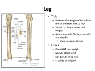
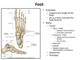
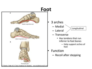
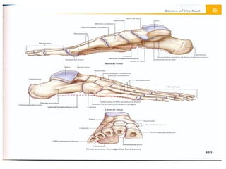
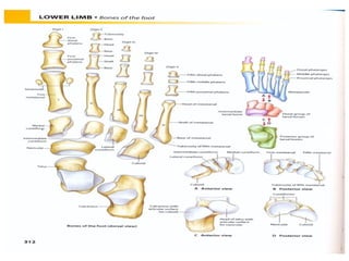
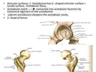
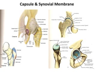
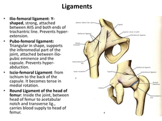
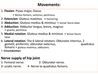
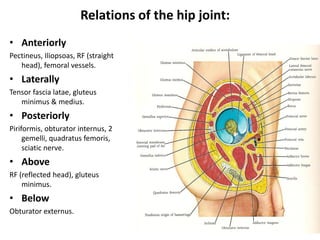
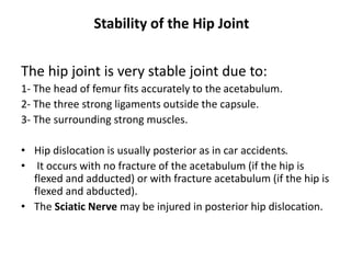
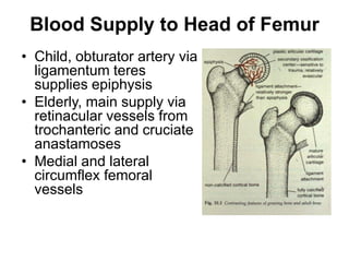
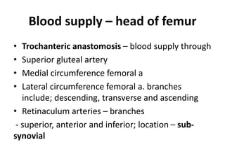
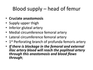
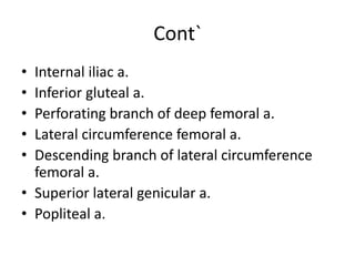
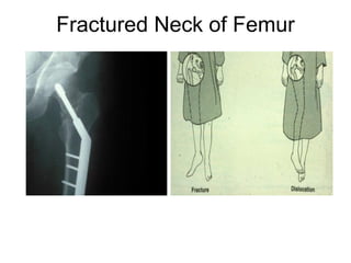
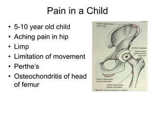
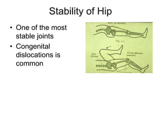
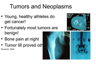
Lower Limb Skeleton and the Hip
Joint
MR SAISHA
Bones of the Lower Limb
• Function:
– Locomotion
– Carry weight of entire erect body
– Support
– Points for muscular attachments
• Components:
– Thigh
• Femur
– Knee
• Patella
– Leg
• Tibia (medial)
• Fibula (lateral)
– Foot
• Tarsals (7)
• Metatarsals (5)
• Phalanges (14)
greater sciatic
notch
ischial spine
ischial tuberosity
lesser sciatic notch
iliac crest
anterior superior
iliac spine
HIP BONE
Thigh
• Femur
– Largest, longest,
strongest bone in the
body!!
– Receives a lot of stress
– Courses medially
• More in women!
– Articulates with
acetabulum proximally
– Articulates with tibia and
patella distally
Knee
• Patella
– Triangular sesamoid
bone
– Protects knee joint
– Improves leverage of
thigh muscles acting
across the knee
– Contained within
patellar ligament
Leg
• Tibia
– Receives the weight of body from
femur and transmits to foot
– Second to femur in size and
weight
– Articulates with fibula proximally
and distally
• Interosseous membrane
• Fibula
– Does NOT bear weight
– Muscle attachment
– Not part of knee joint
– Stabilize ankle joint
Foot
• Function:
– Supports the weight of the
body
– Act as a lever to propel the
body forward
• Parts:
– Tarsals
• Talus = ankle
– Between tibia and fibula
– Articulates with both
• Calcaneus = heel
– Attachment for Calcaneal
tendon
– Carries talus
• Navicular
• Cuboid
• Medial, lateral and
intermediate cuneiforms
– Metatarsals
– Phalanges
Foot
• 3 arches
– Medial
– Lateral
– Transverse
• Has tendons that run
inferior to foot bones
– Help support arches of
foot
• Function
– Recoil after stepping
Longitudinal
The Hip Joint
• Type: Synovial (Ball & Socket)
• Articular Surfaces: head of femur & acetabulum
• Articular surfaces: 1- Acetabulumhas C- shaped articular surface =
lunate surface, Acetabular fossa,
• Acetabular notch -----► converted into acetabular foramen by
transverse ligament of the acetabulum
• Labrum acetabulare deepens the acetabular cavity.
• 2- Head of femur
Capsule & Synovial Membrane
Ligaments
• Ilio-femoral ligament: Y-
shaped, strong, attached
between AIIS and both ends of
trochantric line. Prevents hyper-
extension.
• Pubo-femoral ligament:
Triangular in shape, supports
the inferomedial part of the
joint, attached between ilio-
pubic eminence and the
capsule. Prevents hyper-
abduction.
• Iscio-femoral Ligament: from
ischium to the back of the
capsule. It becomes tense in
medial rotation.
• Round Ligament of the head of
femur: Inside the joint, between
head of femur to acetabular
notch and transverse lig.,
carries blood supply to head of
femur.
Movements:
1- Flexion: Psoas major, Iliacus
+ Rectus femoris, sartorius, pectineus.
2- Extension: Gluteus maximus + hamstring
3- Abduction: Gluteus medius & minimus + tensor fascia latae
4- Adduction: Adductor longus, brevis, magnus
+ gracilis, pectineus
5- Medial rotation: Gluteus medius & minimus + tensor fascia
latae
6- Lateral rotation: The 6 lateral rotators: Obturator internus, 2
gemelli, piriformis, obturator externus, quadratus
femoris + gluteus maximus, adductors.
7- Circumduction:
Nerve supply of hip joint
1- Femoral nerve. 2- Obturator nerve.
3- sciatic nerve. 4- Nerve to quadratus femoris.
Relations of the hip joint:
• Anteriorly
Pectineus, Iliopsoas, RF (straight
head), femoral vessels.
• Laterally
Tensor fascia latae, gluteus
minimus & medius.
• Posteriorly
Piriformis, obturator internus, 2
gemelli, quadratus femoris,
sciatic nerve.
• Above
RF (reflected head), gluteus
minimus.
• Below
Obturator externus.
Stability of the Hip Joint
The hip joint is very stable joint due to:
1- The head of femur fits accurately to the acetabulum.
2- The three strong ligaments outside the capsule.
3- The surrounding strong muscles.
• Hip dislocation is usually posterior as in car accidents.
• It occurs with no fracture of the acetabulum (if the hip is
flexed and adducted) or with fracture acetabulum (if the hip is
flexed and abducted).
• The Sciatic Nerve may be injured in posterior hip dislocation.
Blood Supply to Head of Femur
• Child, obturator artery via
ligamentum teres
supplies epiphysis
• Elderly, main supply via
retinacular vessels from
trochanteric and cruciate
anastamoses
• Medial and lateral
circumflex femoral
vessels
Blood supply – head of femur
• Trochanteric anastomosis – blood supply through
• Superior gluteal artery
• Medial circumference femoral a
• Lateral circumference femoral a. branches
include; descending, transverse and ascending
• Retinaculum arteries – branches
- superior, anterior and inferior; location – sub-
synovial
Blood supply – head of femur
• Cruciate anastomosis
• Supply upper thigh
• Inferior gluteal artery
• Medial circumference femoral artery
• Lateral circumference femoral artery
• 1st Perforating branch of profunda femoris artery
• If there is blockage in the femoral and external
iliac artery blood will reach the popliteal artery
through this anastomosis and blood flows
through;
Cont`
• Internal iliac a.
• Inferior gluteal a.
• Perforating branch of deep femoral a.
• Lateral circumference femoral a.
• Descending branch of lateral circumference
femoral a.
• Superior lateral genicular a.
• Popliteal a.
Blood Supply to the Head of Femur
Blood supply to Acetabular Fossa and
Ligament of the Head of Femur
X-Ray of the Hip Joint
MRI of the Hip Joint
Fractured Neck of Femur
Pain in a Child
• 5-10 year old child
• Aching pain in hip
• Limp
• Limitation of movement
• Perthe’s
• Osteochondritis of head
of femur
Stability of Hip
• One of the most
stable joints
• Congenital
dislocations is
common
Tumors and Neoplasms
• Young, healthy athletes do
get cancer!
• Fortunately most tumors are
benign!
• Bone pain at night
• Tumor till proved otherwise
Renström, 2008
Hip Joint Labral Tear
• Chronic
• Secondary to
acetabular dysplasia
• Part of “rim lesion”
complex
Renström, 2008
MOB TCD
Muscles of the anterior compartment
of the thigh
• The musculature of the thigh can be split into
three sections; anterior, medial and posterior.
• The muscles in the anterior compartment of the
thigh are innervated by the femoral nerve (L2-L4),
and as a general rule, act to extend the leg at the
knee joint.
• There are three major muscles in the anterior
thigh, the pectineus, sartorius and quadriceps
femoris. In addition to these, the end of the
iliopsoas passes into the anterior compartment.
Muscles of the anterior compartment
of the thigh
iliopsoas MUSCLE
• The iliopsoas muscle is a
combination of two
muscles the iliacus muscle
and the psoas major
muscle. They originate
from different areas, but
come together to form a
tendon hence why they are
commonly referred to as
one muscle.
Quadriceps femoris muscles
• The quadriceps femoris consists of four individual
muscles; three vastus muscles which are ( vastus
lateralis, vastus intermedius and vastus medialis) and
the rectus femoris. They form the main bulk of the
thigh, and collectively are one of the most powerful
muscles in the body.
• the function of these muscles is to extend the knee and
stabilize the knee bone or patella.
• All the quadriceps muscles insert into the knee bone or
patella via a tendon called the quadriceps tendon.
However the origination of the quadriceps tendons
varies and is as follows;
Quad muscles
Vastus lateralis
Origin: originates from the greater trochanter of the femur and
lateral aspect of the linea aspera.
Vastus intermedius
Vastus intermedius: originates at the anterior and lateral aspects of
the femur.
Vastus medialis
Vastus medialis: originates at the intertrochanteric line and medial
aspect of the linea aspera.
Nerve supply: branches of the femoral nerve
The quads
IN CONCLUSION OF HIP FLEXORS
• We have provided a concise review of the
anatomical aspect of the hip flexors. The
approach to a better understanding of the hip
flexors gives credence to current and future
complications and solutions of the hip as a
physiotherapist.
THANK YOU
FOR LISTENING

Lower Limb Skeleton and the Hip Joint.pdf

  • 1.
    Lower Limb Skeletonand the Hip Joint MR SAISHA
  • 2.
    Bones of theLower Limb • Function: – Locomotion – Carry weight of entire erect body – Support – Points for muscular attachments • Components: – Thigh • Femur – Knee • Patella – Leg • Tibia (medial) • Fibula (lateral) – Foot • Tarsals (7) • Metatarsals (5) • Phalanges (14)
  • 3.
    greater sciatic notch ischial spine ischialtuberosity lesser sciatic notch iliac crest anterior superior iliac spine HIP BONE
  • 4.
    Thigh • Femur – Largest,longest, strongest bone in the body!! – Receives a lot of stress – Courses medially • More in women! – Articulates with acetabulum proximally – Articulates with tibia and patella distally
  • 5.
    Knee • Patella – Triangularsesamoid bone – Protects knee joint – Improves leverage of thigh muscles acting across the knee – Contained within patellar ligament
  • 6.
    Leg • Tibia – Receivesthe weight of body from femur and transmits to foot – Second to femur in size and weight – Articulates with fibula proximally and distally • Interosseous membrane • Fibula – Does NOT bear weight – Muscle attachment – Not part of knee joint – Stabilize ankle joint
  • 8.
    Foot • Function: – Supportsthe weight of the body – Act as a lever to propel the body forward • Parts: – Tarsals • Talus = ankle – Between tibia and fibula – Articulates with both • Calcaneus = heel – Attachment for Calcaneal tendon – Carries talus • Navicular • Cuboid • Medial, lateral and intermediate cuneiforms – Metatarsals – Phalanges
  • 10.
    Foot • 3 arches –Medial – Lateral – Transverse • Has tendons that run inferior to foot bones – Help support arches of foot • Function – Recoil after stepping Longitudinal
  • 13.
    The Hip Joint •Type: Synovial (Ball & Socket) • Articular Surfaces: head of femur & acetabulum
  • 14.
    • Articular surfaces:1- Acetabulumhas C- shaped articular surface = lunate surface, Acetabular fossa, • Acetabular notch -----► converted into acetabular foramen by transverse ligament of the acetabulum • Labrum acetabulare deepens the acetabular cavity. • 2- Head of femur
  • 15.
  • 16.
    Ligaments • Ilio-femoral ligament:Y- shaped, strong, attached between AIIS and both ends of trochantric line. Prevents hyper- extension. • Pubo-femoral ligament: Triangular in shape, supports the inferomedial part of the joint, attached between ilio- pubic eminence and the capsule. Prevents hyper- abduction. • Iscio-femoral Ligament: from ischium to the back of the capsule. It becomes tense in medial rotation. • Round Ligament of the head of femur: Inside the joint, between head of femur to acetabular notch and transverse lig., carries blood supply to head of femur.
  • 17.
    Movements: 1- Flexion: Psoasmajor, Iliacus + Rectus femoris, sartorius, pectineus. 2- Extension: Gluteus maximus + hamstring 3- Abduction: Gluteus medius & minimus + tensor fascia latae 4- Adduction: Adductor longus, brevis, magnus + gracilis, pectineus 5- Medial rotation: Gluteus medius & minimus + tensor fascia latae 6- Lateral rotation: The 6 lateral rotators: Obturator internus, 2 gemelli, piriformis, obturator externus, quadratus femoris + gluteus maximus, adductors. 7- Circumduction: Nerve supply of hip joint 1- Femoral nerve. 2- Obturator nerve. 3- sciatic nerve. 4- Nerve to quadratus femoris.
  • 18.
    Relations of thehip joint: • Anteriorly Pectineus, Iliopsoas, RF (straight head), femoral vessels. • Laterally Tensor fascia latae, gluteus minimus & medius. • Posteriorly Piriformis, obturator internus, 2 gemelli, quadratus femoris, sciatic nerve. • Above RF (reflected head), gluteus minimus. • Below Obturator externus.
  • 19.
    Stability of theHip Joint The hip joint is very stable joint due to: 1- The head of femur fits accurately to the acetabulum. 2- The three strong ligaments outside the capsule. 3- The surrounding strong muscles. • Hip dislocation is usually posterior as in car accidents. • It occurs with no fracture of the acetabulum (if the hip is flexed and adducted) or with fracture acetabulum (if the hip is flexed and abducted). • The Sciatic Nerve may be injured in posterior hip dislocation.
  • 20.
    Blood Supply toHead of Femur • Child, obturator artery via ligamentum teres supplies epiphysis • Elderly, main supply via retinacular vessels from trochanteric and cruciate anastamoses • Medial and lateral circumflex femoral vessels
  • 21.
    Blood supply –head of femur • Trochanteric anastomosis – blood supply through • Superior gluteal artery • Medial circumference femoral a • Lateral circumference femoral a. branches include; descending, transverse and ascending • Retinaculum arteries – branches - superior, anterior and inferior; location – sub- synovial
  • 22.
    Blood supply –head of femur • Cruciate anastomosis • Supply upper thigh • Inferior gluteal artery • Medial circumference femoral artery • Lateral circumference femoral artery • 1st Perforating branch of profunda femoris artery • If there is blockage in the femoral and external iliac artery blood will reach the popliteal artery through this anastomosis and blood flows through;
  • 23.
    Cont` • Internal iliaca. • Inferior gluteal a. • Perforating branch of deep femoral a. • Lateral circumference femoral a. • Descending branch of lateral circumference femoral a. • Superior lateral genicular a. • Popliteal a.
  • 24.
    Blood Supply tothe Head of Femur
  • 25.
    Blood supply toAcetabular Fossa and Ligament of the Head of Femur
  • 26.
    X-Ray of theHip Joint
  • 27.
    MRI of theHip Joint
  • 28.
  • 29.
    Pain in aChild • 5-10 year old child • Aching pain in hip • Limp • Limitation of movement • Perthe’s • Osteochondritis of head of femur
  • 30.
    Stability of Hip •One of the most stable joints • Congenital dislocations is common
  • 31.
    Tumors and Neoplasms •Young, healthy athletes do get cancer! • Fortunately most tumors are benign! • Bone pain at night • Tumor till proved otherwise Renström, 2008
  • 32.
    Hip Joint LabralTear • Chronic • Secondary to acetabular dysplasia • Part of “rim lesion” complex Renström, 2008 MOB TCD
  • 34.
    Muscles of theanterior compartment of the thigh • The musculature of the thigh can be split into three sections; anterior, medial and posterior. • The muscles in the anterior compartment of the thigh are innervated by the femoral nerve (L2-L4), and as a general rule, act to extend the leg at the knee joint. • There are three major muscles in the anterior thigh, the pectineus, sartorius and quadriceps femoris. In addition to these, the end of the iliopsoas passes into the anterior compartment.
  • 35.
    Muscles of theanterior compartment of the thigh
  • 36.
    iliopsoas MUSCLE • Theiliopsoas muscle is a combination of two muscles the iliacus muscle and the psoas major muscle. They originate from different areas, but come together to form a tendon hence why they are commonly referred to as one muscle.
  • 37.
    Quadriceps femoris muscles •The quadriceps femoris consists of four individual muscles; three vastus muscles which are ( vastus lateralis, vastus intermedius and vastus medialis) and the rectus femoris. They form the main bulk of the thigh, and collectively are one of the most powerful muscles in the body. • the function of these muscles is to extend the knee and stabilize the knee bone or patella. • All the quadriceps muscles insert into the knee bone or patella via a tendon called the quadriceps tendon. However the origination of the quadriceps tendons varies and is as follows;
  • 38.
    Quad muscles Vastus lateralis Origin:originates from the greater trochanter of the femur and lateral aspect of the linea aspera. Vastus intermedius Vastus intermedius: originates at the anterior and lateral aspects of the femur. Vastus medialis Vastus medialis: originates at the intertrochanteric line and medial aspect of the linea aspera. Nerve supply: branches of the femoral nerve
  • 39.
  • 40.
    IN CONCLUSION OFHIP FLEXORS • We have provided a concise review of the anatomical aspect of the hip flexors. The approach to a better understanding of the hip flexors gives credence to current and future complications and solutions of the hip as a physiotherapist.
  • 41.