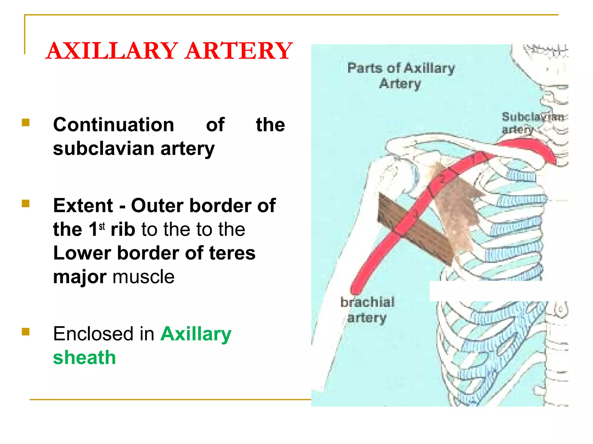
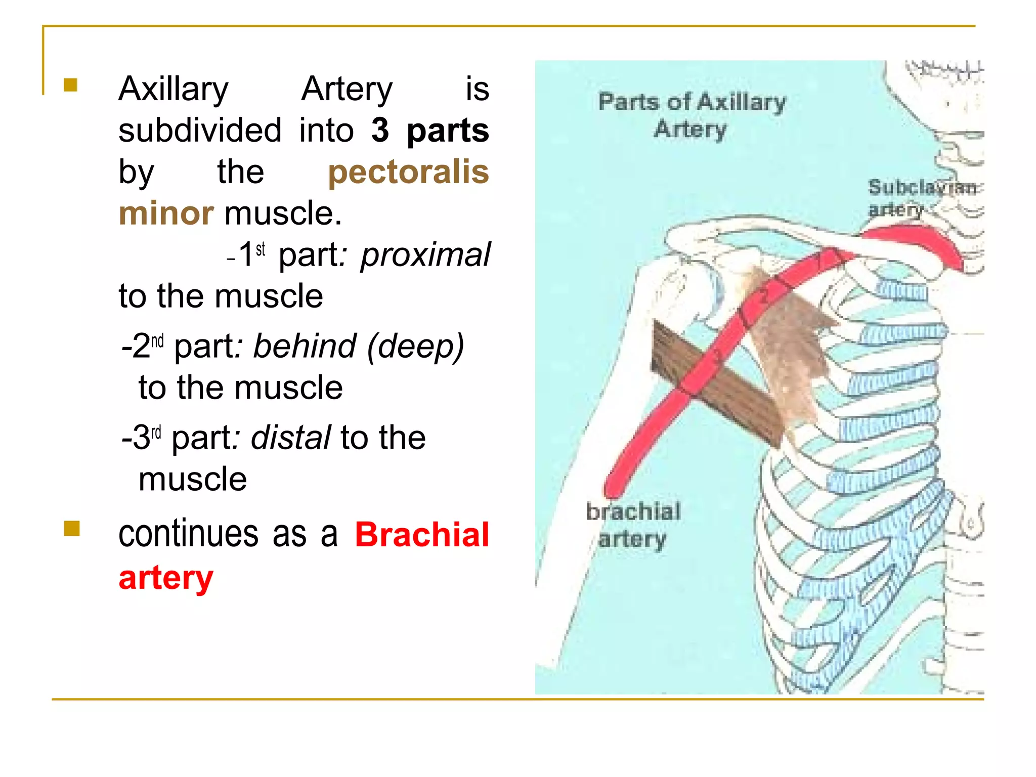
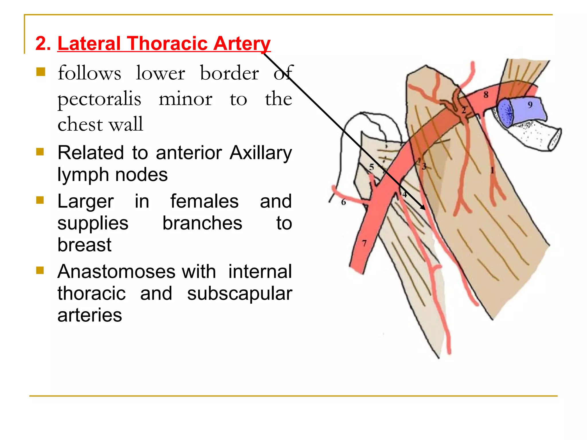
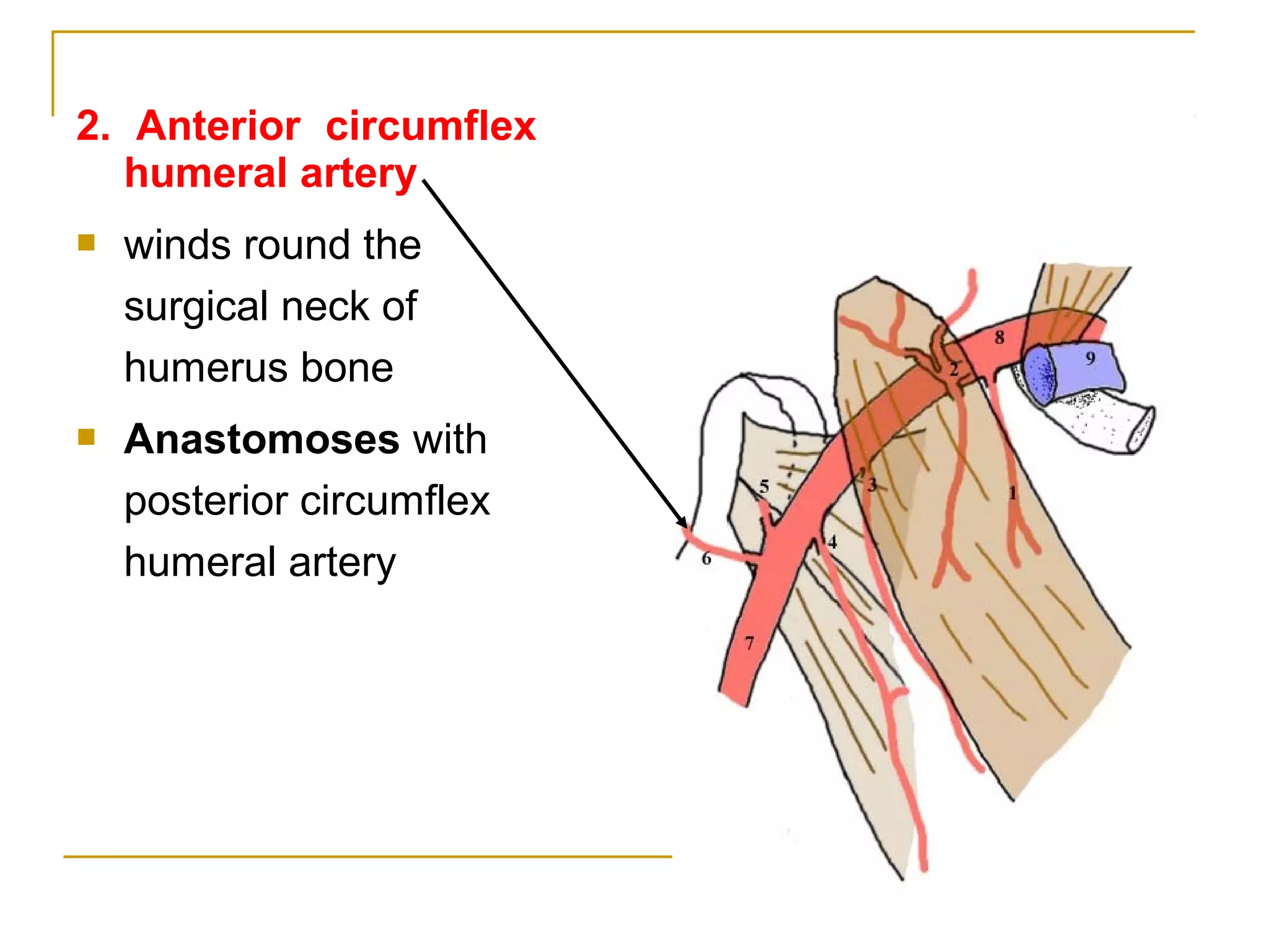
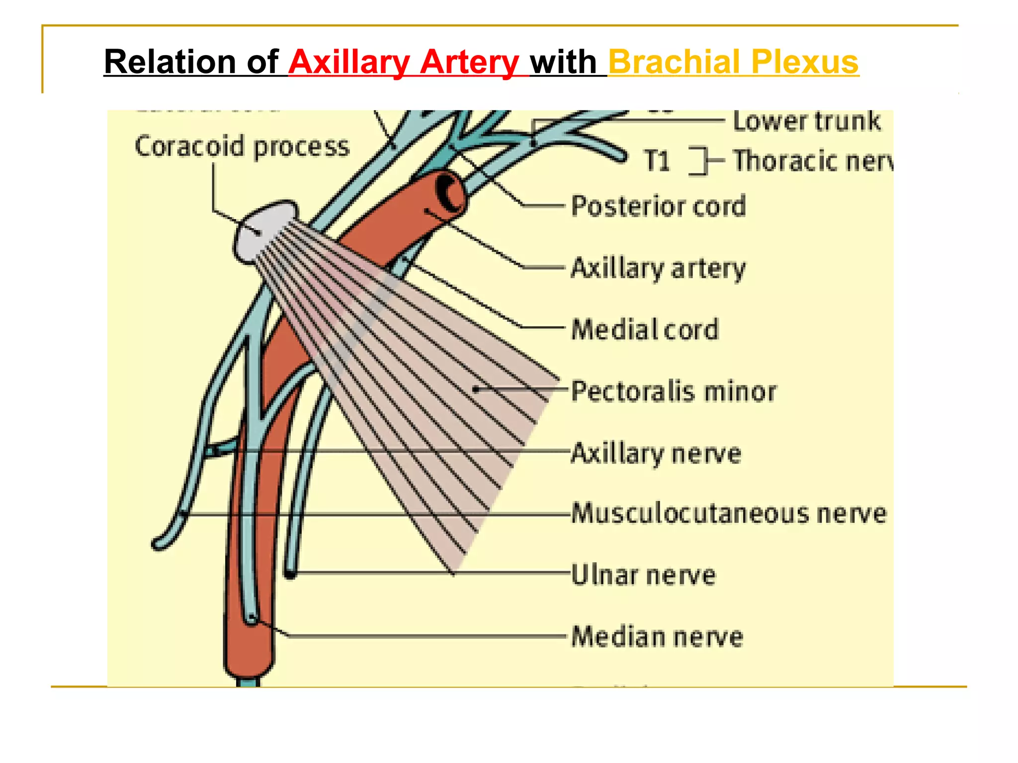
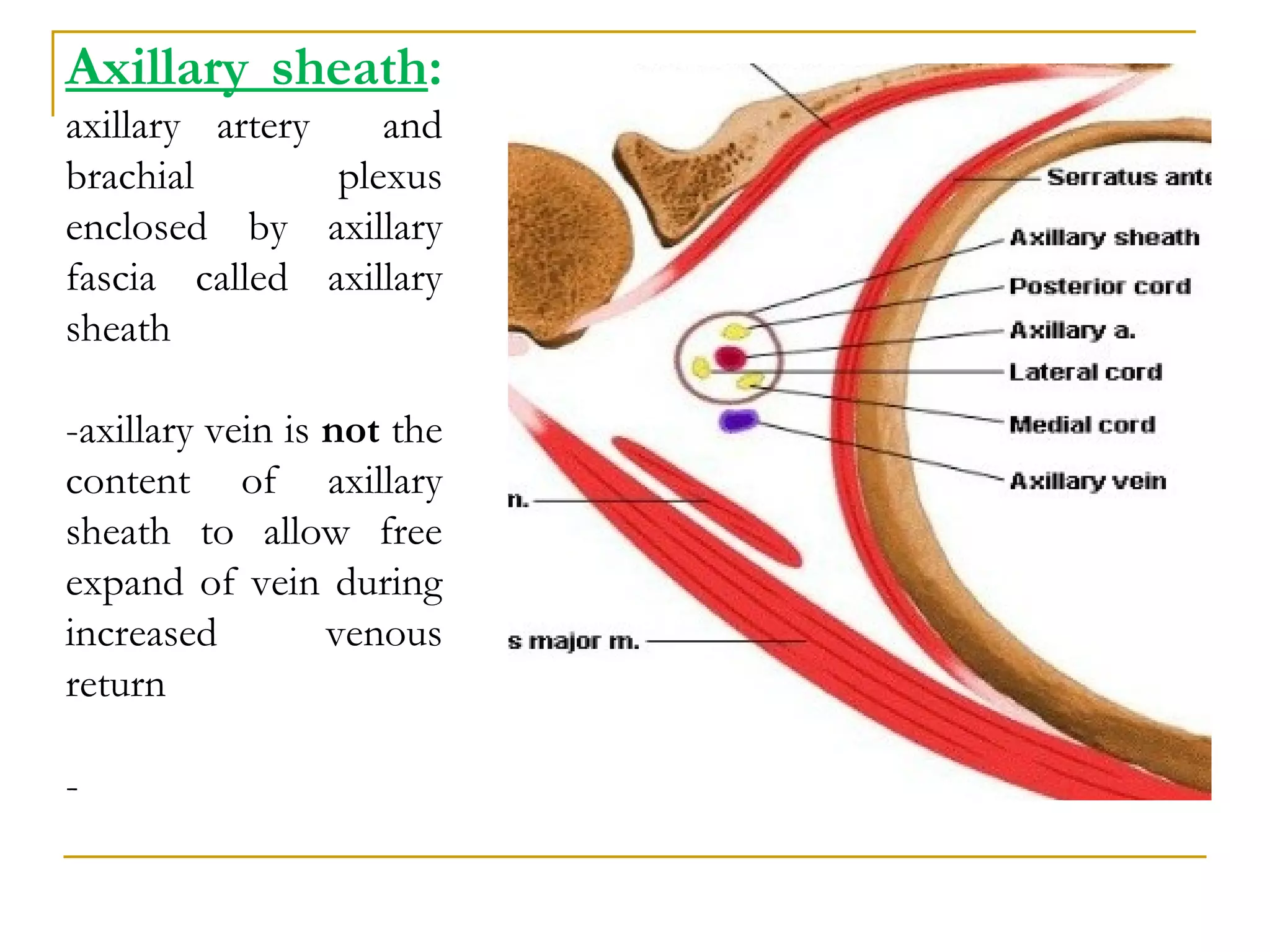
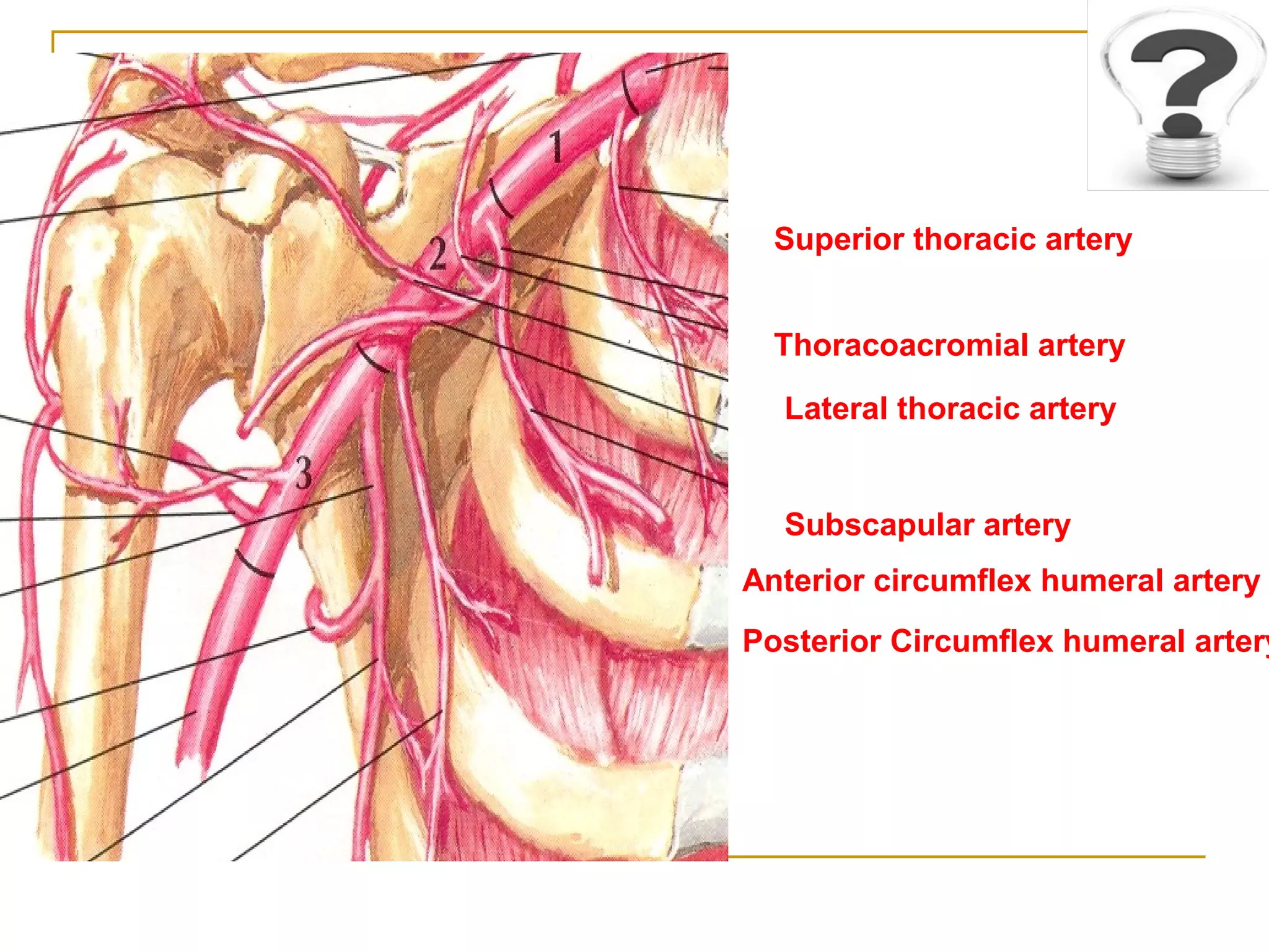
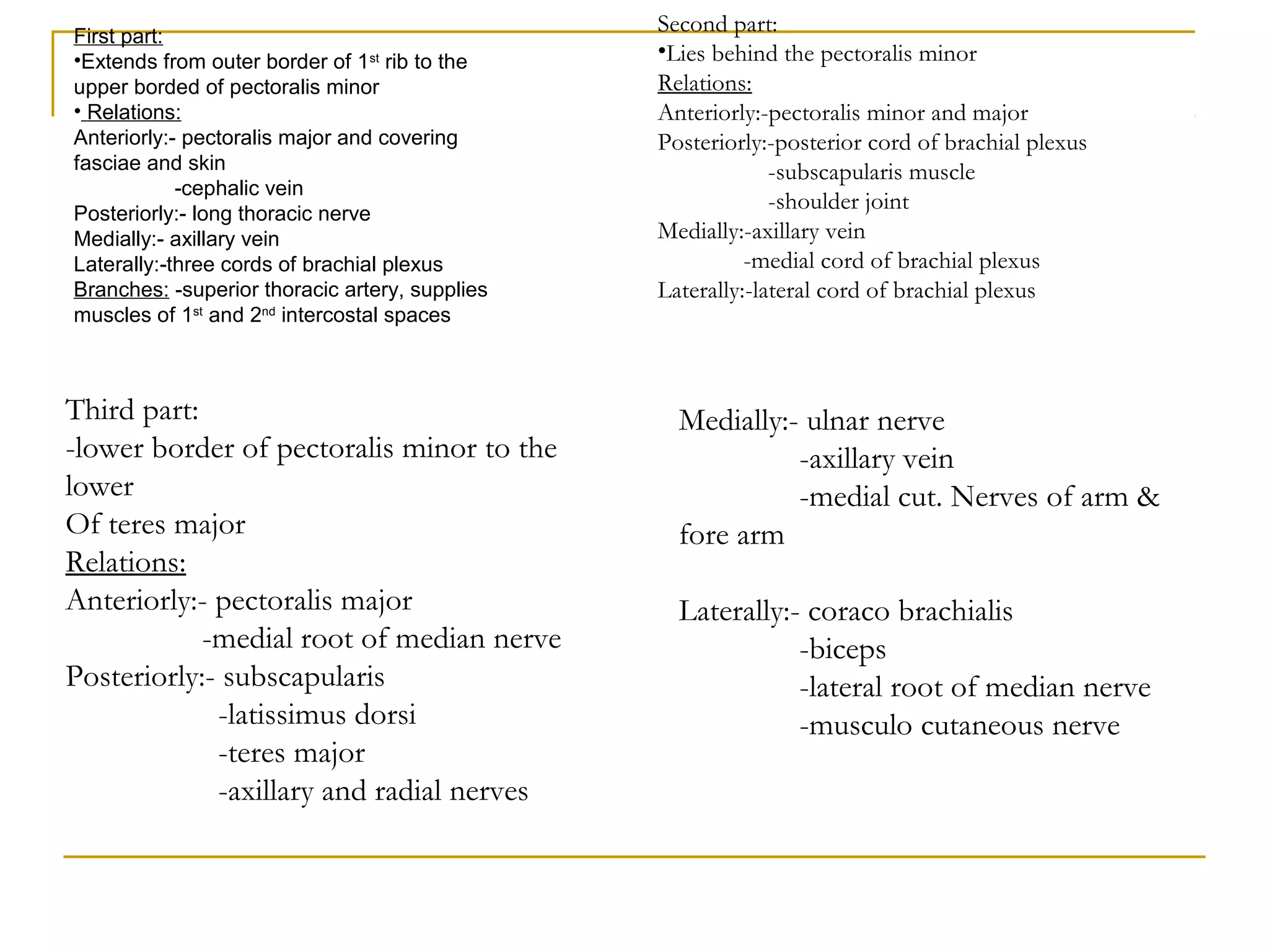
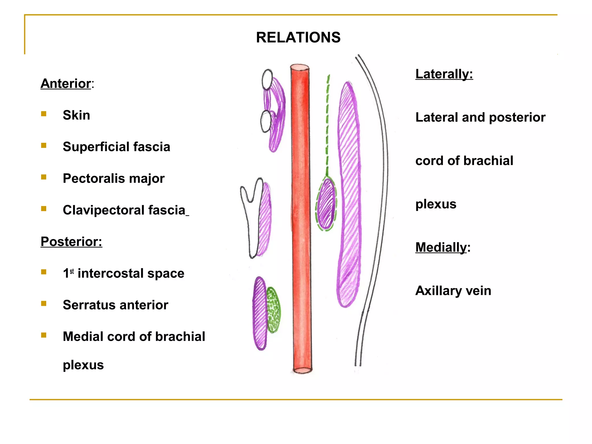
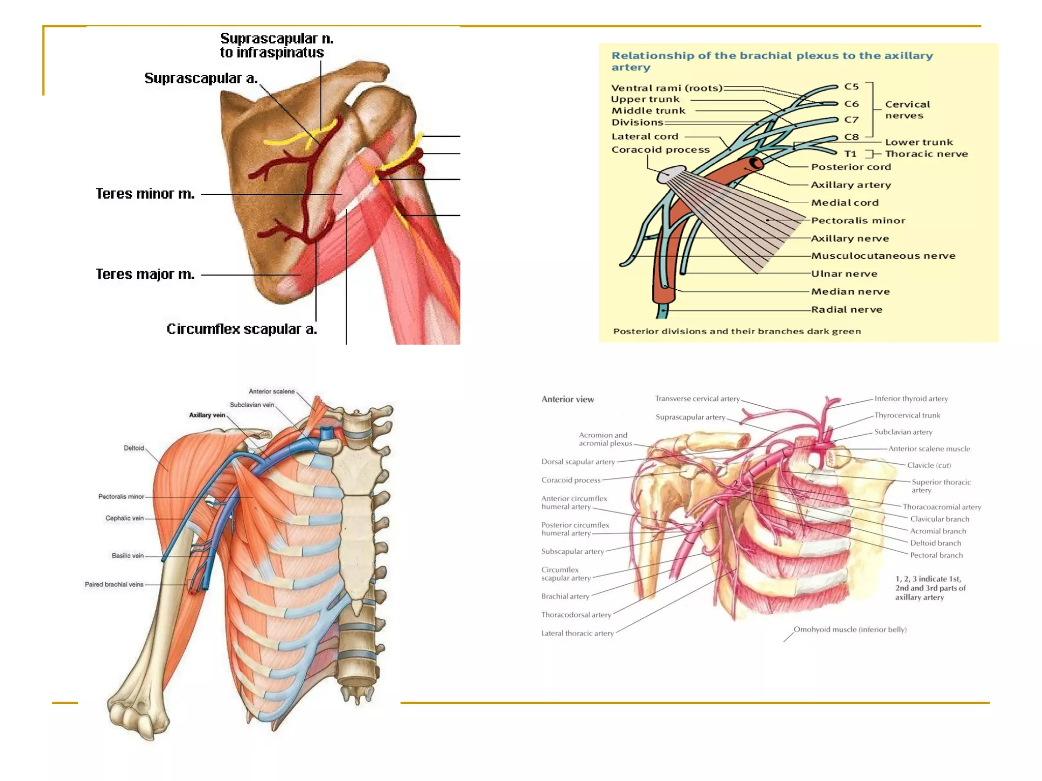
Axillary Artery
& Axillary Vein
Dr. Kamil Khan
ARTERIES OF UPPER LIMB
Axillary artery
A) Brachial artery
B) Radial artery
C) Ulnar artery
AXILLARY ARTERY
 Continuation of the
subclavian artery
 Extent - Outer border of
the 1st
rib to the to the
Lower border of teres
major muscle
 Enclosed in Axillary
sheath
 Axillary Artery is
subdivided into 3 parts
by the pectoralis
minor muscle.
-1st
part: proximal
to the muscle
-2nd
part: behind (deep)
to the muscle
-3rd
part: distal to the
muscle
 continues as a Brachial
artery
Superior thoracic
artery
1st
part of axillary artery : extends from outer border of first
rib to the upper border of pectoralis minor
Branches(1)
1. Superior Thoracic Artery- Arises near lower border of
subclavius muscle & Supplies thoracic wall and breast
Lateral
thoracic
artery
Thoracoacromial
artery
2nd
part of Axillary artery : lies deep to
pectoralis minor
Branches(2)
1.Thoracoacromaial Artery
-Pierces clavipectoral
fascia
Branches:
a. Pectoral
b. Deltoid
c. Acromial
d. Clavicular
a. Deltoid - supplies Pectoralis
major and deltoid muscles
b. Clavicular- Sternoclavicular
joint and subclavius
c. Acromial - anastomosis with
deltoid branch
d. Pectoral - supplies Pectoral
muscles and breast
2. Lateral Thoracic Artery
 follows lower border of
pectoralis minor to the
chest wall
 Related to anterior Axillary
lymph nodes
 Larger in females and
supplies branches to
breast
 Anastomoses with internal
thoracic and subscapular
arteries
3rd
part of Axillary artery : extends from lower
border of pectoralis minor upto the level of lower
border of teres major muscle.
Branches(3)
1. Subscapular artery
2. Anterior circumflex humeral artery
3. Posterior Circumflex humeral artery
1. Subscapular artery
 Largest branch of Axillary
artery
 divides into 2 branches
 a. circumflex scapular artery:
content of upper
triangular space
participate in scapular
anastomosis
 b. thoraco dorsal artery:
runs with the thoraco dorsal
nerve
 Reaches inferior angle
of scapula and
anastomoses
2. Anterior circumflex
humeral artery
 winds round the
surgical neck of
humerus bone
 Anastomoses with
posterior circumflex
humeral artery
3. Posterior Circumflex
humeral artery
 Larger than anterior
circumflex humeral
artery
 pass through the
Quadrangular Space
accompanied by
Axillary nerve
 Anastomoses with
anterior circumflex
around surgical neck
of humerus.
Relation of Axillary Artery with Brachial Plexus
Applied Anatomy of Axillary artery
 Axillary artery pulsation can be felt against lower part of
lat wall of axilla
 In order to check bleeding from distal part of upper limb
the artery can be compressed against humerus against
lower part of lat wall of axilla
 Next to popliteal artery , axill art 2nd
most common art to
be lacerated by violent movements
 Aneurysm of the Axillary artery:-
- Compresses trunks of brachial plexus causing pain
and anesthesia in the areas of the skin supplied by the
affected nerves
Scapular Anastomosis
1. In each of subscapular,supraspinous & infraspinous fossae
Arterial anastomosis takes place around scapula between the branches of the
1st
part of subclavian artery and principally 3rd
part of axillary Artery
 2. over the Acromion Process
Applied Anatomy of Scapular Anastomosis
Collateral circulation
Axillary vein:
Formation: union of venae comitantes of the brachial
artery & basilic vein at the lower border of teres major
-Ends at outer border of 1st
rib by becoming the
subclavian vein
-runs upwards on the medial side of the axillary artery
Tributaries: -cephalic vein
-veins corresponding to the branches of
axillary artery
Vena comitans
•Frequently multiple(2 or3)
•Run with their corresponding
Arteries
Applied Anatomy of axillary vein
Axillary venous thrombosis
Axillary sheath:
axillary artery and
brachial plexus
enclosed by axillary
fascia called axillary
sheath
-axillary vein is not the
content of axillary
sheath to allow free
expand of vein during
increased venous
return
-
 continuous with the prevertebral fascia at the
root of the neck
 Inferiorly, axillary sheath extend upto elbow
Applied-
cold abscess originating from cervical vertebrae
may track down through this sheath
Superior thoracic artery
Thoracoacromial artery
Lateral thoracic artery
Subscapular artery
Anterior circumflex humeral artery
Posterior Circumflex humeral artery
First part:
•Extends from outer border of 1st
rib to the
upper borded of pectoralis minor
• Relations:
Anteriorly:- pectoralis major and covering
fasciae and skin
-cephalic vein
Posteriorly:- long thoracic nerve
Medially:- axillary vein
Laterally:-three cords of brachial plexus
Branches: -superior thoracic artery, supplies
muscles of 1st
and 2nd
intercostal spaces
Second part:
•Lies behind the pectoralis minor
Relations:
Anteriorly:-pectoralis minor and major
Posteriorly:-posterior cord of brachial plexus
-subscapularis muscle
-shoulder joint
Medially:-axillary vein
-medial cord of brachial plexus
Laterally:-lateral cord of brachial plexus
Third part:
-lower border of pectoralis minor to the
lower
Of teres major
Relations:
Anteriorly:- pectoralis major
-medial root of median nerve
Posteriorly:- subscapularis
-latissimus dorsi
-teres major
-axillary and radial nerves
Medially:- ulnar nerve
-axillary vein
-medial cut. Nerves of arm &
fore arm
Laterally:- coraco brachialis
-biceps
-lateral root of median nerve
-musculo cutaneous nerve
Anterior:
 Skin
 Superficial fascia
 Pectoralis major
 Clavipectoral fascia
Posterior:
 1st
intercostal space
 Serratus anterior
 Medial cord of brachial
plexus
Laterally:
Lateral and posterior
cord of brachial
plexus
Medially:
Axillary vein
RELATIONS
SKIN
SUPERFICIAL
FASCIA
CLAVIPECTORAL
FASCIA
PECTORALIS
MAJOR
PECTORALIS
MINOR
AXILLARY ARTERY
1ST
RIB
2ND RIB
SUBSCAPULARIS
TERES MAJOR
LATISSIMUS DORSI
SERRATUS ANTERIOR
Branches :AXILLARY
A
Axillary artery and axillary vein

Axillary artery and axillary vein

  • 1.
    Axillary Artery & AxillaryVein Dr. Kamil Khan
  • 2.
    ARTERIES OF UPPERLIMB Axillary artery A) Brachial artery B) Radial artery C) Ulnar artery
  • 3.
    AXILLARY ARTERY  Continuationof the subclavian artery  Extent - Outer border of the 1st rib to the to the Lower border of teres major muscle  Enclosed in Axillary sheath
  • 4.
     Axillary Arteryis subdivided into 3 parts by the pectoralis minor muscle. -1st part: proximal to the muscle -2nd part: behind (deep) to the muscle -3rd part: distal to the muscle  continues as a Brachial artery
  • 5.
    Superior thoracic artery 1st part ofaxillary artery : extends from outer border of first rib to the upper border of pectoralis minor Branches(1) 1. Superior Thoracic Artery- Arises near lower border of subclavius muscle & Supplies thoracic wall and breast
  • 6.
    Lateral thoracic artery Thoracoacromial artery 2nd part of Axillaryartery : lies deep to pectoralis minor Branches(2) 1.Thoracoacromaial Artery -Pierces clavipectoral fascia Branches: a. Pectoral b. Deltoid c. Acromial d. Clavicular
  • 7.
    a. Deltoid -supplies Pectoralis major and deltoid muscles b. Clavicular- Sternoclavicular joint and subclavius c. Acromial - anastomosis with deltoid branch d. Pectoral - supplies Pectoral muscles and breast
  • 8.
    2. Lateral ThoracicArtery  follows lower border of pectoralis minor to the chest wall  Related to anterior Axillary lymph nodes  Larger in females and supplies branches to breast  Anastomoses with internal thoracic and subscapular arteries
  • 9.
    3rd part of Axillaryartery : extends from lower border of pectoralis minor upto the level of lower border of teres major muscle. Branches(3) 1. Subscapular artery 2. Anterior circumflex humeral artery 3. Posterior Circumflex humeral artery
  • 10.
    1. Subscapular artery Largest branch of Axillary artery  divides into 2 branches  a. circumflex scapular artery: content of upper triangular space participate in scapular anastomosis  b. thoraco dorsal artery: runs with the thoraco dorsal nerve  Reaches inferior angle of scapula and anastomoses
  • 11.
    2. Anterior circumflex humeralartery  winds round the surgical neck of humerus bone  Anastomoses with posterior circumflex humeral artery
  • 12.
    3. Posterior Circumflex humeralartery  Larger than anterior circumflex humeral artery  pass through the Quadrangular Space accompanied by Axillary nerve  Anastomoses with anterior circumflex around surgical neck of humerus.
  • 13.
    Relation of AxillaryArtery with Brachial Plexus
  • 14.
    Applied Anatomy ofAxillary artery  Axillary artery pulsation can be felt against lower part of lat wall of axilla  In order to check bleeding from distal part of upper limb the artery can be compressed against humerus against lower part of lat wall of axilla  Next to popliteal artery , axill art 2nd most common art to be lacerated by violent movements  Aneurysm of the Axillary artery:- - Compresses trunks of brachial plexus causing pain and anesthesia in the areas of the skin supplied by the affected nerves
  • 15.
    Scapular Anastomosis 1. Ineach of subscapular,supraspinous & infraspinous fossae Arterial anastomosis takes place around scapula between the branches of the 1st part of subclavian artery and principally 3rd part of axillary Artery
  • 16.
     2. overthe Acromion Process
  • 17.
    Applied Anatomy ofScapular Anastomosis Collateral circulation
  • 18.
    Axillary vein: Formation: unionof venae comitantes of the brachial artery & basilic vein at the lower border of teres major -Ends at outer border of 1st rib by becoming the subclavian vein
  • 19.
    -runs upwards onthe medial side of the axillary artery Tributaries: -cephalic vein -veins corresponding to the branches of axillary artery
  • 20.
    Vena comitans •Frequently multiple(2or3) •Run with their corresponding Arteries
  • 21.
    Applied Anatomy ofaxillary vein Axillary venous thrombosis
  • 22.
    Axillary sheath: axillary arteryand brachial plexus enclosed by axillary fascia called axillary sheath -axillary vein is not the content of axillary sheath to allow free expand of vein during increased venous return -
  • 23.
     continuous withthe prevertebral fascia at the root of the neck  Inferiorly, axillary sheath extend upto elbow Applied- cold abscess originating from cervical vertebrae may track down through this sheath
  • 24.
    Superior thoracic artery Thoracoacromialartery Lateral thoracic artery Subscapular artery Anterior circumflex humeral artery Posterior Circumflex humeral artery
  • 26.
    First part: •Extends fromouter border of 1st rib to the upper borded of pectoralis minor • Relations: Anteriorly:- pectoralis major and covering fasciae and skin -cephalic vein Posteriorly:- long thoracic nerve Medially:- axillary vein Laterally:-three cords of brachial plexus Branches: -superior thoracic artery, supplies muscles of 1st and 2nd intercostal spaces Second part: •Lies behind the pectoralis minor Relations: Anteriorly:-pectoralis minor and major Posteriorly:-posterior cord of brachial plexus -subscapularis muscle -shoulder joint Medially:-axillary vein -medial cord of brachial plexus Laterally:-lateral cord of brachial plexus Third part: -lower border of pectoralis minor to the lower Of teres major Relations: Anteriorly:- pectoralis major -medial root of median nerve Posteriorly:- subscapularis -latissimus dorsi -teres major -axillary and radial nerves Medially:- ulnar nerve -axillary vein -medial cut. Nerves of arm & fore arm Laterally:- coraco brachialis -biceps -lateral root of median nerve -musculo cutaneous nerve
  • 27.
    Anterior:  Skin  Superficialfascia  Pectoralis major  Clavipectoral fascia Posterior:  1st intercostal space  Serratus anterior  Medial cord of brachial plexus Laterally: Lateral and posterior cord of brachial plexus Medially: Axillary vein RELATIONS
  • 28.
  • 29.