SlideShare a Scribd company logo
Ojvensha E learning Resources-Prepared by Dr.B.B.Gosai
Ventricles of Brain
Introduction: Cavities in the brain containing cerebrospinal fluid is known as Ventricles.
Cavities of Brain:
Cavities of Cerebrum: Lateral Ventricles (Right and Left Lateral ventricle in right and left cerebral
hemispheres.
Cavity of Diencepahlon: Third Ventricle.
Cavity of Midbrain: Cerebral aqueduct connecting third ventricle with fourth ventricle.
Cavity of Hindbrain (Pons-Medulla oblongata and Cerebellum): Fourth ventricle.
Lateral Ventricle (Cavity of cerebrum)
(*****Most Important Short Note)
Ojvensha E learning Resources-Prepared by Dr.B.B.Gosai
FEATURES SEEN IN LATERAL VENTRICLE
 Cavity of Telencephalon-cerebrum
 Shape: “C” shaped
 Parts with features seen in each part:
 Anterior Horn: Located in frontal lobe.
 Features and boundaries:
 Floor contains head of caudate nucleus.
 Anterior wall, floor and roof formed by corpus callosum.
 Medial wall by septum pellucidum.
 Lateral wall and floor is sloping and formed by head of caudate nucleus.
 Central Part: Located in parietal lobe.
 Features and boundaries:
 Floor contain: Body of caudate nucleus and thalamus.
 Roof by trunk of corpus callosum.
Ojvensha E learning Resources-Prepared by Dr.B.B.Gosai
 Medial wall by septum pellucidum.
 Lateral wall and floor is sloping and formed by head of caudate nucleus.
 Posterior horn: Located in occipital lobe. Small and inconstant.
 Features: If this horn is present it shows following features:
 Bulb of posterior horn raised by forceps major.
 Calcar avis raised by calcarine sulcus is seen into it.
 Inferior horn: Located in temporal lobe.
 Features seen:
 Features in the floor: Hippocampus and pes hippocampi seen its floor covered
by alveus and fimbria.
 Roof formed by tail of caudate nucleus and amygdaloid body.
 Communication with third ventricle by interventricular foramen of Monroe
 Function: Contain choroid plexus responsible for secretion of cerebrospinal fluid.
 Applied Anatomy: Blockage of interventricular foramen leads to dilatation of lateral ventricle.
This condition is known as Hydrocephalus. It leads to mental retardation.
Third Ventricle (Cavity of Diencephalon)
(***Important Short Note)
 Cavity of diencephalon located between diencephalon i.e. between two thalami.
 Shape: Slit like
 Anterior wall: Lamina terminalis, anterior commissure and anterior column of fornix.
 Posterior wall: Pineal gland with pineal recess and habenular and posterior commissure.
 Lateral wall: Thalamus and Hypothalamus separated by hypothalamic sulcus
 Superior wall (Roof): Ependyma, tela choroidea of third ventricle, internal cerebral veins,
fornix and corpus callosum.
 Inferior wall (Floor): Optic chiasma, tuber cinerium, infundibulum with its recess, mammilary
bodies, tegmentum of cerebral peduncles of midbrain.
Ojvensha E learning Resources-Prepared by Dr.B.B.Gosai
 Recesses:
 Pineal recess
 Supraoptic recess
 Infundibular recess
 Communications:
 Anterosuperiorly with lateral ventricle by interventricular foramen of Monroe.
 Posteroinferiorly with fourth ventricle by cerebral aqueduct.
 Function: Contain choroid plexus responsible for secretion of cerebrospinal fluid.
 Applied Anatomy: Blockage of cerebral aqueduct leads to dilatation of third ventricle and both
lateral ventricle. This condition is known as Hydrocephalus. It leads to mental retardation.
Ojvensha E learning Resources-Prepared by Dr.B.B.Gosai
Fourth Ventricle (Cavity of Hindbrain)
(*****Most Important Short Note)
 Cavity of hindbrain located between pons and medulla anteriorly and cerebellum posteriorly.
 Shape: “Tent: shape in sagittal section.
 Lateral Boundaries: Caudal part by inferior cerebellar peduncle and cranial part by superior
cerebellar peduncle.
 Posterior wall (Roof): tent shaped projects into cerebellum. Related to superior and nferior
medullary velum. Contains Median aperture (Foramen of Megendie) anf Lateral openings
(Foramen of Luschka).
 Rhomboid Fossa (Floor of fourth ventricle): Diamond shaped. Formed by posterior surface of
pons and medulla oblongata.
 Boundaries: Superolaterlly by superior cerebellar peduncle and inferolaterally by
Inferior cerebellar peduncle.
 Features seen:
Ojvensha E learning Resources-Prepared by Dr.B.B.Gosai
 Hypoglossal and vagal triangles
 facial colliculus
 Vestibular area
 Median sulcus
 Medial eminence
 Sulcus limitans
 Recesses:
 Dorsal median recess
 Two lateral recesses with lateral aperture.
 Communications:
 Superiorly with third ventricle by cerebral
 Inferiorly with central canal of spinal cord, and subarachnoid space by Median
aperture of Megendie.
Ojvensha E learning Resources-Prepared by Dr.B.B.Gosai
 Laterally with subarachnoid space by lateral aperture of Lushka.
 Function: Contain choroid plexus responsible for secretion of cerebrospinal fluid.
 Applied Anatomy: Blockage of openings leads to dilatation of all ventricles. This condition is
known as Hydrocephalus. It leads to mental retardation.
Applied Anatomy of Ventricles:
 HYDROCEPHALUS: Abnormal increase in the volume of cerebrospinal fluid within skull.
 Types:
 Non-communicating hydrocephalus: raised pressure is due to blockage between
choroid plexus and exit foramina.
 Communicating hydrocephalus: No obstruction. Causes can be excessive formation
or diminished absorption.
 INTRACRANIAL HYPERTENSION AND PAPILLEDEMA: congestion of retinal veins,
bulging forward of optic disc and edema of the disc.
Hydrocephalus
(Photo for understanding dilatation of ventricles and thinning of brain tissue)
==================X================

More Related Content

What's hot

Lateral ventricle of Brain. By Dr.N.Mugunthan.M.S
Lateral ventricle of Brain. By Dr.N.Mugunthan.M.SLateral ventricle of Brain. By Dr.N.Mugunthan.M.S
Lateral ventricle of Brain. By Dr.N.Mugunthan.M.S
mgmcri1234
 
Third ventricle
Third ventricleThird ventricle
Third ventricle
PGINeurosurgery
 
Facial artery dr gosai
Facial artery dr gosaiFacial artery dr gosai
Facial artery dr gosai
Dr.B.B. Gosai
 
Anterior triangle of the neck
Anterior triangle of the neckAnterior triangle of the neck
Anterior triangle of the neck
Moamer Gabsa
 
4th ventricle
4th ventricle4th ventricle
4th ventricle
Rati Tandon
 
Ansa cervicalis
Ansa cervicalisAnsa cervicalis
Ansa cervicalis
GeethaHari3
 
Anatomy of midbrain
Anatomy of midbrainAnatomy of midbrain
Anatomy of midbrain
Dr. Mohammad Mahmoud
 
Anatomy of face
Anatomy of faceAnatomy of face
Anatomy of face
Yogha Padhma Asokan
 
Scalp
Scalp Scalp
Scalp
mgmcri1234
 
Norma basalis
Norma basalis  Norma basalis
Norma basalis
Dr. Devi Shankar
 
The brainstem
The brainstemThe brainstem
The brainstem
Vyjayanthi Kadambi
 
Anatomy of medulla oblongata
Anatomy of medulla oblongataAnatomy of medulla oblongata
Anatomy of medulla oblongata
Dr. Mohammad Mahmoud
 
Diencephalon
DiencephalonDiencephalon
Diencephalon
Med Study
 
White matter of cerebrum dr.gosai
White matter of cerebrum dr.gosaiWhite matter of cerebrum dr.gosai
White matter of cerebrum dr.gosai
Dr.B.B. Gosai
 
Anatomy of scalp
Anatomy of scalp Anatomy of scalp
Anatomy of scalp
Arjun Shenoy
 
Temporal & Infratemporal Region -I
Temporal &  Infratemporal  Region -ITemporal &  Infratemporal  Region -I
Temporal & Infratemporal Region -I
Prabhakar Yadav
 
Anatomy (meninges of brain)
Anatomy (meninges of brain)Anatomy (meninges of brain)
Anatomy (meninges of brain)
Usmantahir78
 
Ventricles of the brain
Ventricles of the brainVentricles of the brain
Ventricles of the brain
Dr. Maimuna Sayeed
 
Dural folds and cavernous sinus
Dural  folds and cavernous sinusDural  folds and cavernous sinus
Dural folds and cavernous sinusAravind Murugesan
 
White fibers of cerebrum 1
White fibers of cerebrum 1White fibers of cerebrum 1
White fibers of cerebrum 1
Dr. sana yaseen
 

What's hot (20)

Lateral ventricle of Brain. By Dr.N.Mugunthan.M.S
Lateral ventricle of Brain. By Dr.N.Mugunthan.M.SLateral ventricle of Brain. By Dr.N.Mugunthan.M.S
Lateral ventricle of Brain. By Dr.N.Mugunthan.M.S
 
Third ventricle
Third ventricleThird ventricle
Third ventricle
 
Facial artery dr gosai
Facial artery dr gosaiFacial artery dr gosai
Facial artery dr gosai
 
Anterior triangle of the neck
Anterior triangle of the neckAnterior triangle of the neck
Anterior triangle of the neck
 
4th ventricle
4th ventricle4th ventricle
4th ventricle
 
Ansa cervicalis
Ansa cervicalisAnsa cervicalis
Ansa cervicalis
 
Anatomy of midbrain
Anatomy of midbrainAnatomy of midbrain
Anatomy of midbrain
 
Anatomy of face
Anatomy of faceAnatomy of face
Anatomy of face
 
Scalp
Scalp Scalp
Scalp
 
Norma basalis
Norma basalis  Norma basalis
Norma basalis
 
The brainstem
The brainstemThe brainstem
The brainstem
 
Anatomy of medulla oblongata
Anatomy of medulla oblongataAnatomy of medulla oblongata
Anatomy of medulla oblongata
 
Diencephalon
DiencephalonDiencephalon
Diencephalon
 
White matter of cerebrum dr.gosai
White matter of cerebrum dr.gosaiWhite matter of cerebrum dr.gosai
White matter of cerebrum dr.gosai
 
Anatomy of scalp
Anatomy of scalp Anatomy of scalp
Anatomy of scalp
 
Temporal & Infratemporal Region -I
Temporal &  Infratemporal  Region -ITemporal &  Infratemporal  Region -I
Temporal & Infratemporal Region -I
 
Anatomy (meninges of brain)
Anatomy (meninges of brain)Anatomy (meninges of brain)
Anatomy (meninges of brain)
 
Ventricles of the brain
Ventricles of the brainVentricles of the brain
Ventricles of the brain
 
Dural folds and cavernous sinus
Dural  folds and cavernous sinusDural  folds and cavernous sinus
Dural folds and cavernous sinus
 
White fibers of cerebrum 1
White fibers of cerebrum 1White fibers of cerebrum 1
White fibers of cerebrum 1
 

Similar to Ventricles of brain dr.gosai

Brain ventricles
Brain ventriclesBrain ventricles
Brain ventricles
Zainab&Sons
 
ventricle
ventricleventricle
ventricle
RathiNivedhana
 
Ventricles
VentriclesVentricles
Ventricles
Med Study
 
The Ventricular System
The Ventricular SystemThe Ventricular System
The Ventricular System
Imad Ullah
 
Copy of Brain Surgery XL by Slidesgo.pdf
Copy of Brain Surgery XL by Slidesgo.pdfCopy of Brain Surgery XL by Slidesgo.pdf
Copy of Brain Surgery XL by Slidesgo.pdf
honeyhada18
 
VENTRICULAR SYSTEM by Dr. Rabia Inam Gandapore.pptx
VENTRICULAR SYSTEM by Dr. Rabia Inam Gandapore.pptxVENTRICULAR SYSTEM by Dr. Rabia Inam Gandapore.pptx
VENTRICULAR SYSTEM by Dr. Rabia Inam Gandapore.pptx
Dr. Rabia Inam Gandapore
 
Ventricle anatomy and CSF physiology.pptx
Ventricle anatomy and CSF physiology.pptxVentricle anatomy and CSF physiology.pptx
Ventricle anatomy and CSF physiology.pptx
Michael Sintayehu
 
Ventricular system of brain & the CSF
Ventricular system of brain & the CSFVentricular system of brain & the CSF
Ventricular system of brain & the CSF
ABDUL QADEER MEMON
 
Ventricular system of brain final
Ventricular system of brain finalVentricular system of brain final
Ventricular system of brain finalfarhan_aq91
 
Ventricles,csf and dural sinuses anudeep singh
Ventricles,csf and dural sinuses anudeep singhVentricles,csf and dural sinuses anudeep singh
Ventricles,csf and dural sinuses anudeep singh
ANUDEEP SINGH
 
Anatomy ear
Anatomy earAnatomy ear
Anatomy of Cerebellum - Dr.Sajith MD RD
Anatomy of Cerebellum - Dr.Sajith MD RD Anatomy of Cerebellum - Dr.Sajith MD RD
Anatomy of Cerebellum - Dr.Sajith MD RD Sajith Selvaganesan
 
Ventricles of the brain
Ventricles of the brain Ventricles of the brain
Ventricles of the brain
RAVINDRANATH GANDRAKOTA
 
14. Less Dienceph,mesenceph.pptx
14. Less Dienceph,mesenceph.pptx14. Less Dienceph,mesenceph.pptx
14. Less Dienceph,mesenceph.pptx
SachinSaharan9
 
Brain stem
Brain stemBrain stem
Brain stem
Zainab&Sons
 
3rd ventricle n pineal gland
3rd ventricle n pineal gland3rd ventricle n pineal gland
3rd ventricle n pineal gland
Rati Tandon
 
Ventricles_of_the_brain_The ventricles of the brain are a communicating netwo...
Ventricles_of_the_brain_The ventricles of the brain are a communicating netwo...Ventricles_of_the_brain_The ventricles of the brain are a communicating netwo...
Ventricles_of_the_brain_The ventricles of the brain are a communicating netwo...
Ahmed Karim
 
2. brain
2. brain2. brain
2. brain
Abitigenet
 
Anatomy of nose
Anatomy of noseAnatomy of nose
Anatomy of nose
Priyanka Shastri
 
Nervous systems in vertebrates: T.Y.B.Sc. Sem VI Notes
Nervous systems in vertebrates: T.Y.B.Sc. Sem VI NotesNervous systems in vertebrates: T.Y.B.Sc. Sem VI Notes
Nervous systems in vertebrates: T.Y.B.Sc. Sem VI Notes
Dr. Sudesh D. Rathod, B N Bandodkar College of Science
 

Similar to Ventricles of brain dr.gosai (20)

Brain ventricles
Brain ventriclesBrain ventricles
Brain ventricles
 
ventricle
ventricleventricle
ventricle
 
Ventricles
VentriclesVentricles
Ventricles
 
The Ventricular System
The Ventricular SystemThe Ventricular System
The Ventricular System
 
Copy of Brain Surgery XL by Slidesgo.pdf
Copy of Brain Surgery XL by Slidesgo.pdfCopy of Brain Surgery XL by Slidesgo.pdf
Copy of Brain Surgery XL by Slidesgo.pdf
 
VENTRICULAR SYSTEM by Dr. Rabia Inam Gandapore.pptx
VENTRICULAR SYSTEM by Dr. Rabia Inam Gandapore.pptxVENTRICULAR SYSTEM by Dr. Rabia Inam Gandapore.pptx
VENTRICULAR SYSTEM by Dr. Rabia Inam Gandapore.pptx
 
Ventricle anatomy and CSF physiology.pptx
Ventricle anatomy and CSF physiology.pptxVentricle anatomy and CSF physiology.pptx
Ventricle anatomy and CSF physiology.pptx
 
Ventricular system of brain & the CSF
Ventricular system of brain & the CSFVentricular system of brain & the CSF
Ventricular system of brain & the CSF
 
Ventricular system of brain final
Ventricular system of brain finalVentricular system of brain final
Ventricular system of brain final
 
Ventricles,csf and dural sinuses anudeep singh
Ventricles,csf and dural sinuses anudeep singhVentricles,csf and dural sinuses anudeep singh
Ventricles,csf and dural sinuses anudeep singh
 
Anatomy ear
Anatomy earAnatomy ear
Anatomy ear
 
Anatomy of Cerebellum - Dr.Sajith MD RD
Anatomy of Cerebellum - Dr.Sajith MD RD Anatomy of Cerebellum - Dr.Sajith MD RD
Anatomy of Cerebellum - Dr.Sajith MD RD
 
Ventricles of the brain
Ventricles of the brain Ventricles of the brain
Ventricles of the brain
 
14. Less Dienceph,mesenceph.pptx
14. Less Dienceph,mesenceph.pptx14. Less Dienceph,mesenceph.pptx
14. Less Dienceph,mesenceph.pptx
 
Brain stem
Brain stemBrain stem
Brain stem
 
3rd ventricle n pineal gland
3rd ventricle n pineal gland3rd ventricle n pineal gland
3rd ventricle n pineal gland
 
Ventricles_of_the_brain_The ventricles of the brain are a communicating netwo...
Ventricles_of_the_brain_The ventricles of the brain are a communicating netwo...Ventricles_of_the_brain_The ventricles of the brain are a communicating netwo...
Ventricles_of_the_brain_The ventricles of the brain are a communicating netwo...
 
2. brain
2. brain2. brain
2. brain
 
Anatomy of nose
Anatomy of noseAnatomy of nose
Anatomy of nose
 
Nervous systems in vertebrates: T.Y.B.Sc. Sem VI Notes
Nervous systems in vertebrates: T.Y.B.Sc. Sem VI NotesNervous systems in vertebrates: T.Y.B.Sc. Sem VI Notes
Nervous systems in vertebrates: T.Y.B.Sc. Sem VI Notes
 

More from Dr.B.B. Gosai

AN 1.1 introduction anatomical terminology-Dr.Gosai
AN 1.1 introduction anatomical terminology-Dr.GosaiAN 1.1 introduction anatomical terminology-Dr.Gosai
AN 1.1 introduction anatomical terminology-Dr.Gosai
Dr.B.B. Gosai
 
Sexual differentiation-Dr.B.B.Gosai
Sexual differentiation-Dr.B.B.GosaiSexual differentiation-Dr.B.B.Gosai
Sexual differentiation-Dr.B.B.Gosai
Dr.B.B. Gosai
 
Vestibular and auditory apparatus-Dr.B.B.Gosai
Vestibular and auditory apparatus-Dr.B.B.GosaiVestibular and auditory apparatus-Dr.B.B.Gosai
Vestibular and auditory apparatus-Dr.B.B.Gosai
Dr.B.B. Gosai
 
Brachial and lumbosacral plexus-Dr.B.B.Gosai
Brachial and lumbosacral plexus-Dr.B.B.GosaiBrachial and lumbosacral plexus-Dr.B.B.Gosai
Brachial and lumbosacral plexus-Dr.B.B.Gosai
Dr.B.B. Gosai
 
Lower limb bones-joints-muscles-Dr.B.B.Gosai
Lower limb bones-joints-muscles-Dr.B.B.GosaiLower limb bones-joints-muscles-Dr.B.B.Gosai
Lower limb bones-joints-muscles-Dr.B.B.Gosai
Dr.B.B. Gosai
 
Upper limb bones-joints-muscles-Dr.B.B.Gosai
Upper limb bones-joints-muscles-Dr.B.B.GosaiUpper limb bones-joints-muscles-Dr.B.B.Gosai
Upper limb bones-joints-muscles-Dr.B.B.Gosai
Dr.B.B. Gosai
 
Introduction human movement apparatus-Dr.B.B.Gosai
Introduction human movement apparatus-Dr.B.B.GosaiIntroduction human movement apparatus-Dr.B.B.Gosai
Introduction human movement apparatus-Dr.B.B.Gosai
Dr.B.B. Gosai
 
Spinal cord dr.gosai
Spinal cord dr.gosaiSpinal cord dr.gosai
Spinal cord dr.gosai
Dr.B.B. Gosai
 
Classification of joints dr.gosai
Classification of joints dr.gosaiClassification of joints dr.gosai
Classification of joints dr.gosai
Dr.B.B. Gosai
 
Anatomy of Breast in clinical perspective-Dr.Gosai
Anatomy of Breast in clinical perspective-Dr.GosaiAnatomy of Breast in clinical perspective-Dr.Gosai
Anatomy of Breast in clinical perspective-Dr.Gosai
Dr.B.B. Gosai
 
Pericardium and heart
Pericardium and heartPericardium and heart
Pericardium and heart
Dr.B.B. Gosai
 
Extraocular muscles dr.gosai
Extraocular muscles dr.gosaiExtraocular muscles dr.gosai
Extraocular muscles dr.gosai
Dr.B.B. Gosai
 
Blood supply of brain dr.gosai
Blood supply of brain dr.gosaiBlood supply of brain dr.gosai
Blood supply of brain dr.gosai
Dr.B.B. Gosai
 
Cerebellum dr.gosai
Cerebellum dr.gosaiCerebellum dr.gosai
Cerebellum dr.gosai
Dr.B.B. Gosai
 
Basal ganglia dr.gosai
Basal ganglia dr.gosaiBasal ganglia dr.gosai
Basal ganglia dr.gosai
Dr.B.B. Gosai
 
Cerebrum
CerebrumCerebrum
Cerebrum
Dr.B.B. Gosai
 
Introduction abdomen-dr.gosai
Introduction abdomen-dr.gosaiIntroduction abdomen-dr.gosai
Introduction abdomen-dr.gosai
Dr.B.B. Gosai
 
Esophagus stomach-dr.gosai
Esophagus stomach-dr.gosaiEsophagus stomach-dr.gosai
Esophagus stomach-dr.gosaiDr.B.B. Gosai
 

More from Dr.B.B. Gosai (20)

AN 1.1 introduction anatomical terminology-Dr.Gosai
AN 1.1 introduction anatomical terminology-Dr.GosaiAN 1.1 introduction anatomical terminology-Dr.Gosai
AN 1.1 introduction anatomical terminology-Dr.Gosai
 
Sexual differentiation-Dr.B.B.Gosai
Sexual differentiation-Dr.B.B.GosaiSexual differentiation-Dr.B.B.Gosai
Sexual differentiation-Dr.B.B.Gosai
 
Vestibular and auditory apparatus-Dr.B.B.Gosai
Vestibular and auditory apparatus-Dr.B.B.GosaiVestibular and auditory apparatus-Dr.B.B.Gosai
Vestibular and auditory apparatus-Dr.B.B.Gosai
 
Brachial and lumbosacral plexus-Dr.B.B.Gosai
Brachial and lumbosacral plexus-Dr.B.B.GosaiBrachial and lumbosacral plexus-Dr.B.B.Gosai
Brachial and lumbosacral plexus-Dr.B.B.Gosai
 
Lower limb bones-joints-muscles-Dr.B.B.Gosai
Lower limb bones-joints-muscles-Dr.B.B.GosaiLower limb bones-joints-muscles-Dr.B.B.Gosai
Lower limb bones-joints-muscles-Dr.B.B.Gosai
 
Upper limb bones-joints-muscles-Dr.B.B.Gosai
Upper limb bones-joints-muscles-Dr.B.B.GosaiUpper limb bones-joints-muscles-Dr.B.B.Gosai
Upper limb bones-joints-muscles-Dr.B.B.Gosai
 
Introduction human movement apparatus-Dr.B.B.Gosai
Introduction human movement apparatus-Dr.B.B.GosaiIntroduction human movement apparatus-Dr.B.B.Gosai
Introduction human movement apparatus-Dr.B.B.Gosai
 
Spinal cord dr.gosai
Spinal cord dr.gosaiSpinal cord dr.gosai
Spinal cord dr.gosai
 
Classification of joints dr.gosai
Classification of joints dr.gosaiClassification of joints dr.gosai
Classification of joints dr.gosai
 
Anatomy of Breast in clinical perspective-Dr.Gosai
Anatomy of Breast in clinical perspective-Dr.GosaiAnatomy of Breast in clinical perspective-Dr.Gosai
Anatomy of Breast in clinical perspective-Dr.Gosai
 
Pericardium and heart
Pericardium and heartPericardium and heart
Pericardium and heart
 
Larynx
LarynxLarynx
Larynx
 
Pharynx dr.gosai
Pharynx dr.gosaiPharynx dr.gosai
Pharynx dr.gosai
 
Extraocular muscles dr.gosai
Extraocular muscles dr.gosaiExtraocular muscles dr.gosai
Extraocular muscles dr.gosai
 
Blood supply of brain dr.gosai
Blood supply of brain dr.gosaiBlood supply of brain dr.gosai
Blood supply of brain dr.gosai
 
Cerebellum dr.gosai
Cerebellum dr.gosaiCerebellum dr.gosai
Cerebellum dr.gosai
 
Basal ganglia dr.gosai
Basal ganglia dr.gosaiBasal ganglia dr.gosai
Basal ganglia dr.gosai
 
Cerebrum
CerebrumCerebrum
Cerebrum
 
Introduction abdomen-dr.gosai
Introduction abdomen-dr.gosaiIntroduction abdomen-dr.gosai
Introduction abdomen-dr.gosai
 
Esophagus stomach-dr.gosai
Esophagus stomach-dr.gosaiEsophagus stomach-dr.gosai
Esophagus stomach-dr.gosai
 

Recently uploaded

Mule 4.6 & Java 17 Upgrade | MuleSoft Mysore Meetup #46
Mule 4.6 & Java 17 Upgrade | MuleSoft Mysore Meetup #46Mule 4.6 & Java 17 Upgrade | MuleSoft Mysore Meetup #46
Mule 4.6 & Java 17 Upgrade | MuleSoft Mysore Meetup #46
MysoreMuleSoftMeetup
 
TESDA TM1 REVIEWER FOR NATIONAL ASSESSMENT WRITTEN AND ORAL QUESTIONS WITH A...
TESDA TM1 REVIEWER  FOR NATIONAL ASSESSMENT WRITTEN AND ORAL QUESTIONS WITH A...TESDA TM1 REVIEWER  FOR NATIONAL ASSESSMENT WRITTEN AND ORAL QUESTIONS WITH A...
TESDA TM1 REVIEWER FOR NATIONAL ASSESSMENT WRITTEN AND ORAL QUESTIONS WITH A...
EugeneSaldivar
 
Operation Blue Star - Saka Neela Tara
Operation Blue Star   -  Saka Neela TaraOperation Blue Star   -  Saka Neela Tara
Operation Blue Star - Saka Neela Tara
Balvir Singh
 
CLASS 11 CBSE B.St Project AIDS TO TRADE - INSURANCE
CLASS 11 CBSE B.St Project AIDS TO TRADE - INSURANCECLASS 11 CBSE B.St Project AIDS TO TRADE - INSURANCE
CLASS 11 CBSE B.St Project AIDS TO TRADE - INSURANCE
BhavyaRajput3
 
Synthetic Fiber Construction in lab .pptx
Synthetic Fiber Construction in lab .pptxSynthetic Fiber Construction in lab .pptx
Synthetic Fiber Construction in lab .pptx
Pavel ( NSTU)
 
2024.06.01 Introducing a competency framework for languag learning materials ...
2024.06.01 Introducing a competency framework for languag learning materials ...2024.06.01 Introducing a competency framework for languag learning materials ...
2024.06.01 Introducing a competency framework for languag learning materials ...
Sandy Millin
 
Unit 8 - Information and Communication Technology (Paper I).pdf
Unit 8 - Information and Communication Technology (Paper I).pdfUnit 8 - Information and Communication Technology (Paper I).pdf
Unit 8 - Information and Communication Technology (Paper I).pdf
Thiyagu K
 
special B.ed 2nd year old paper_20240531.pdf
special B.ed 2nd year old paper_20240531.pdfspecial B.ed 2nd year old paper_20240531.pdf
special B.ed 2nd year old paper_20240531.pdf
Special education needs
 
Overview on Edible Vaccine: Pros & Cons with Mechanism
Overview on Edible Vaccine: Pros & Cons with MechanismOverview on Edible Vaccine: Pros & Cons with Mechanism
Overview on Edible Vaccine: Pros & Cons with Mechanism
DeeptiGupta154
 
Palestine last event orientationfvgnh .pptx
Palestine last event orientationfvgnh .pptxPalestine last event orientationfvgnh .pptx
Palestine last event orientationfvgnh .pptx
RaedMohamed3
 
June 3, 2024 Anti-Semitism Letter Sent to MIT President Kornbluth and MIT Cor...
June 3, 2024 Anti-Semitism Letter Sent to MIT President Kornbluth and MIT Cor...June 3, 2024 Anti-Semitism Letter Sent to MIT President Kornbluth and MIT Cor...
June 3, 2024 Anti-Semitism Letter Sent to MIT President Kornbluth and MIT Cor...
Levi Shapiro
 
The French Revolution Class 9 Study Material pdf free download
The French Revolution Class 9 Study Material pdf free downloadThe French Revolution Class 9 Study Material pdf free download
The French Revolution Class 9 Study Material pdf free download
Vivekanand Anglo Vedic Academy
 
Instructions for Submissions thorugh G- Classroom.pptx
Instructions for Submissions thorugh G- Classroom.pptxInstructions for Submissions thorugh G- Classroom.pptx
Instructions for Submissions thorugh G- Classroom.pptx
Jheel Barad
 
Additional Benefits for Employee Website.pdf
Additional Benefits for Employee Website.pdfAdditional Benefits for Employee Website.pdf
Additional Benefits for Employee Website.pdf
joachimlavalley1
 
Acetabularia Information For Class 9 .docx
Acetabularia Information For Class 9  .docxAcetabularia Information For Class 9  .docx
Acetabularia Information For Class 9 .docx
vaibhavrinwa19
 
678020731-Sumas-y-Restas-Para-Colorear.pdf
678020731-Sumas-y-Restas-Para-Colorear.pdf678020731-Sumas-y-Restas-Para-Colorear.pdf
678020731-Sumas-y-Restas-Para-Colorear.pdf
CarlosHernanMontoyab2
 
Supporting (UKRI) OA monographs at Salford.pptx
Supporting (UKRI) OA monographs at Salford.pptxSupporting (UKRI) OA monographs at Salford.pptx
Supporting (UKRI) OA monographs at Salford.pptx
Jisc
 
Adversarial Attention Modeling for Multi-dimensional Emotion Regression.pdf
Adversarial Attention Modeling for Multi-dimensional Emotion Regression.pdfAdversarial Attention Modeling for Multi-dimensional Emotion Regression.pdf
Adversarial Attention Modeling for Multi-dimensional Emotion Regression.pdf
Po-Chuan Chen
 
The Challenger.pdf DNHS Official Publication
The Challenger.pdf DNHS Official PublicationThe Challenger.pdf DNHS Official Publication
The Challenger.pdf DNHS Official Publication
Delapenabediema
 
Language Across the Curriculm LAC B.Ed.
Language Across the  Curriculm LAC B.Ed.Language Across the  Curriculm LAC B.Ed.
Language Across the Curriculm LAC B.Ed.
Atul Kumar Singh
 

Recently uploaded (20)

Mule 4.6 & Java 17 Upgrade | MuleSoft Mysore Meetup #46
Mule 4.6 & Java 17 Upgrade | MuleSoft Mysore Meetup #46Mule 4.6 & Java 17 Upgrade | MuleSoft Mysore Meetup #46
Mule 4.6 & Java 17 Upgrade | MuleSoft Mysore Meetup #46
 
TESDA TM1 REVIEWER FOR NATIONAL ASSESSMENT WRITTEN AND ORAL QUESTIONS WITH A...
TESDA TM1 REVIEWER  FOR NATIONAL ASSESSMENT WRITTEN AND ORAL QUESTIONS WITH A...TESDA TM1 REVIEWER  FOR NATIONAL ASSESSMENT WRITTEN AND ORAL QUESTIONS WITH A...
TESDA TM1 REVIEWER FOR NATIONAL ASSESSMENT WRITTEN AND ORAL QUESTIONS WITH A...
 
Operation Blue Star - Saka Neela Tara
Operation Blue Star   -  Saka Neela TaraOperation Blue Star   -  Saka Neela Tara
Operation Blue Star - Saka Neela Tara
 
CLASS 11 CBSE B.St Project AIDS TO TRADE - INSURANCE
CLASS 11 CBSE B.St Project AIDS TO TRADE - INSURANCECLASS 11 CBSE B.St Project AIDS TO TRADE - INSURANCE
CLASS 11 CBSE B.St Project AIDS TO TRADE - INSURANCE
 
Synthetic Fiber Construction in lab .pptx
Synthetic Fiber Construction in lab .pptxSynthetic Fiber Construction in lab .pptx
Synthetic Fiber Construction in lab .pptx
 
2024.06.01 Introducing a competency framework for languag learning materials ...
2024.06.01 Introducing a competency framework for languag learning materials ...2024.06.01 Introducing a competency framework for languag learning materials ...
2024.06.01 Introducing a competency framework for languag learning materials ...
 
Unit 8 - Information and Communication Technology (Paper I).pdf
Unit 8 - Information and Communication Technology (Paper I).pdfUnit 8 - Information and Communication Technology (Paper I).pdf
Unit 8 - Information and Communication Technology (Paper I).pdf
 
special B.ed 2nd year old paper_20240531.pdf
special B.ed 2nd year old paper_20240531.pdfspecial B.ed 2nd year old paper_20240531.pdf
special B.ed 2nd year old paper_20240531.pdf
 
Overview on Edible Vaccine: Pros & Cons with Mechanism
Overview on Edible Vaccine: Pros & Cons with MechanismOverview on Edible Vaccine: Pros & Cons with Mechanism
Overview on Edible Vaccine: Pros & Cons with Mechanism
 
Palestine last event orientationfvgnh .pptx
Palestine last event orientationfvgnh .pptxPalestine last event orientationfvgnh .pptx
Palestine last event orientationfvgnh .pptx
 
June 3, 2024 Anti-Semitism Letter Sent to MIT President Kornbluth and MIT Cor...
June 3, 2024 Anti-Semitism Letter Sent to MIT President Kornbluth and MIT Cor...June 3, 2024 Anti-Semitism Letter Sent to MIT President Kornbluth and MIT Cor...
June 3, 2024 Anti-Semitism Letter Sent to MIT President Kornbluth and MIT Cor...
 
The French Revolution Class 9 Study Material pdf free download
The French Revolution Class 9 Study Material pdf free downloadThe French Revolution Class 9 Study Material pdf free download
The French Revolution Class 9 Study Material pdf free download
 
Instructions for Submissions thorugh G- Classroom.pptx
Instructions for Submissions thorugh G- Classroom.pptxInstructions for Submissions thorugh G- Classroom.pptx
Instructions for Submissions thorugh G- Classroom.pptx
 
Additional Benefits for Employee Website.pdf
Additional Benefits for Employee Website.pdfAdditional Benefits for Employee Website.pdf
Additional Benefits for Employee Website.pdf
 
Acetabularia Information For Class 9 .docx
Acetabularia Information For Class 9  .docxAcetabularia Information For Class 9  .docx
Acetabularia Information For Class 9 .docx
 
678020731-Sumas-y-Restas-Para-Colorear.pdf
678020731-Sumas-y-Restas-Para-Colorear.pdf678020731-Sumas-y-Restas-Para-Colorear.pdf
678020731-Sumas-y-Restas-Para-Colorear.pdf
 
Supporting (UKRI) OA monographs at Salford.pptx
Supporting (UKRI) OA monographs at Salford.pptxSupporting (UKRI) OA monographs at Salford.pptx
Supporting (UKRI) OA monographs at Salford.pptx
 
Adversarial Attention Modeling for Multi-dimensional Emotion Regression.pdf
Adversarial Attention Modeling for Multi-dimensional Emotion Regression.pdfAdversarial Attention Modeling for Multi-dimensional Emotion Regression.pdf
Adversarial Attention Modeling for Multi-dimensional Emotion Regression.pdf
 
The Challenger.pdf DNHS Official Publication
The Challenger.pdf DNHS Official PublicationThe Challenger.pdf DNHS Official Publication
The Challenger.pdf DNHS Official Publication
 
Language Across the Curriculm LAC B.Ed.
Language Across the  Curriculm LAC B.Ed.Language Across the  Curriculm LAC B.Ed.
Language Across the Curriculm LAC B.Ed.
 

Ventricles of brain dr.gosai

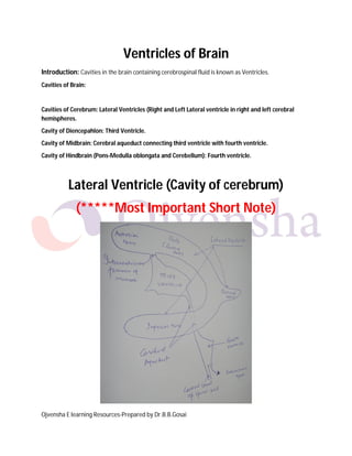
  • 1. Ojvensha E learning Resources-Prepared by Dr.B.B.Gosai Ventricles of Brain Introduction: Cavities in the brain containing cerebrospinal fluid is known as Ventricles. Cavities of Brain: Cavities of Cerebrum: Lateral Ventricles (Right and Left Lateral ventricle in right and left cerebral hemispheres. Cavity of Diencepahlon: Third Ventricle. Cavity of Midbrain: Cerebral aqueduct connecting third ventricle with fourth ventricle. Cavity of Hindbrain (Pons-Medulla oblongata and Cerebellum): Fourth ventricle. Lateral Ventricle (Cavity of cerebrum) (*****Most Important Short Note)
  • 2. Ojvensha E learning Resources-Prepared by Dr.B.B.Gosai FEATURES SEEN IN LATERAL VENTRICLE  Cavity of Telencephalon-cerebrum  Shape: “C” shaped  Parts with features seen in each part:  Anterior Horn: Located in frontal lobe.  Features and boundaries:  Floor contains head of caudate nucleus.  Anterior wall, floor and roof formed by corpus callosum.  Medial wall by septum pellucidum.  Lateral wall and floor is sloping and formed by head of caudate nucleus.  Central Part: Located in parietal lobe.  Features and boundaries:  Floor contain: Body of caudate nucleus and thalamus.  Roof by trunk of corpus callosum.
  • 3. Ojvensha E learning Resources-Prepared by Dr.B.B.Gosai  Medial wall by septum pellucidum.  Lateral wall and floor is sloping and formed by head of caudate nucleus.  Posterior horn: Located in occipital lobe. Small and inconstant.  Features: If this horn is present it shows following features:  Bulb of posterior horn raised by forceps major.  Calcar avis raised by calcarine sulcus is seen into it.  Inferior horn: Located in temporal lobe.  Features seen:  Features in the floor: Hippocampus and pes hippocampi seen its floor covered by alveus and fimbria.  Roof formed by tail of caudate nucleus and amygdaloid body.  Communication with third ventricle by interventricular foramen of Monroe  Function: Contain choroid plexus responsible for secretion of cerebrospinal fluid.  Applied Anatomy: Blockage of interventricular foramen leads to dilatation of lateral ventricle. This condition is known as Hydrocephalus. It leads to mental retardation. Third Ventricle (Cavity of Diencephalon) (***Important Short Note)  Cavity of diencephalon located between diencephalon i.e. between two thalami.  Shape: Slit like  Anterior wall: Lamina terminalis, anterior commissure and anterior column of fornix.  Posterior wall: Pineal gland with pineal recess and habenular and posterior commissure.  Lateral wall: Thalamus and Hypothalamus separated by hypothalamic sulcus  Superior wall (Roof): Ependyma, tela choroidea of third ventricle, internal cerebral veins, fornix and corpus callosum.  Inferior wall (Floor): Optic chiasma, tuber cinerium, infundibulum with its recess, mammilary bodies, tegmentum of cerebral peduncles of midbrain.
  • 4. Ojvensha E learning Resources-Prepared by Dr.B.B.Gosai  Recesses:  Pineal recess  Supraoptic recess  Infundibular recess  Communications:  Anterosuperiorly with lateral ventricle by interventricular foramen of Monroe.  Posteroinferiorly with fourth ventricle by cerebral aqueduct.  Function: Contain choroid plexus responsible for secretion of cerebrospinal fluid.  Applied Anatomy: Blockage of cerebral aqueduct leads to dilatation of third ventricle and both lateral ventricle. This condition is known as Hydrocephalus. It leads to mental retardation.
  • 5. Ojvensha E learning Resources-Prepared by Dr.B.B.Gosai Fourth Ventricle (Cavity of Hindbrain) (*****Most Important Short Note)  Cavity of hindbrain located between pons and medulla anteriorly and cerebellum posteriorly.  Shape: “Tent: shape in sagittal section.  Lateral Boundaries: Caudal part by inferior cerebellar peduncle and cranial part by superior cerebellar peduncle.  Posterior wall (Roof): tent shaped projects into cerebellum. Related to superior and nferior medullary velum. Contains Median aperture (Foramen of Megendie) anf Lateral openings (Foramen of Luschka).  Rhomboid Fossa (Floor of fourth ventricle): Diamond shaped. Formed by posterior surface of pons and medulla oblongata.  Boundaries: Superolaterlly by superior cerebellar peduncle and inferolaterally by Inferior cerebellar peduncle.  Features seen:
  • 6. Ojvensha E learning Resources-Prepared by Dr.B.B.Gosai  Hypoglossal and vagal triangles  facial colliculus  Vestibular area  Median sulcus  Medial eminence  Sulcus limitans  Recesses:  Dorsal median recess  Two lateral recesses with lateral aperture.  Communications:  Superiorly with third ventricle by cerebral  Inferiorly with central canal of spinal cord, and subarachnoid space by Median aperture of Megendie.
  • 7. Ojvensha E learning Resources-Prepared by Dr.B.B.Gosai  Laterally with subarachnoid space by lateral aperture of Lushka.  Function: Contain choroid plexus responsible for secretion of cerebrospinal fluid.  Applied Anatomy: Blockage of openings leads to dilatation of all ventricles. This condition is known as Hydrocephalus. It leads to mental retardation. Applied Anatomy of Ventricles:  HYDROCEPHALUS: Abnormal increase in the volume of cerebrospinal fluid within skull.  Types:  Non-communicating hydrocephalus: raised pressure is due to blockage between choroid plexus and exit foramina.  Communicating hydrocephalus: No obstruction. Causes can be excessive formation or diminished absorption.  INTRACRANIAL HYPERTENSION AND PAPILLEDEMA: congestion of retinal veins, bulging forward of optic disc and edema of the disc. Hydrocephalus (Photo for understanding dilatation of ventricles and thinning of brain tissue) ==================X================