Downloaded 347 times


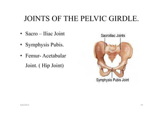
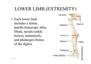


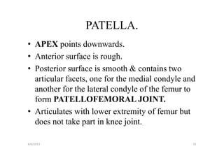


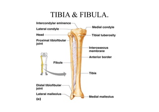
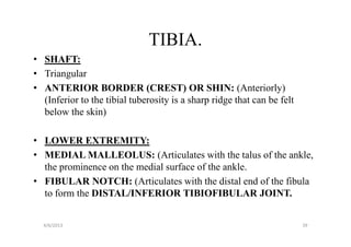


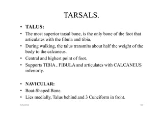


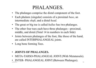


This document provides information about the bones of the pelvic girdle and lower limbs. It begins by listing the objectives, which are to identify the bones and their markings, describe the divisions of the pelvis and differences between male and female pelvis. It then lists the bones of the lower limbs and provides details about the pelvic girdle bones including the ilium, ischium, pubis, acetabulum, and gender differences in pelvis shape. It also describes some of the major lower limb bones like the femur, patella, tibia, and fibula as well as joints like the hip and sacroiliac joints.


![2 Osteology_of_the_lower_limb[1].1111111pptx](https://cdn.slidesharecdn.com/ss_thumbnails/2osteologyofthelowerlimb1-240303214000-c583864f-thumbnail.jpg?width=640&height=640&fit=bounds)


![Eye presentation [compatibility mode]](https://cdn.slidesharecdn.com/ss_thumbnails/eyepresentationcompatibilitymode-130123020537-phpapp02-thumbnail.jpg?width=640&height=640&fit=bounds)
![Nervous anatomy sem2 [compatibility mode]](https://cdn.slidesharecdn.com/ss_thumbnails/nervousanatomysem2compatibilitymode-130123021342-phpapp01-thumbnail.jpg?width=640&height=640&fit=bounds)
![Generic metabolism [compatibility mode]](https://cdn.slidesharecdn.com/ss_thumbnails/genericmetabolismcompatibilitymode-130123021245-phpapp01-thumbnail.jpg?width=640&height=640&fit=bounds)
