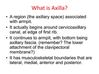
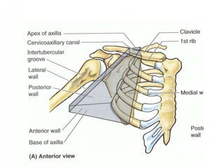
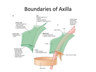
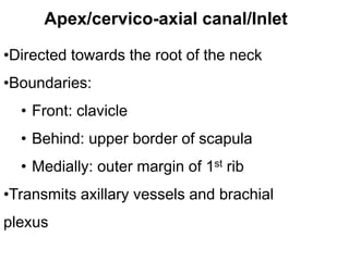
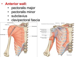
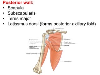
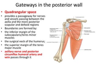
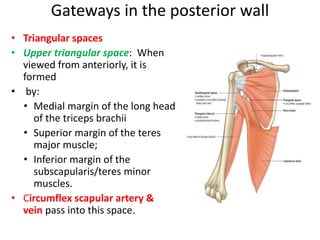
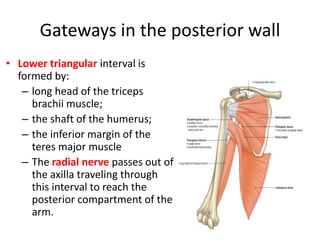
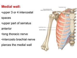
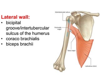
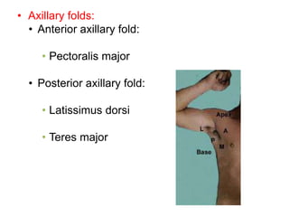
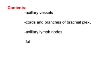
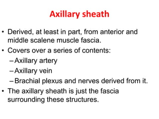
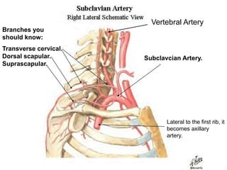
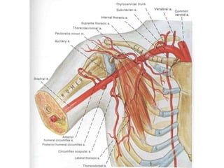
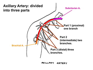
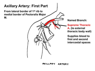
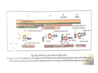
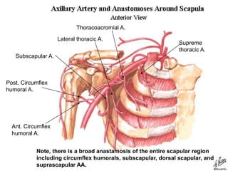
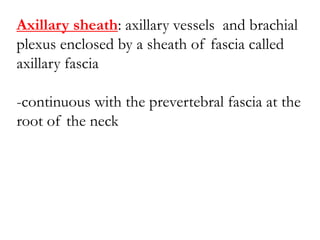
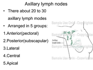
Dr. K. S. Ravi
M.B.B.S.,M.D.,MNAMS,FAIMER(M-FIILIPE),D.Litt.
Additional Professor
Department of Anatomy
AIIMS Rishikesh
1. Definition of Axilla
2. Boundaries
3. Cervico-Axillary Canal
4. Spaces/ Gateways in posterior wall
5. Axillary folds & Sheath
6. Contents of Axilla
7. Axillary artery- Parts & Branches
8. Axillary Vein & Lymph nodes
9. Applied Anatomy
Axilla (arm pit)
• Axilla is gateway to upper limb,
providing an area of transition
between neck & arm
• Between upper arm & thoracic
wall
• Boundaries
-Apex
-Base
-Anterior wall
-Posterior wall
-Medial wall
-Lateral wall
What is Axilla?
• A region (the axillary space) associated
with armpit.
• It actually begins around cervicoaxillary
canal, at edge of first rib.
• It continues to armpit, with bottom being
axillary fascia. (remember? The lower
attachment of the clavipectoral
membrane?)
• It has musculoskeletal boundaries that are
lateral, medial, anterior and posterior.
AXILLARY SPACE
Boundaries of Axilla
•Directed towards the root of the neck
•Boundaries:
• Front: clavicle
• Behind: upper border of scapula
• Medially: outer margin of 1st rib
•Transmits axillary vessels and brachial
plexus
Apex/cervico-axial canal/Inlet
• Anterior wall:
• pectoralis major
• pectoralis minor
• subclavius
• clavipectoral fascia
Posterior wall:
• Scapula
• Subscapularis
• Teres major
• Latissmus dorsi (forms posterior axillary fold)
Gateways in the posterior wall
• Quadrangular space
• provides a passageway for nerves
and vessels passing between the
axilla and the more posterior
scapular and deltoid regions
• Boundaries are formed by:
• the inferior margin of the
subscapularis/teres minor
muscles
• the surgical neck of the humerus;
• the superior margin of the teres
major muscle
• axillary nerve and posterior
circumflex humeral artery and
vein passes through it
Gateways in the posterior wall
• Triangular spaces
• Upper triangular space: When
viewed from anteriorly, it is
formed
• by:
• Medial margin of the long head
of the triceps brachii
• Superior margin of the teres
major muscle;
• Inferior margin of the
subscapularis/teres minor
muscles.
• Circumflex scapular artery &
vein pass into this space.
Gateways in the posterior wall
• Lower triangular interval is
formed by:
– long head of the triceps
brachii muscle;
– the shaft of the humerus;
– the inferior margin of the
teres major muscle
– The radial nerve passes out of
the axilla traveling through
this interval to reach the
posterior compartment of the
arm.
Medial wall:
•upper 3 or 4 intercostal
spaces
•upper part of serratus
anterior
•long thoracic nerve
•intercosto brachial nerve
pierces the medial wall
Lateral wall:
• bicipital
groove/intertubercular
sulcus of the humerus
• coraco brachialis
• biceps brachii
Base:
-skin
-superficial fascia
-axillary fascia (deep fascia)
• Axillary folds:
• Anterior axillary fold:
• Pectoralis major
• Posterior axillary fold:
• Latissimus dorsi
• Teres major
Contents:
-axillary vessels
-cords and branches of brachial plexu
-axillary lymph nodes
-fat
Axillary sheath
• Derived, at least in part, from anterior and
middle scalene muscle fascia.
• Covers over a series of contents:
–Axillary artery
–Axillary vein
–Brachial plexus and nerves derived from it.
• The axillary sheath is just the fascia
surrounding these structures.
Subclavcian Artery.
Lateral to the first rib, it
becomes axillary
artery.
Branches you
should know:
Transverse cervical.
Dorsal scapular.
Suprascapular.
Vertebral Artery
Axillary Artery: divided
into three parts
Part 1 (proximal)
one branch
Part 2
(intermediate) two
branches.
Part 3 (distal) three
branches.
Subclavian A.
Brachial A.
Axillary Artery: First Part
From lateral border of 1st rib to
medial border of Pectoralis Major
M.
Named Branch:
Supreme Thoracic
A. (to external
thoracic body wall)
Supplies blood to
first and second
intercostal spaces
Axillary Artery: Second part
Deep to the pectoralis minor M.
Thoracoacromial trunk
Branches to:
Clavicular area
Pectoralis region
Acromion of Scapula
Deltoid Muscle.
Lateral Thoracic Artery
Bbr. to Serratus Ant. M.
Axillary Artery: third part
Lateral border of Pectoralis minor M. to
lateral border of Teres major M.
Subscapular A.:
Branches:
Circumflex scapular
A. (to multiple
muscles associated
with the scapula)
1.
Thoracodorsal A.
(to Latissimus
dorsi M.)
2.
Posterior circumflex
humeral A.
Anterior circumflex
humeral A.
How it will look in lab
Supreme
thoracic A.
Thoracoacromial A.
Lateral thoracic A.
Subscapular A.
Ant. Circumflex
humoral A.
Post. Circumflex
humoral A.
Note, there is a broad anastamosis of the entire scapular region
including circumflex humorals, subscapular, dorsal scapular, and
suprascapular AA.
Axillary vein:
Formation:
Continuation of basilic vein at
the lower border of teres major
- runs upwards on the medial
side of the axillary artery
-Ends at outer border of 1st rib
by becoming the subclavian vein
Tributaries:
-cephalic vein
-veins corresponding to the
branches of axillary artery
Vena comitans
•Frequently multiple(2 or3)
•Run with their corresponding
Arteries
Axillary sheath: axillary vessels and brachial
plexus enclosed by a sheath of fascia called
axillary fascia
-continuous with the prevertebral fascia at the
root of the neck
Axillary lymph nodes
• There about 20 to 30
axillary lymph modes
• Arranged in 5 groups:
1.Anterior(pectoral)
2.Posterior(subscapular)
3.Lateral
4.Central
5.Apical
Regional Lymph Nodes for Breast
A: Pectoralis major muscle
B: Axillary lymph nodes level I
Ant,, Post & Lat
(Below & Lateral)
C: Axillary lymph nodes level II
Central- Behind, I
nterpectoral- In front
D: Axillary lymph nodes level III
Apical- Above
E: Supraclavicular lymph nodes
F: Internal mammary lymph nodes
??
1. What are the structures that make up
anterior wall of the axilla?
a.Pectoralis major, clavipectoral fascia and
pectoralis minor
b.Subscapularis, teres major and latissimus dorsi
c. Pectoralis major, clavipectoral fascia and
latissimus dorsi
d.Subscapularis, teres major and pectoralis
minor
2. What are the structures that make up the
posterior wall of the axilla?
a.Pectoralis major, clavipectoral fascia and
pectoralis minor
b.Subscapularis, teres major and latissimus dorsi
c. Pectoralis major, clavipectoral fascia and
latissimus dorsi
d.Subscapularis, teres major and pectoralis
minor
3. The anterior group of axillary lymph nodes
lies along the:
a.Below & lateral to lower margin of pectoralis
minor
b.Lower margin of the posterior wall
c. Posteromedial to the axillary vein
d.In the fat of axilla
e.Behind and above the pectoralis minor, medial
to the axillary vein
4. The apical group of axillary lymph nodes lies
along the:
a.Lower border of the pectoralis minor
b.Lower margin of the posterior wall
c. Posteromedial to the axillary vein
d.In the fat of axilla
e.above the upper margin of pectoralis minor,
medial to the axillary vein
5. Which of the following branches of the
axillary artery supplies the pectoral muscles
and the thoracic wall?
a.Anterior circumflex humeral artery
b.Lateral thoracic artery
c. Posterior circumflex humeral artery
d.Superior thoracic artery
6. Which of the following branches of the
axillary artery supplies the head of the
humerus and the shoulder joint?
a.Anterior circumflex humeral artery
b.Lateral thoracic artery
c. Posterior circumflex humeral artery
d.Superior thoracic artery
Axilla - Dr.K.S.Ravi

Axilla - Dr.K.S.Ravi

  • 1.
    Dr. K. S.Ravi M.B.B.S.,M.D.,MNAMS,FAIMER(M-FIILIPE),D.Litt. Additional Professor Department of Anatomy AIIMS Rishikesh
  • 2.
    1. Definition ofAxilla 2. Boundaries 3. Cervico-Axillary Canal 4. Spaces/ Gateways in posterior wall 5. Axillary folds & Sheath 6. Contents of Axilla 7. Axillary artery- Parts & Branches 8. Axillary Vein & Lymph nodes 9. Applied Anatomy
  • 3.
    Axilla (arm pit) •Axilla is gateway to upper limb, providing an area of transition between neck & arm • Between upper arm & thoracic wall • Boundaries -Apex -Base -Anterior wall -Posterior wall -Medial wall -Lateral wall
  • 4.
    What is Axilla? •A region (the axillary space) associated with armpit. • It actually begins around cervicoaxillary canal, at edge of first rib. • It continues to armpit, with bottom being axillary fascia. (remember? The lower attachment of the clavipectoral membrane?) • It has musculoskeletal boundaries that are lateral, medial, anterior and posterior.
  • 5.
  • 6.
  • 7.
    •Directed towards theroot of the neck •Boundaries: • Front: clavicle • Behind: upper border of scapula • Medially: outer margin of 1st rib •Transmits axillary vessels and brachial plexus Apex/cervico-axial canal/Inlet
  • 8.
    • Anterior wall: •pectoralis major • pectoralis minor • subclavius • clavipectoral fascia
  • 9.
    Posterior wall: • Scapula •Subscapularis • Teres major • Latissmus dorsi (forms posterior axillary fold)
  • 10.
    Gateways in theposterior wall • Quadrangular space • provides a passageway for nerves and vessels passing between the axilla and the more posterior scapular and deltoid regions • Boundaries are formed by: • the inferior margin of the subscapularis/teres minor muscles • the surgical neck of the humerus; • the superior margin of the teres major muscle • axillary nerve and posterior circumflex humeral artery and vein passes through it
  • 11.
    Gateways in theposterior wall • Triangular spaces • Upper triangular space: When viewed from anteriorly, it is formed • by: • Medial margin of the long head of the triceps brachii • Superior margin of the teres major muscle; • Inferior margin of the subscapularis/teres minor muscles. • Circumflex scapular artery & vein pass into this space.
  • 12.
    Gateways in theposterior wall • Lower triangular interval is formed by: – long head of the triceps brachii muscle; – the shaft of the humerus; – the inferior margin of the teres major muscle – The radial nerve passes out of the axilla traveling through this interval to reach the posterior compartment of the arm.
  • 13.
    Medial wall: •upper 3or 4 intercostal spaces •upper part of serratus anterior •long thoracic nerve •intercosto brachial nerve pierces the medial wall
  • 14.
    Lateral wall: • bicipital groove/intertubercular sulcusof the humerus • coraco brachialis • biceps brachii
  • 15.
  • 16.
    • Axillary folds: •Anterior axillary fold: • Pectoralis major • Posterior axillary fold: • Latissimus dorsi • Teres major
  • 17.
    Contents: -axillary vessels -cords andbranches of brachial plexu -axillary lymph nodes -fat
  • 18.
    Axillary sheath • Derived,at least in part, from anterior and middle scalene muscle fascia. • Covers over a series of contents: –Axillary artery –Axillary vein –Brachial plexus and nerves derived from it. • The axillary sheath is just the fascia surrounding these structures.
  • 19.
    Subclavcian Artery. Lateral tothe first rib, it becomes axillary artery. Branches you should know: Transverse cervical. Dorsal scapular. Suprascapular. Vertebral Artery
  • 21.
    Axillary Artery: divided intothree parts Part 1 (proximal) one branch Part 2 (intermediate) two branches. Part 3 (distal) three branches. Subclavian A. Brachial A.
  • 22.
    Axillary Artery: FirstPart From lateral border of 1st rib to medial border of Pectoralis Major M. Named Branch: Supreme Thoracic A. (to external thoracic body wall) Supplies blood to first and second intercostal spaces
  • 23.
    Axillary Artery: Secondpart Deep to the pectoralis minor M. Thoracoacromial trunk Branches to: Clavicular area Pectoralis region Acromion of Scapula Deltoid Muscle. Lateral Thoracic Artery Bbr. to Serratus Ant. M.
  • 24.
    Axillary Artery: thirdpart Lateral border of Pectoralis minor M. to lateral border of Teres major M. Subscapular A.: Branches: Circumflex scapular A. (to multiple muscles associated with the scapula) 1. Thoracodorsal A. (to Latissimus dorsi M.) 2. Posterior circumflex humeral A. Anterior circumflex humeral A. How it will look in lab
  • 26.
    Supreme thoracic A. Thoracoacromial A. Lateralthoracic A. Subscapular A. Ant. Circumflex humoral A. Post. Circumflex humoral A. Note, there is a broad anastamosis of the entire scapular region including circumflex humorals, subscapular, dorsal scapular, and suprascapular AA.
  • 27.
    Axillary vein: Formation: Continuation ofbasilic vein at the lower border of teres major - runs upwards on the medial side of the axillary artery -Ends at outer border of 1st rib by becoming the subclavian vein Tributaries: -cephalic vein -veins corresponding to the branches of axillary artery
  • 28.
    Vena comitans •Frequently multiple(2or3) •Run with their corresponding Arteries
  • 29.
    Axillary sheath: axillaryvessels and brachial plexus enclosed by a sheath of fascia called axillary fascia -continuous with the prevertebral fascia at the root of the neck
  • 30.
    Axillary lymph nodes •There about 20 to 30 axillary lymph modes • Arranged in 5 groups: 1.Anterior(pectoral) 2.Posterior(subscapular) 3.Lateral 4.Central 5.Apical
  • 31.
    Regional Lymph Nodesfor Breast A: Pectoralis major muscle B: Axillary lymph nodes level I Ant,, Post & Lat (Below & Lateral) C: Axillary lymph nodes level II Central- Behind, I nterpectoral- In front D: Axillary lymph nodes level III Apical- Above E: Supraclavicular lymph nodes F: Internal mammary lymph nodes
  • 32.
    ?? 1. What arethe structures that make up anterior wall of the axilla? a.Pectoralis major, clavipectoral fascia and pectoralis minor b.Subscapularis, teres major and latissimus dorsi c. Pectoralis major, clavipectoral fascia and latissimus dorsi d.Subscapularis, teres major and pectoralis minor
  • 33.
    2. What arethe structures that make up the posterior wall of the axilla? a.Pectoralis major, clavipectoral fascia and pectoralis minor b.Subscapularis, teres major and latissimus dorsi c. Pectoralis major, clavipectoral fascia and latissimus dorsi d.Subscapularis, teres major and pectoralis minor
  • 34.
    3. The anteriorgroup of axillary lymph nodes lies along the: a.Below & lateral to lower margin of pectoralis minor b.Lower margin of the posterior wall c. Posteromedial to the axillary vein d.In the fat of axilla e.Behind and above the pectoralis minor, medial to the axillary vein
  • 35.
    4. The apicalgroup of axillary lymph nodes lies along the: a.Lower border of the pectoralis minor b.Lower margin of the posterior wall c. Posteromedial to the axillary vein d.In the fat of axilla e.above the upper margin of pectoralis minor, medial to the axillary vein
  • 36.
    5. Which ofthe following branches of the axillary artery supplies the pectoral muscles and the thoracic wall? a.Anterior circumflex humeral artery b.Lateral thoracic artery c. Posterior circumflex humeral artery d.Superior thoracic artery
  • 37.
    6. Which ofthe following branches of the axillary artery supplies the head of the humerus and the shoulder joint? a.Anterior circumflex humeral artery b.Lateral thoracic artery c. Posterior circumflex humeral artery d.Superior thoracic artery